. 2013 Jul 1:4:221.
doi: 10.3389/fpls.2013.00221. eCollection 2013.

Intersection of transfer cells with phloem biology-broad evolutionary trends, function, and induction

Affiliations

Intersection of transfer cells with phloem biology-broad evolutionary trends, function, and induction

Felicity A Andriunas et al. Front Plant Sci. .

Abstract

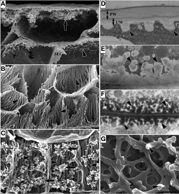
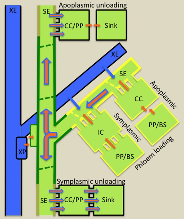
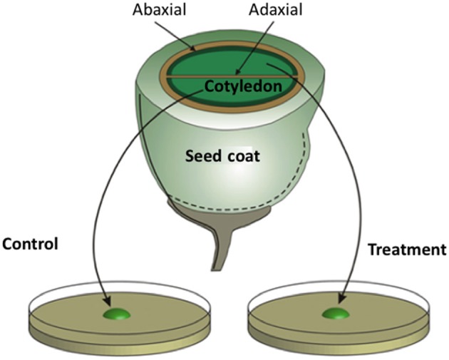
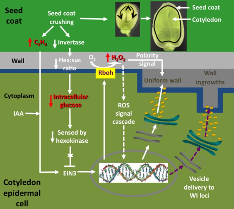
Transfer cells (TCs) are ubiquitous throughout the plant kingdom. Their unique ingrowth wall labyrinths, supporting a plasma membrane enriched in transporter proteins, provides these cells with an enhanced membrane transport capacity for resources. In certain plant species, TCs have been shown to function to facilitate phloem loading and/or unloading at cellular sites of intense resource exchange between symplasmic/apoplasmic compartments. Within the phloem, the key cellular locations of TCs are leaf minor veins of collection phloem and stem nodes of transport phloem. In these locations, companion and phloem parenchyma cells trans-differentiate to a TC morphology consistent with facilitating loading and re-distribution of resources, respectively. At a species level, occurrence of TCs is significantly higher in transport than in collection phloem. TCs are absent from release phloem, but occur within post-sieve element unloading pathways and particularly at interfaces between generations of developing Angiosperm seeds. Experimental accessibility of seed TCs has provided opportunities to investigate their inductive signaling, regulation of ingrowth wall formation and membrane transport function. This review uses this information base to explore current knowledge of phloem transport function and inductive signaling for phloem-associated TCs. The functional role of collection phloem and seed TCs is supported by definitive evidence, but no such information is available for stem node TCs that present an almost intractable experimental challenge. There is an emerging understanding of inductive signals and signaling pathways responsible for initiating trans-differentiation to a TC morphology in developing seeds. However, scant information is available to comment on a potential role for inductive signals (auxin, ethylene and reactive oxygen species) that induce seed TCs, in regulating induction of phloem-associated TCs. Biotic phloem invaders have been used as a model to speculate on involvement of these signals.

Keywords: inductive signals; ingrowth wall architecture; phloem transport; transfer cell.


Figures

Figure 1
Figure 1
Images of transfer cells of developing seeds during their storage phase illustrating ingrowth wall morphologies. (A–C) Scanning (A,B) and field emission scanning (C) electron microscope images of cells following freeze-fracture, removal of their cytoplasm and fixation [for method see Talbot et al. (2002)]. (A) Epidermal transfer cells (ETC) of a Vicia faba cotyledon with an extensive reticulate ingrowth wall labyrinth (arrow) polarized to the outer periclinal wall. Ingrowth wall deposition (dart) is restricted to wall portions abutting intercellular spaces adjacent to the sub-epidermal cells (SEC) [modified after Talbot et al. (2001)]. (B) Basal endosperm transfer cells of Zea mays exhibiting flange wall ingrowth morphology [modified after Talbot et al. (2002)]. The wall ingrowth ribs (darts) extend the length of each cell and are more extensive at their outer periclinal walls. (C) Thin-walled parenchyma transfer cells located at the inner surface of the inner seed coat of Gossypium hirsutum with wall ingrowth flanges (darts) extending the length of each cell on which are deposited groups of reticulate wall ingrowths (arrows) [modified after Pugh et al. (2010)]. (D–F) Transmission electron microscope images of portions of transverse sections of transfer cells: (D) The outer periclinal wall of an adaxial epidermal cell of a V. faba cotyledon induced to trans-differentiate to a transfer cell morphology. A uniform wall (UW), distinguishable from the original primary wall (PW) by a different electron opaqueness, is deposited against the primary wall and small papillate wall ingrowths (darts) arise from it. (E) Small papillate ingrowths (darts) of a seed coat transfer cell of V. faba exhibiting reticulate architecture. (F) Antler-shaped reticulate wall ingrowths (darts) of a nucellar projection transfer cell of a developing Triticum turgidum var. durum seed [modified after Wang et al. (1994)]. (G) Field emission scanning electron microscope image of the cytoplasmic face of the reticulate ingrowth wall labyrinth of an abaxial epidermal transfer cell of a V. faba cotyledon following removal of the cytoplasm and dry cleaving [for method see Talbot et al. (2001), image modified after Talbot et al. (2001)]. Note the multi-layered fenestrated sheets of wall material (numbered) and the small wall ingrowth papillae arising from the most recently deposited layer (darts). Single scale bar for (A,B) = 2.5 μm; for (C) = 5 μm; for (D,E) = 1 μm; for (F) = 0.25 μm; for (G) = 0.5 μm.
Figure 2
Figure 2
Diagrammatic illustration of cellular pathways of water (blue arrows) and photoassimilate (brown arrows) loading into collection phloem (yellow border) in source leaves, of mineral nutrient exchange from xylem (blue) to transport phloem (green border) at nodes and of resource (water, mineral nutrients and photoassimilates) unloading from release phloem (kaki borders) in sinks. Phloem loading mechanisms are defined according to cellular routes followed by photoassimilates moving from phloem parenchyma (PP) or bundle sheath (BS) cells to intermediary cells (IC; modified companion cells) or companion cells (CC) of collection phloem. Photoassimilate movement may occur from PP/BS cells through interconnecting plasmodesmata (symplasmic loading) to ICs or, in the absence of an adequate plasmodesmal connectivity, photoassimilates are effluxed from PP or BS cells into the cell wall matrix from which they are loaded into adjacent CCs (apoplasmic loading). Thereafter, for either loading mechanism, photoassimilates (primarily sugars) move symplasmically into adjacent sieve elements (SEs) of collection phloem. Accumulation of sugars to high concentrations in SEs causes an osmotic uptake of water from adjacent xylem elements (XE and see blue arrows) to generate large hydrostatic pressures (1–2 MPa). These drive a bulk flow of resources (brown superimposed on blue arrows) through the sieve tube system from source leaves to sinks. At nodes, mineral nutrients are extracted from XEs by xylem parenchyma (XP) cells and retrieved from the nodal apoplasm into transport phloem SEs. Resource exit from SEs of release phloem in sinks commonly follows a symplasmic route. Thereafter, further transport through the post-SE unloading pathway can continue through interconnecting plasmodesmata (symplasmic unloading) or, in certain sink types, a symplasmic discontinuity diverts resource flow through the intervening apoplasm (apoplasmic unloading—e.g., developing seeds).
Figure 3
Figure 3
Schematic of a transfer cell inductive system using Vicia faba cotyledons. An equatorial view of a V. faba seed sliced transversely in half. A maternal seed coat encloses two large cotyledons. At the maternal/filial interface, thin-walled parenchyma cells of the seed coat and abaxial epidermal cells of the cotyledons (solid dark green lines) trans-differentiate to a transfer cell morphology. In contrast, adaxial epidermal cells of the cotyledons do not trans-differentiate to a transfer cell morphology. Upon surgical removal of cotyledons from their enclosing seed coats and transfer to a MS medium, cotyledon adaxial epidermal cells undergo a synchronous trans-differentiation to transfer cells that are structurally and functionally identical to their abaxial counterparts. Sister cotyledons of each seed are randomly allocated to MS medium alone or a MS medium carrying a pharmacological agent that interferes with developmental signals or wall building machinery.
Figure 4
Figure 4
Interrelationships between signals and signal cascades leading to induction, organization, and construction of polarized ingrowth walls in epidermal transfer cells of Vicia faba cotyledons. In cultured cotyledons, a combination of declining intracellular glucose levels and an auxin (IAA) regulated burst in ethylene production, initiates ingrowth wall formation within adaxial epidermal cells. The glucose/ethylene sequence is reproduced in planta and downstream events are deduced from findings obtained using cultured cotyledons. Prior to the onset of cotyledon growth, an extracellular invertase, localized to inner cells of the seed coat (dark green), hydrolyzes sucrose and the glucose product, sensed by a cotyledon epidermal cell (light green) hexokinase, blocks an ethylene signal cascade by down regulating ethylene insensitive 3 (EIN3), a key ethylene signal cascade transcription factor. Upon initiating expansion growth, the cotyledons crush inner cells of the seed coat (see images of seeds cut through their longitudinal plan illustrating cotyledon growth at two stages). Cell crushing results in a loss of extracellular invertase activity and hence the glucose signal. This is accompanied by an amplified wound-induced ethylene (C2H4) signal cascade, mediated through EIN3, driving expression of respiratory burst oxidases (Rbohs), trafficked to portions of the plasma membrane (blue) that line the outer periclinal walls of cotyledon epidermal cells. The Rbohs catalyze production of extracellular reactive oxygen species (ROS) that are further reduced to form an extracellular hydrogen peroxide (H2O2) signal that serves two known functions. These are activating expression of cell wall building machinery through an unknown signal cascade (arrows with broken shafts) and serving as a positional signal directing construction of the uniform wall (dark gray) polarized to the outer periclinal walls (light gray) of the cotyledon epidermal cells. How the polarized wall construction subsequently is constrained to loci from which wall ingrowth papillae arise remains to be determined (purple arrows with broken shafts) but likely involves directional control by cytosolic calcium signals.
Figure 5
Figure 5
Speculative model illustrating reactive oxygen species (ROS) mediated regulatory mechanisms that may account for observed patterns of inter- and intracellular formation of ingrowth walls (dark gray) in collection phloem companion cells (CC) and phloem parenchyma cells (PP) undergoing trans-differentiation to a transfer cell (TC) morphology. In all cases, developing sieve elements (SEs), undergoing partial programmed cell death, generate an intracellular ROS signal. (A) CC/TC trans-differentiation: SE-produced intracellular ROS (red dots) diffuses (thick red arrows) into CC, through interconnecting plasmodesmata, to induce formation of a non-polarized ingrowth wall covering the entire CC wall. (B) CC/TC and PP/TC trans-differentiation: an intracellular ROS signal induces ingrowth wall formation as described in (A). In addition, intracellular ROS, released from developing SEs through plasma membrane aquaporins abutting adjacent PP (thin red arrows) into the shared SE/PP cell wall space, elicits a polarized formation of ingrowth walls proximal to SEs. (C) PP/TC trans-differentiation: A transient and excessive intracellular generation of ROS by developing SEs gates plasmodesmata interconnecting CCs closed (filled in) thus preventing transmission of the ROS signal (curved red arrows) across the developmental period in which CCs are otherwise ROS-responsive. Ingrowth wall formation in PPs occurs as described in (B).

References

    1. Aloni R. (2010). The induction of vascular tissues by auxin in ‘Plant Hormones. Biosynthesis, Signal Transduction, Action! ed Davies P. J. (Amsterdam: Springer; ), 485–518
    1. Aloni R., Schwalm K., Langhans M., Ullrich C. L. (2003). Gradual shifts in sites of free-auxin production during leaf-primordium development and their role in vascular differentiation and leaf morphogenesis in Arabidopsis. Planta 216, 841–853 - PubMed
    1. Amiard V., Mueh K., Demming-Adams B., Ebbert V., Turgeon R. (2005). Anatomical and photosynthetic acclimation to the light environment in species with differing mechanisms of phloem loading. Proc. Natl. Acad. Sci. U.S.A. 102, 12968–12973 - PMC - PubMed
    1. Amiard V., Mueh K., Demming-Adams B., Mueh K. E., Turgeon R., Combs A. F., et al. (2007). Role of light and jasmonic acid signaling in regulating foliar phloem cell wall ingrowth development. New Phytol. 173, 722–731 10.1111/j.1469-8137.2006.01954.x - DOI - PubMed
    1. Andriunas F. A., Zhang H. M., Weber H., McCurdy D. W., Offler C. E., Patrick J. W. (2011). Glucose and ethylene signalling pathways converge to regulate trans-differentiation of epidermal transfer cells in Vicia narbonensis cotyledons. Plant J. 68, 987–998 10.1111/j.1365-313X.2011.04749.x - DOI - PubMed

LinkOut - more resources