

A case of traumatic bilateral abducens and unilateral hypoglossal nerve palsy
- PMID: 23847710
- PMCID: PMC3702690
- DOI: 10.12659/AJCR.889065
A case of traumatic bilateral abducens and unilateral hypoglossal nerve palsy
Abstract
Patient: Female, 47 FINAL DIAGNOSIS: Traumatic bilateral abducens • unilateral hypoglossal nerve palsy
Symptoms: Diplopia Medication: - Clinical Procedure: - Specialty: Neurology Objective: Rare disease.
Background: Incidence of unilateral abducens palsy from head trauma has been reported to be as high as 1% to 2.7%, but bilateral abducens nerve palsy is extremely rare.
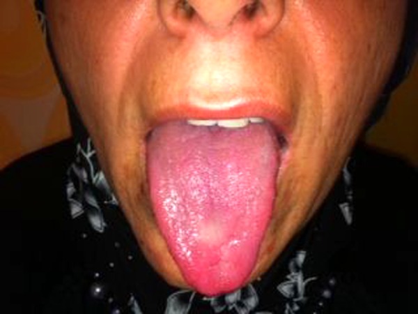
Case report: We present a case in which bilateral abducens nerve and unilateral hypoglossal nerve palsy developed with a high Glasgow Coma Score (GCS) 3 hours after head trauma due to a motor vehicle crash.
Conclusions: This case highlights the occurrence and management of posttraumatic bilateral sixth nerve palsy.
Keywords: abducens nerve injury; hypoglossal nerve injury; trauma.
Figures


Similar articles
-
Delayed bilateral abducens nerve palsy after head trauma.J Korean Neurosurg Soc. 2008 Dec;44(6):396-8. doi: 10.3340/jkns.2008.44.6.396. Epub 2008 Dec 31. J Korean Neurosurg Soc. 2008. PMID: 19137087 Free PMC article.
-
Bilateral Abducens Nerve Palsy after Closed Head Trauma without Acute Intracranial Pathology.J Emerg Trauma Shock. 2019 Jul-Sep;12(3):222-224. doi: 10.4103/JETS.JETS_98_18. J Emerg Trauma Shock. 2019. PMID: 31543647 Free PMC article.
-
Transient Bilateral Sixth Nerve Palsy: A Rare Sequela of Head Trauma.Cureus. 2021 Jan 20;13(1):e12805. doi: 10.7759/cureus.12805. Cureus. 2021. PMID: 33628673 Free PMC article.
-
Bilateral traumatic abducens nerve avulsion: A case series and literature review.J Clin Neurosci. 2017 Oct;44:30-33. doi: 10.1016/j.jocn.2017.06.023. Epub 2017 Jun 30. J Clin Neurosci. 2017. PMID: 28673673 Review.
-
[A case of crushing head injury showing bilateral abducens, facial and acoustic nerve palsies].No Shinkei Geka. 1988 Jun;16(7):869-73. No Shinkei Geka. 1988. PMID: 3065670 Review. Japanese.
Cited by
-
Post-traumatic Unilateral Avulsion of the Abducens Nerve with Damage to Cranial Nerves VII and VIII: Case Report.NMC Case Rep J. 2016 May 16;3(3):81-83. doi: 10.2176/nmccrj.cr.2015-0272. eCollection 2016 Jul. NMC Case Rep J. 2016. PMID: 28664004 Free PMC article.
-
Bilateral Abducent Nerve Palsy After Neck Trauma: A Case Report.Trauma Mon. 2016 Feb 6;21(1):e31984. doi: 10.5812/traumamon.31984. eCollection 2016 Feb. Trauma Mon. 2016. PMID: 27218062 Free PMC article.
References
-
- Katsuno M, Yokota H, Yamamoto Y, Teramoto A. Bilateral traumatic abducens nerve palsy associated with skull base fracture – case report. Neurol Med Chir (Tokyo) 2007;47(7):307–9. - PubMed
-
- Yanamadala V, Walcott BP, Nahed BV, Coumans JV. Delayed post-traumatic bilateral abducens nerve palsy with complete recovery. J Clin Neurosci. 2012;19(4):585–86. - PubMed
-
- Ayberk G, Ozveren MF, Yildirim T, et al. Review of a series with abducens nerve palsy. Turk Neurosurg. 2008;18(4):366–73. - PubMed
-
- Ozveren MF, Uchida K, Aiso S, Kawase T. Meningovenous structures of the petroclival region: clinical importance for surgery and intravascular surgery. Neurosurgery. 2002;50(4):829–36. discussion 836–37. - PubMed
-
- Dwarakanath S, Ravichandra Gopal S, Venkataramana NK. Post-traumatic bilateral abducens nerve palsy. Neurol India. 2006;54(2):221–22. - PubMed
LinkOut - more resources
Full Text Sources
Other Literature Sources