. 2013 Jul 9:3:180.
doi: 10.3389/fonc.2013.00180. eCollection 2013.

Detection of Human Endogenous Retrovirus K (HERV-K) Transcripts in Human Prostate Cancer Cell Lines

Affiliations

Detection of Human Endogenous Retrovirus K (HERV-K) Transcripts in Human Prostate Cancer Cell Lines

Lorenzo Agoni et al. Front Oncol. .

Abstract

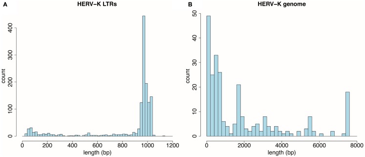
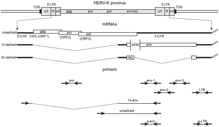
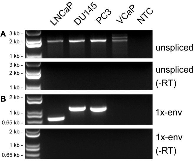
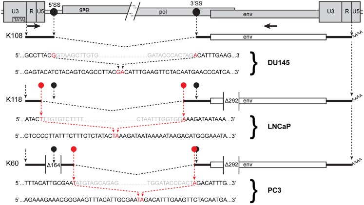
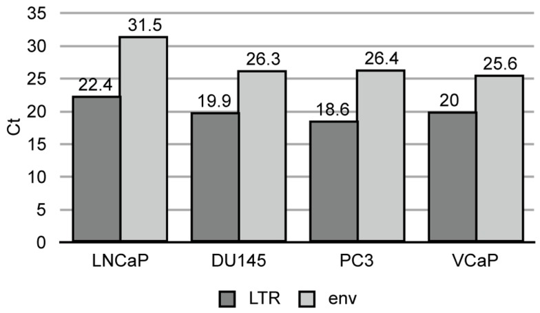
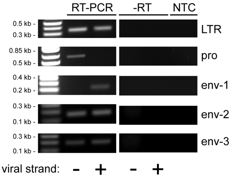
Human endogenous retroviruses (HERVs) are transcribed in many cancers including prostate cancer. Human endogenous retrovirus K (HERV-K) of the HML2 subtype is the most recently integrated and most intact retrovirus in the human genome, with many of the viral genomes encoding full- or partial-length viral proteins. To assess transcripts of HERV-K in prostate cancer cell lines and identify the specific HERV-K elements in the human genome that are transcribed, reverse transcriptase-PCR (RT-PCR) and cDNA sequencing were undertaken. Strand-specific RT-PCR, plasmid subcloning, and cDNA sequencing detected the presence of HERV-K(HML2) coding strand transcripts within four prostate cell lines (LNCaP, DU145, PC3, and VCaP). RT-PCR across splice junctions revealed splicing variants for env gene mRNA in three cell lines, two involving previously undescribed alternative splice sites. To determine the HERV-K loci from which the transcripts arose, RepeatMasker was used to compile a list of over 200 HERV-K internal genome segment fragments and over 1,000 HERV-K solo long terminal repeat (LTR) fragments in the human genome. Surprisingly, the sequences identified from internal positions of the viral genome were mostly smaller segments, while the LTRs were relatively intact. Possible reasons for this are discussed. The transcripts in the cell lines tested, arose from several HERV-K loci, with some proviruses being detected in multiple cell lines and others in only one of the four used. In some instances, transcripts from viral antisense strands was also detected. In addition, transcripts from both strands of solo LTRs were detected. These data show that transcripts from HERV-K loci commonly occur in prostate cancer cell lines and that transcription of either strand can occur. They also emphasize the importance of single nucleotide level analysis to identify the specific, individual HERV-K loci that are transcribed, and indicate that HERV-K expression in prostate cancer warrants further study.

Keywords: HERV-K; RT-PCR; cancer; endogenous retroviruses; evolution; prostate cancer; unconventional splicing.


Figures

Figure 1
Figure 1
Distribution by size of HERV-K components detected by RepeatMasker in the UCSC Genome Browser, GRCh37/hg19 human genome assembly. (A) Thousand three hundred and sixty-one HERV-K LTR fragments are plotted as function of 20 bp length windows. (B) Two hundred fifty-five HERV-K provirus internal genome segments are plotted as a function of 200 bp length windows.
Figure 2
Figure 2
Structure of the HERV-K genome and spliced mRNAs showing the primers used for reverse transcription and PCR. A genetic map of a HERV-K provirus (darker gray) inserted into host genome (lighter gray) is shown with target duplicated sequence (TDS) indicated as black boxes. The unspliced primary viral transcript, 1×-spliced env mRNA and 2×-spliced rec mRNAs are shown below the viral genome with 3′ poly(A) tails indicated (AAAA). The env mRNA shows the position of the 292-nt deletion (Δ292) of type I HERV-K proviruses that spans the pol-env junction. Type II HERV-K provirus transcripts contain these nucleotides. Positions of the PCR primer pairs are shown at the bottom with the names by which they are identified throughout the paper. The dashed, angled line shows the excised intronic sequences that the 1×-env primer pair was designed to cross.
Figure 3
Figure 3
Detection of HERV-K transcripts in four prostate cancer cell lines. RT-PCR was performed to detect viral sense strand transcripts specifically, and the products were resolved by electrophoresis. (A) RT-PCR products from a primer pair designed to detect unspliced viral RNAs (unspliced). (B) RT-PCR products from primers designed to detect RNAs spliced at the conventional env mRNA splice junction (1×-env). Genomic positions of the primers are shown in Figure 2. Parallel controls were performed without reverse transcriptase (−RT) and with no added template (NTC, no template control) as controls to exclude DNA contamination. DNA size markers are shown on the left.
Figure 4
Figure 4
Human endogenous retrovirus K RNA splice sites determined by sequencing of RT-PCR products. The 5′ and 3′ portions of a HERV-K provirus are shown at the top separated by / and /. 5′SS and 3′SS indicate the conventional splicing sites of HERV-K, and their positions are marked as black circles. The positions of the outer PCR primers used for the nested PCR are shown as arrows. Structures of the env spliced RT-PCR products from the cell lines and proviruses indicated are diagrammed below the viral genome. Dashed angled lines show the excised introns. Red circles show the positions of the sequences where unconventional splicing occurred. For each spliced product, the top sequence shows the inferred primary transcript sequence determined from that of the cognate genomic locus, and the bottom sequence shows the sequence of the RT-PCR product. Red nucleotides were joined in the spliced product. Gray nucleotides show the ends of the excised sequences. The positions of the 292-nt deletion definitive of type I proviruses and a 164-nt deletion in the K60 provirus relative to other HERV-K proviruses are shown.
Figure 5
Figure 5
Quantitative RT-PCR of HERV-K LTR and env sequences in prostate cancer cell lines. Threshold cycle is shown for the viral components in each cell line.
Figure 6
Figure 6
Reverse transcriptase-PCR analysis of HERV-K transcripts in LNCaP cells. Strand-specific, RT-PCR was performed on RNA isolated from LNCaP cells for five different positions in the HERV-K genome using the primer pairs identified in Figure 2, and the products were resolved by electrophoresis. Viral strand indicates RT-PCR products that originated from viral sense (+) or antisense (−) RNAs. Parallel controls were performed without reverse transcriptase (−RT) and without adding any RNA template (NTC, no template control) to exclude DNA contamination. DNA size markers are shown on the left.

References

    1. Blikstad V, Benachenhou F, Sperber GO, Blomberg J. Evolution of human endogenous retroviral sequences: a conceptual account. Cell Mol Life Sci (2008) 65:3348–6510.1007/s00018-008-8495-2 - DOI - PMC - PubMed
    1. Barbulescu M, Turner G, Seaman MI, Deinard AS, Kidd KK, Lenz J. Many human endogenous retrovirus K (HERV-K) proviruses are unique to humans. Curr Biol (1999) 9:861–810.1016/S0960-9822(99)80390-X - DOI - PubMed
    1. Medstrand P, Mager DL. Human-specific integrations of the HERV-K endogenous retrovirus family. J Virol (1998) 72:9782–7 - PMC - PubMed
    1. Hughes JF, Coffin JM. Evidence for genomic rearrangements mediated by human endogenous retroviruses during primate evolution. Nat Genet (2001) 29:487–910.1038/ng775 - DOI - PubMed
    1. Belshaw R, Dawson ALA, Woolven-Allen J, Redding J, Burt A, Tristem M. Genomewide screening reveals high levels of insertional polymorphism in the human endogenous retrovirus family HERV-K(HML2): implications for present-day activity. J Virol (2005) 79:12507–1410.1128/JVI.79.19.12507-12514.2005 - DOI - PMC - PubMed

LinkOut - more resources