. 2013 Jul 8:3:28.
doi: 10.3389/fcimb.2013.00028. eCollection 2013.

BtpB, a novel Brucella TIR-containing effector protein with immune modulatory functions

Affiliations

BtpB, a novel Brucella TIR-containing effector protein with immune modulatory functions

Suzana P Salcedo et al. Front Cell Infect Microbiol. .

Abstract

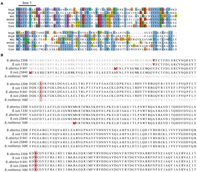
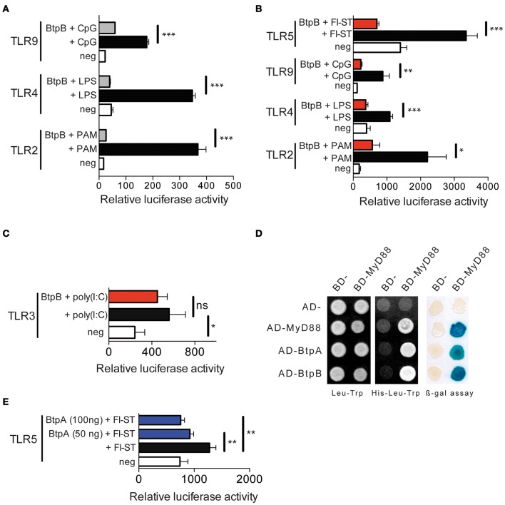
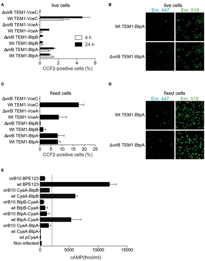
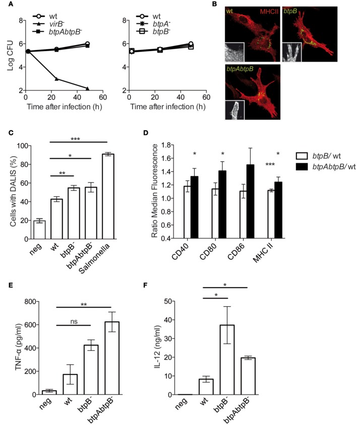
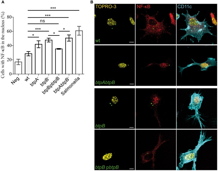
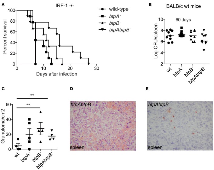
Several bacterial pathogens have TIR domain-containing proteins that contribute to their pathogenesis. We identified a second TIR-containing protein in Brucella spp. that we have designated BtpB. We show it is a potent inhibitor of TLR signaling, probably via MyD88. BtpB is a novel Brucella effector that is translocated into host cells and interferes with activation of dendritic cells. In vivo mouse studies revealed that BtpB is contributing to virulence and control of local inflammatory responses with relevance in the establishment of chronic brucellosis. Together, our results show that BtpB is a novel Brucella effector that plays a major role in the modulation of host innate immune response during infection.

Keywords: Brucella; Btp1/BtpA; DC; NF-κB; TIR domain; TLR.


Figures

Figure 1
Figure 1
Identification of BtpB. (A) Identification of BtpB as bacterial member of TLR/IL-1R (TIR) family. Comparison of the predicted amino acid sequences of the TIR domain of BtpB with BtpA and the human members of the TIR family: MAL, MyD88, TLR2 and TLR4. The alignment was constructed with T-Coffee::advanced server from EMBnet (http://www.ch.embnet.org) and coloring scheme corresponds to standard ClustalX in which each residue in the alignment is assigned a color if the amino acid profile at each position meets a minimum criteria specific for the residue type. Box 1 corresponds to the signature sequence of the TLR family. (B) Alignment of BtpB amino acid sequences for B. abortus 2308 (BAB1_0756), B. suis 1330 (BR0735), B. abortus 9-941 (BruAb1_0752) and B. melitensis 16M (BME1216). The annotated starting codons (Methionine/Valine) are highlighted in red. Amino acid differences are shaded in red.
Figure 2
Figure 2
BtpB interferes with TLR signaling. (A) HEK293 cells were transiently transfected for 24 h with the luciferase reporter vector and either TLR2, TLR4 and TLR9, in the presence or the absence of the 178 amino acid BtpB (50 ng). Cells were then stimulated with the appropriate ligand (PAM, LPS and CpG) for 6 h before measurement of luciferase activity. White bars correspond to negative control, black bars to cells stimulated with the appropriate ligand and grey bars to cells transfected with BtpB and stimulated with the ligand. Data represent the means ± standard errors of relative luciferase activity obtained from triplicates of a representative experiment. (B) Luciferase activity in the presence or absence of the BtpB (1-292) (red bars). TLR5 was also included and stimulated with Flagellin from S. typhimurium (Fl-ST) and (C) TLR3 following stimulation with poly(I:C). (D) Yeast containing Gal4 BD- and Gal4 AD-fusion proteins were selected on synthetic medium lacking leucine (Leu) and tryptophan (Trp) (left panel). Protein interactions were identified on synthetic medium lacking histidine (His) and supplemented with 20 mM 3AT (middle panel). Growth on this medium indicates interaction between fusion proteins. The blue yeast colonies observed in the β-galactosidase expression filter assay indicate interaction between the fusion proteins (right panel). BD and AD indicate empty vectors and were used as negative controls, while MyD88 homodimerization was used as positive control. (E) Luciferase activity in cells transfected with TLR5 in the absence or presence of 100 ng and 50 ng of BtpA (275 aa). P ≤ 0.001 are denoted with ***; P ≤ 0.01 are denoted with ** and P between 0.01 and 0.05 are denoted with *.
Figure 3
Figure 3
BtpB is translocated into host cells during infection. (A) RAW macrophages were infected with wild type (wt) or ΔvirB9 B. abortus strains carrying N-terminal TEM-1 fused VceA, VceC, BtpA, and BtpB for 4 h and 24 h. Data represents the means ± standard errors of the percentage of cells with coumarin fluorescence from 5 independent experiments. (B) Representative confocal images of RAW cells infected with either wilt-type B. abortus (wt) or Δ virB9 mutant carrying TEM-fused BtpA, at 24 h after inoculation. Appearance of blue cells is indicative of translocated TEM lactamase. (C) and (D) Analysis of TEM-1 translocation assay for fixed samples of VceA, VceC, BtpA, and BtpB 24 h after infection. (E) Intracellular cAMP levels in J774.A1 cells infected for 4 h with isogenic strains with a functional (wt) or non-functional VirB system (virB10) expressing Btp proteins fused to CyaA. Non-infected cells and a wild type strain expressing the CyaA domain alone (pCyaA) were included as negative controls. A wild type strain expressing BPE123-CyaA was included as a positive control. Means and SD are shown for one representative out of three independent experiments.
Figure 4
Figure 4
Role of BtpB in control of DC activation. (A) BMDCs infected with wild type B. abortus or the btpAbtpB mutant (left panel) and the single mutants (right panel) were lysed and intracellular CFUs enumerated at different times after inoculation. (B) Representative images of BMDCs infected with either the wild type, btpB or btpAbtpB mutants for 24 h. Cells were labeled for MHC class II (red) and surface expression is of a representative area is shown in zoom inlets. (C) Quantification of the percentage of DCs containing DALIS after 24 h of infection with wild type B. abortus (wt), btpB or btpAbtpB mutant. (D) Flow cytometry of the surface expression of MHC class II, CD40, CD80 and CD86 at 24 h post-infection. Data are normalized to wt values. (E) Analysis of TNF-α and (F) IL-12 (p40/p70) secretion measured by ELISA from the supernatant of DCs 24 h after inoculation. All the results correspond to the means ± standard errors of 4 independent experiments. P ≤ 0.001 are denoted with ***; P ≤ 0.01 are denoted with ** and P between 0.01 and 0.05 are denoted with *.
Figure 5
Figure 5
Modulation of NF-κB translocation to the nucleus during Brucella infection. Bone marrow-derived DCs were infected with wild type (wt) B. abortus, btpA, btpB and btpAbtpB mutants as well as btpB mutant carrying the complementing plasmid (pbtpB) for 2 h and processed for immunofluorescence confocal microscopy. Cells were labeled for CD11c (cyan) and p65 NF-κB (red). Bacteria were labeled with anti-LPS antibody followed by FITC secondary and nuclei with TOPRO3. Salmonella infected cells were used as a positive control. (A) Data corresponds to means ± standard errors of 4 independent experiments. (B) Representative images obtained by confocal microscopy are shown for DCs infected with wild type, btpB mutant, btpBpbtpB complemented strain and btpAbtpB mutant. Scale bars correspond to 5 μm. P ≤ 0.001 are denoted with ***; P ≤ 0.01 are denoted with ** and P between 0.01 and 0.05 are denoted with *.
Figure 6
Figure 6
Role of BtpB during Brucella infection in the mouse model of brucellosis. (A) Susceptibility of IRF-1−/− to B. abortus 2308 (wt), btpA, btpB and btpAbtpB mutant (n = 9 per group). Infected mice were monitored daily for survival. Mice infected with btpB and tpAbtpB survived longer than wild type Brucella infected mice (P = 0.0433 and P = 0.0152, respectively). (B) Persistence of B. abortus 2308 (wt), btpA, btpB or btpAbtpB mutants in spleens of wild type BALB/c infected mice at 60 days p.i. Each symbol represents an animal and the median values are marked by horizontal bold lines. (C) Analysis of granuloma formation in the spleens of wild type BALB/c mice infected for 60 days with wild type B. abortus, btpA, btpB or btpAbtpB mutants. Data represent means ± standard deviations of 4 or 5 mice. (D) Representative image from the spleen of a mouse infected with btpAbtpB mutant (hematoxylin-eosin, original magnification ×400). (E) Bacteria were revealed by immunostaining in the spleen of wild type BALB/c mice infected by btpAbtpB mutant of B. abortus. Macrophages present in inflammatory granulomas in the red pulp are packed with coarse immunopositive material (hemalun counterstain, original magnification ×400). P ≤ 0.01 are denoted with **.

Similar articles

Cited by

References

    1. Celli J., de Chastellier C., Franchini D.-M., Pizarro-Cerda J., Moreno E., Gorvel J.-P. (2003). Brucella evades macrophage killing via VirB-dependent sustained interactions with the endoplasmic reticulum. J. Exp. Med. 198, 545–556 10.1084/jem.20030088 - DOI - PMC - PubMed
    1. Celli J., Salcedo S. P., Gorvel J.-P. (2005). Brucella coopts the small GTPase Sar1 for intracellular replication. Proc. Natl. Acad. Sci. U.S.A. 102, 1673–1678 10.1073/pnas.0406873102 - DOI - PMC - PubMed
    1. Chaudhary A., Ganguly K., Cabantous S., Waldo G. S., Micheva-Viteva S. N., Nag K., et al. (2011). The Brucella TIR-like protein TcpB interacts with the death domain of MyD88. Biochem. Biophys. Res. Commun. 417, 1–6 - PMC - PubMed
    1. Cirl C., Wieser A., Yadav M., Duerr S., Schubert S., Fischer H., et al. (2008). Subversion of Toll-like receptor signaling by a unique family of bacterial Toll/interleukin-1 receptor domain-containing proteins. Nat. Med. 14, 399–406 10.1038/nm1734 - DOI - PubMed
    1. de Barsy M., Jamet A., Filopon D., Nicolas C., Laloux G., Rual J.-F., et al. (2011). Identification of a Brucella spp. secreted effector specifically interacting with human small GTPase Rab2. Cell Microbiol. 13, 1044–1058 10.1111/j.1462-5822.2011.01601.x - DOI - PubMed

MeSH terms