Comment
. 2014 Jan;171(2):294-313.
doi: 10.1111/bph.12296.

Orexin/hypocretin receptor signalling: a functional perspective

Affiliations
Comment

Orexin/hypocretin receptor signalling: a functional perspective

C S Leonard et al. Br J Pharmacol. 2014 Jan.

Abstract

Multiple homeostatic systems are regulated by orexin (hypocretin) peptides and their two known GPCRs. Activation of orexin receptors promotes waking and is essential for expression of normal sleep and waking behaviour, with the sleep disorder narcolepsy resulting from the absence of orexin signalling. Orexin receptors also influence systems regulating appetite/metabolism, stress and reward, and are found in several peripheral tissues. Nevertheless, much remains unknown about the signalling pathways and targets engaged by native receptors. In this review, we integrate knowledge about the orexin receptor signalling capabilities obtained from studies in expression systems and various native cell types (as presented in Kukkonen and Leonard, this issue of British Journal of Pharmacology) with knowledge of orexin signalling in different tissues. The tissues reviewed include the CNS, the gastrointestinal tract, the pituitary gland, pancreas, adrenal gland, adipose tissue and the male reproductive system. We also summarize the findings in different native and recombinant cell lines, especially focusing on the different cascades in CHO cells, which is the most investigated cell line. This reveals that while a substantial gap exists between what is known about orexin receptor signalling and effectors in recombinant systems and native systems, mounting evidence suggests that orexin receptor signalling is more diverse than originally thought. Moreover, rather than being restricted to orexin receptor 'overexpressing' cells, this signalling diversity may be utilized by native receptors in a site-specific manner.

Keywords: adipose tissue; adrenal gland; cell death; depolarization; hormone release; hypocretin; neuron; orexin; plasticity; second messenger.


Figures

Figure 1
Figure 1
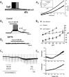
Orexin-mediated depolarizations in the CNS. (A) Orexins close leak-like K+ channels. The orexin-B-mediated (100 nM) depolarization in layer 6b neocortical pyramidal neurons, which causes firing, is associated with an increase in membrane resistance (note the larger negative-going voltage deflections produced by constant current pulses during the depolarization (A1). The I-V curve for the orexin-activated current reverses near the predicted K+ equilibrium potential in an extracellular [K+] of 6.25 mM (upper I-V curve) and 12 mM (lower I-V curve) (A2). The inset shows the mean reversal potential measured at each extracellular [K+]. (B) Orexins stimulate electrogenic NCX. Orexin-A produces a suprathreshold depolarization in type C arcuate nucleus neurons of the hypothalamus (B1). The depolarizing current is insensitive to lowering extracellular [Ca2+] (not shown) but is completely blocked by strong buffering of intracellular Ca2+ (BAPTA, 10 mM), consistent with it being triggered by the release of Ca2+ from intracellular stores (B2). The reversal potential of the orexin-activated exchanger current shifts with changes of Na+ and Ca2+ gradients (B3). (C) Orexins activate a ‘noisy’ non-selective cation current that is not reduced by low extracellular [Ca2+] or strong buffering of intracellular [Ca2+]. The orexin-A-activated current in dorsal raphé neurons was initiated, but was almost absent in low extracellular [Na+] (26 mM; NMDG). The current was reinstated by switching back to normal extracellular [Na+] (150 mM; C1). The reversal potential of this cation current is near 0 mV in normal extracellular [Na+] (C2, upper subfigure) but shifts to near −60 mV in low extracellular [Na+] (C2, lower subfigure). Subfigures (A), (B) and (C) are adapted from Bayer et al. (2004); Burdakov et al. (2003); and Brown et al. (2002), respectively and are reproduced with permission.
Figure 2
Figure 2
In addition to producing depolarization, orexins also modulate the integrative properties of neurons in the CNS. (A) Orexin-A (ORX-A, 10 nM) slowed action potential repolarization and broadened the spike without altering the magnitude of AHP in nucleus of the solitary tract neurons. The duration of the action potential at both 50% (APD50) and 90% (APD90) recovery was longer following orexin. This was associated with a reduction in non-inactivating voltage-dependent outward currents (not shown). (B) In dissociated locus coeruleus neurons, orexin-A and orexin-B (ORX-B) reduced the magnitude and duration of the AHP. This too was associated with a reduction in a non-inactivating, voltage-dependent outward current (not shown), which may be different from that reduced in (A). Reprinted from Murai and Akaike (2005), with permission from Elsevier. (C) In paraventricular thalamic neurons, orexins reduce an IsAHP and decrease spike-frequency adaptation. These neurons have a classical IsAHP, which is mediated mainly by a Ca2+-dependent K+ current and a Na+-dependent K+ current, which is activated with stronger stimuli. IsAHP was strongly reduced by orexin-A (200 nM; C1) and by stimulating either the PKA and PKC pathways (not shown). The orexin-A-mediated reduction was blocked by an inhibitor of PKC but not an inhibitor of PKA(C2). The reduction of this current by orexins (and PKA and PKC activators) decreases spike-frequency adaptation and increases the number of spikes fired for the same input current in these cells (C3). (D) Orexin-A (300 nM) depolarizes serotonergic dorsal raphé neurons and also enhances a late AHP. Orexin-A (300 nM) produced a suprathreshold depolarization (D1). The late AHP was evoked (5 spikes at 20 Hz) before orexin application (left arrow) and during orexin (right arrow) after returning to the same baseline membrane potential by current injection (-dc). Orexin made the late AHP larger (198 ± 19 % of control, n = 20) and longer (429 ± 52 % of control half recovery time, n = 20). This orexin-enhanced AHP slowed steady-state firing in response to 100 pA (2 s) injected current pulses (D2) without changing the shape of the action potential (spikes superimposed from before and after orexin; D2 inset). Subfigures (A), (B) and (C) are adapted from Yang and Ferguson (2003), Murai and Akaike (2005), Zhang et al. (2010) and are reproduced with permission. Subfigure (D) was adapted from Ishibashi M, Gumenchuk I, Leonard CS (unpubl. data).
Figure 3
Figure 3
Summary of concentration–response relations for different responses produced by orexin-A at the human OX1 receptor. This illustrates the different potencies with which orexin-A stimulates different signalling cascades under conditions of equal receptor expression levels in the same clone of CHO cells. Thus, cell background and receptor expression levels are equal across cases so potencies are directly comparable. The results are expressed as % of maximal response for each output. The responses measured are: ‘inward current’ (whole-cell patch-clamp recordings), ‘cPLA2’ (pyrrophenone-sensitive and tetrahydrolipstatin-insensitive AA release chromatographically measured in 14C-AA-labelled cells), ‘Ca2+’ (cytosolic Ca2+ elevation measured with fura-2 parallel to the patch-clamp recordings), ‘PLD1’ (PLD transphosphatidylation assay in 14C-oleic acid-labelled cells with chromatographic separation; PLD1 defined by pharmacological PLD1 and PLD2 inhibitors), ‘PKCδ (AC)’ [nPKC (likely PKCδ)-dependent AC stimulation measured chromatographically in 3H-adenine-labelled cells], ‘PKCγ-C1 (transl.; PLD + PLC)’ (DAG generation by both PLC and PLD measured by translocation of the fusion protein of C1-domain of PKCγ and green fluorescent protein to the plasma membrane), ‘PLC total’ (total inositol phosphate release from 3H-inositol-labelled cells determined by chromatography), ‘PKCγ-C1 (transl.; PLC)’ (DAG generation by PLC alone measured by translocation of the fusion protein of C1-domain of PKCγ and green fluorescent protein to the plasma membrane in cells expressing dominant-negative PLD), ‘DGL’ (tetrahydrolipstatin-sensitive and pyrrophenone-insensitive 2-AG release chromatographically measured in 14C-AA-labelled cells), ‘cell death’ (assessed by staining and counting of viable cells), ‘ERK2’ [ERK2 phosphorylation assessed by Western blotting against the phosphorylated (active) ERK1/2 species], ‘PLC-PIP2’ (IP3 release assessed by a IP3-binding protein kit and by translocation of the PH-domain of PLCδ1 from the plasma membrane upon PLC activity towards PIP2), and ‘Gs’ (Gαs-dependent AC stimulation measured chromatographically in 3H-adenine-labelled cells). For the sake of clarity, only curve-fitting results and not the original data points are presented. The curves can be identified with the symbols added on the traces. The measurements originate from the following studies: Holmqvist et al. (2005); Larsson et al. (2005); Ammoun et al. (,b2006b); Johansson et al. (2008); Jäntti et al. (2012); Turunen et al. (2012).
Figure 4
Figure 4
Schematic summary of some of the relationships of human OX1 receptor signalling cascades in CHO cells as described in Figure 3. References are as for Figure 3. While Gq may underlie PLC activation (and possibly other responses), this has not been explicitly assessed and is thus not indicated in the figure. L-GPL, lyso-glycerophospholipid; PA, phosphatidic acid.

Comment on

Similar articles

Cited by

References

    1. Acuna-Goycolea C, van den Pol AN. Neuroendocrine proopiomelanocortin neurons are excited by hypocretin/orexin. J Neurosci. 2009;29:1503–1513. - PMC - PubMed
    1. Adamantidis AR, Zhang F, Aravanis AM, Deisseroth K, de Lecea L. Neural substrates of awakening probed with optogenetic control of hypocretin neurons. Nature. 2007;450:420–424. - PMC - PubMed
    1. Aghajanian GK, Vandermaelen CP. Intracellular recordings from serotonergic dorsal raphe neurons: pacemaker potentials and the effect of LSD. Brain Res. 1982;238:463–469. - PubMed
    1. Ammoun S, Holmqvist T, Shariatmadari R, Oonk HB, Detheux M, Parmentier M, et al. Distinct recognition of OX1 and OX2 receptors by orexin peptides. J Pharmacol Exp Ther. 2003;305:507–514. - PubMed
    1. Ammoun S, Johansson L, Ekholm ME, Holmqvist T, Danis AS, Korhonen L, et al. OX1 orexin receptors activate extracellular signal-regulated kinase (ERK) in CHO cells via multiple mechanisms: the role of Ca2+ influx in OX1 receptor signaling. Mol Endocrinol. 2006a;20:80–99. - PubMed

Publication types

Substances