. 2013 Oct;170(4):807-21.
doi: 10.1111/bph.12302.

Emodin inhibits growth and induces apoptosis in an orthotopic hepatocellular carcinoma model by blocking activation of STAT3

Affiliations

Emodin inhibits growth and induces apoptosis in an orthotopic hepatocellular carcinoma model by blocking activation of STAT3

Aruljothi Subramaniam et al. Br J Pharmacol. 2013 Oct.

Abstract

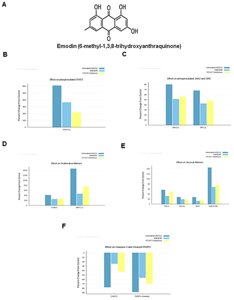
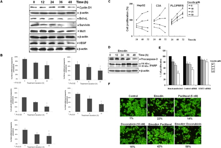
Background and purpose: Aberrant activation of STAT3 is frequently encountered and promotes proliferation, survival, metastasis and angiogenesis in hepatocellular carcinoma (HCC). Here, we have investigated whether emodin mediates its effect through interference with the STAT3 activation pathway in HCC.

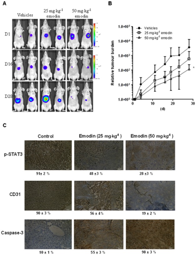
Experimental approach: The effect of emodin on STAT3 activation, associated protein kinases and apoptosis was investigated using various HCC cell lines. Additionally, we also used a predictive tumour technology to analyse the effects of emodin . The in vivo effects of emodin were assessed in an orthotopic mouse model of HCC.

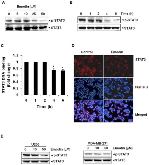
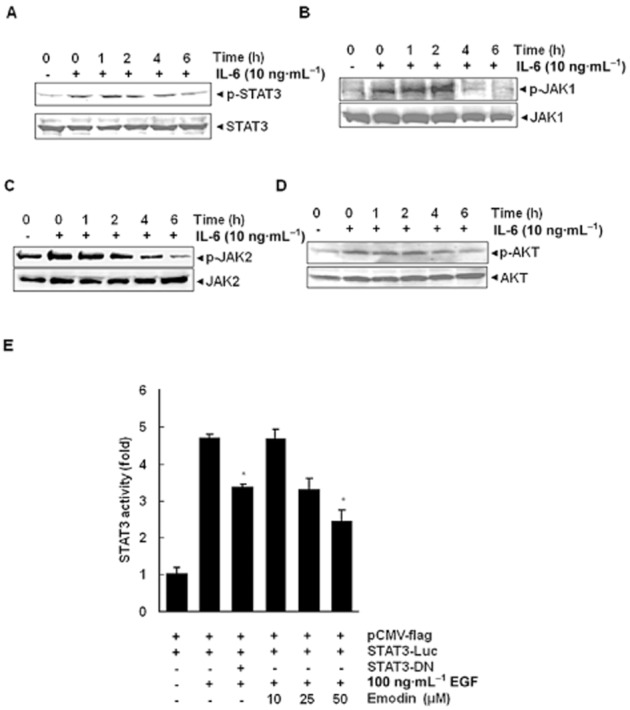
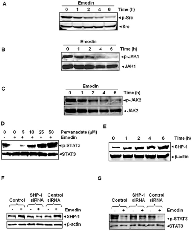
Key results: Emodin suppressed STAT3 activation in a dose- and time-dependent manner in HCC cells, mediated by the modulation of activation of upstream kinases c-Src, JAK1 and JAK2. Vanadate treatment reversed emodin-induced down-regulation of STAT3, suggesting the involvement of a tyrosine phosphatase and emodin induced the expression of the tyrosine phosphatase SHP-1 that correlated with the down-regulation of constitutive STAT3 activation. Interestingly, silencing of the SHP-1 gene by siRNA abolished the ability of emodin to inhibit STAT3 activation. Finally, when administered i.p., emodin inhibited the growth of human HCC orthotopic tumours in male athymic nu/nu mice and STAT3 activation in tumour tissues.

Conclusions and implications: Emodin mediated its effects predominantly through inhibition of the STAT3 signalling cascade and thus has a particular potential for the treatment of cancers expressing constitutively activated STAT3.

Keywords: STAT3; apoptosis; emodin; hepatocellular carcinoma; proliferation.


Figures

Figure 1
Figure 1
Results generated by predictive proteomics in silico using a virtual tumour platform. (A) The chemical structure of emodin. (B) The graph illustrates the percentage reduction in phosphorylated levels of STAT3. STAT3 is showing a reduction of 35% with emodin treatment in the HEPG2 baseline as compared with the untreated placebo. STAT3 activity was also inhibited separately by 40% to compare its effect with emodin treatment. (C) The graph illustrates the percentage reduction in JAK2 and SRC with emodin treatment. The percentage reduction is around 16% for JAK 2 and 15% for SRC with emodin. STAT3 inhibition alone is also showing similar effects on these kinases with the effect being 14% for JAK2 and 12% for SRC. (D) The graph illustrates the percentage change in proliferation markers – CCND1 and cMYC. CCND1 is showing a reduction of 32% from the baseline and the reduction in cMYC is 65% with respect to the untreated baseline. STAT3 inhibition alone is showing a similar reduction of 30% in CCND1 and 50% reduction in cMYC. (E) The graph illustrates the percentage reduction in survival markers with emodin treatment and STAT3 inhibition alone. The percentage reduction is 16% for BCL2, 7% for BCL-xL, 12% for XIAP and 32% for survivin with emodin treatment. The reduction with STAT3 inhibition alone is 5% for Bcl-2, 9% in Bcl-xL, 10% for XIAP and 28% for surviving. (F) The graph illustrates the percentage change in CASP3 and PARP1 cleaved with emodin treatment and STAT3 inhibition alone. An increase of approximately 3.5-fold is seen in the levels of both CASP3 and PARP1 cleaved with emodin treatment. The increase was approximately 2.6-fold with STAT3 inhibition alone.
Figure 2
Figure 2
Emodin inhibits constitutively active STAT3 in HepG2 cells. (A) Emodin suppresses phospho-STAT3 levels in a dose-dependent manner. HepG2 cells (2 × 10⁶·mL−1) were treated with the indicated concentrations of emodin for 6 h, after which whole cell extracts were prepared, and 30 μg of protein was resolved on 7.5% SDS-PAGE gel, electrotransferred onto nitrocellulose membranes, and probed for phospho-STAT3/STAT3 proteins. (B) Emodin suppresses phospho-STAT3 levels in a time-dependent manner. HepG2 cells (2 × 10⁶·mL−1) were treated with 50 μM emodin for the indicated times, after which Western blotting was performed as described for panel B. (C) Emodin suppresses STAT3 DNA-binding ability in HepG2 cells. HepG2 cells were treated with 50 μM emodin for indicated time points; nuclear extracts were prepared, and 20 μg of the nuclear extract protein was used for elisa-based DNA-binding assay. The results shown are representative of two independent experiments. *P < 0.05. (D) Emodin causes inhibition of translocation of STAT3 to the nucleus. HepG2 cells (1 × 10⁵·mL−1) were incubated with or without 50 μM emodin for 6 h and then analysed for the intracelullar distribution of STAT3 by immunocytochemistry. The same slides were counterstained for nuclei with Hoechst (50 ng·mL−1) for 5 min. The results shown are representative of three independent experiments. (E) Multiple myeloma (U266) and breast adenocarcinoma (MDA-MB-231) cells (2 × 10⁶·mL−1) were treated with the indicated concentrations of emodin for 6 h, after which whole cell extracts were prepared, and 30 μg of protein was resolved on 7.5% SDS-PAGE gel, electrotransferred onto nitrocellulose membranes, and probed for phospho-STAT3/STAT3 proteins.
Figure 3
Figure 3
HUH-7 cells (2 × 10⁶·mL−1) were treated with 50 μM emodin for the indicated times and then stimulated with IL-6 (10 ng·mL−1) for 15 min. Whole cell extracts were then prepared and analysed for phospho-STAT3/STAT3 by Western blotting. The results shown are representative of three independent experiments. (B) HUH-7 cells (2 × 10⁶·mL−1) were treated with 50 μM emodin for the indicated times and then stimulated with IL-6 (10 ng·mL−1) for 15 min. Whole cell extracts were then prepared and analysed for phospho-JAK1/JAK1 by Western blotting. (C) HUH-7 cells (2 × 10⁶·mL−1) were treated with 50 μM emodin for the indicated times and then stimulated with IL-6 (10 ng·mL−1) for 15 min. Whole cell extracts were then prepared and analysed for phospho-JAK2/JAK2 proteins. (D) HUH-7 cells (2 × 10⁶·mL−1) were treated with 50 μM emodin for the indicated times and then stimulated with IL-6 (10 ng·mL−1) for 15 min. Whole cell extracts were then prepared and analysed for phospho-Akt by Western blotting. The same blots were stripped and reprobed with Akt antibody to verify equal protein loading. (E) PLC/PRF/5 cells (5 × 10⁵·mL−1) were transfected with STAT3-luciferase (STAT3-Luc) plasmid, incubated for 24 h, and treated with 10, 25 and 50 μM emodin for 6 h and then stimulated with EGF (100 ng·mL−1) for 2 h. Whole cell extracts were then prepared and analysed for luciferase activity. The results shown are representative of three independent experiments. *P < 0.05, significantly different from EGF alone; Student's t-test.
Figure 4
Figure 4
(A) Emodin suppresses phospho-Src levels in a time-dependent manner. HepG2 cells (2 × 10⁶·mL−1) were treated with 50 μM emodin, after which whole cell extracts were prepared and 30 μg of aliquots of those extracts were resolved on 10% SDS-PAGE, electrotransferred onto nitrocellulose membranes, and probed for phospho-Src/Src antibodies. (B) Emodin suppresses phospho-JAK1 levels in a time-dependent manner. HepG2 cells (2 × 10⁶·mL−1) were treated with 50 μM emodin for indicated time intervals, after which whole cell extracts were prepared and 30 μg portions of those extracts were resolved on 10% SDS-PAGE, electrotransferred onto nitrocellulose membranes, and probed with phospho-JAK1/JAK1 antibodies. (C) Emodin suppresses phospho-JAK2 levels in a time-dependent manner. HepG2 cells (2 × 10⁶·mL−1) were treated with 50 μM emodin for indicated time intervals, after which whole cell extracts were prepared and 30 μg portions of those extracts were resolved on 10% SDS-PAGE, electrotransferred onto nitrocellulose membranes, and probed with phospho-JAK2/JAK2 antibodies. (D) Pervanadate reverses the phospho-STAT3 inhibitory effect of emodin. HepG2 cells (2 × 10⁶·mL−1) were treated with the indicated concentrations of pervanadate and 50 μM emodin for 6 h, after which whole cell extracts were prepared and 30 μg portions of those extracts were resolved on 7.5% SDS-PAGE gel, electrotransferred onto nitrocellulose membranes, and probed for phospho-STAT3 and STAT3. (E) Emodin induces the expression of SHP-1 protein in HepG2 cells. HepG2 cells were treated with indicated concentrations of emodin for 6 h, after which Western blotting was performed. (F) Effect of SHP-1 knock-down on emodin-induced expression of SHP-1. HepG2 cells were transfected with either SHP-1 siRNA or scrambled siRNA (50 nM). After 24 h, cells were treated with 50 μM emodin for 6 h and whole cell extracts were subjected to Western blot analysis. (G) HepG2 cells were transfected with either SHP-1 siRNA or scrambled siRNA (50 nM). After 24 h, cells were treated with 50 μM emodin for 6 h and whole cell extracts were subjected to Western blot analysis for phosphorylated STAT3. The results shown are representative of three independent experiments.
Figure 5
Figure 5
Emodin suppresses STAT3-regulated gene products involved in proliferation, survival and angiogenesis. (A) HepG2 cells (2 × 10⁶·mL−1) were treated with 50 μM emodin for indicated time intervals, after which whole cell extracts were prepared and 30 μg portions of those extracts were resolved on 10% SDS-PAGE, membrane sliced according to molecular weight and probed against cyclin D1, Bcl-2, survivin, Mcl-1 and VEGF antibodies. The same blots were stripped and reprobed with β-actin antibody to verify equal protein loading. The results shown are representative of three independent experiments. (B) HepG2 cells (3 × 10⁵·mL−1) were treated with 50 μM emodin for the indicated time intervals, after which cells were harvested after treatment and RNA samples were extracted. About 1 μg portions of the respective RNA extracts were used for reverse transcription to generate corresponding cDNA. Real-time PCR was performed to measure the relative quantities of mRNA. Each RT product was targeted against cyclin D1, Bcl-2, Bcl-XL, Mcl-1 and VEGF TaqMan probes, with HuGAPDH as endogenous control for measurement of equal loading of RNA samples. Results were analysed using Sequence Detection Software version 1.3 provided by Applied Biosystems (Foster City, CA, USA). Relative gene expression was obtained after normalization with endogenous HuGAPDH and determination of the difference in threshold cycle (Ct) between treated and untreated cells using 2-ΔΔCt method. (C) HepG2, C3A and PLC/PRF/5 cells (5 × 10³·mL−1) were plated in triplicate, treated with indicated concentrations of emodin, and then subjected to MTT assay after 24, 48 and 72 h to measure the proliferation of cells. SDs between the triplicates are indicated. (D) HepG2 cells were treated with 50 μM emodin for the indicated times, whole cell extracts were prepared, separated on SDS-PAGE, and subjected to Western blotting against pro-caspase-3 and PARP antibodies. The same blot was stripped and reprobed with β-actin antibody to show equal protein loading. The results shown are representative of three independent experiments. (E) Knock-down of STAT3 siRNA reduces the anti-proliferative effect of emodin. HepG2 cells were transfected with either STAT3-specific or control siRNA (50 nM). After 48 h, cells were treated with indicated concentrations of emodin for 72 h and then subjected to MTT assay, *P ≤ 0.05; **P ≤ 0.005, significant effect of emodin; Student's t-test. (F) Emodin potentiates the apoptotic effect of doxorubicin and paclitaxel. HepG2 cells (1 × 10⁶·mL−1) were treated with 10 μM emodin and 10 nM doxorubicin or 5 nM paclitaxel alone or in combination for 24 h at 37°C. Cells were stained with a live/dead assay reagent for 30 min and then the percentage of apoptotic cells was determined, using a fluorescence microscope.
Figure 6
Figure 6
Emodin inhibits the growth of human HCC in vivo. (A) Representative images of mice from bioluminescent imaging. D = day. (B) Relative tumour burden in athymic mice bearing orthotopically implanted HCCLM3-Luc2 tumours treated with vehicle alone (n = 9), 25 mg·kg−1 (n = 10) or 50 mg·kg−1 (n = 9) of emodin. Points, mean; bars, SE. * P < 0.05, significant effect of emodin; one-way anova. (C) Immunohistochemical analysis of p-STAT3, CD31 and caspase-3 showed the inhibition in expression of p-STAT3, and CD31 and increased levels of caspase-3 expression in emodin-treated samples as compared with control group. Percentage indicates positive staining for the biomarker shown. The photographs were taken at magnification 40×.

References

    1. Alexander SPH, Mathie A, Peters JA. Guide to Receptors and Channels (GRAC), 5th edition. Br J Pharmacol. 2011;164(Suppl. 1):S1–324. - PMC - PubMed
    1. Battistutta R, Sarno S, De Moliner E, Papinutto E, Zanotti G, Pinna LA. The replacement of ATP by the competitive inhibitor emodin induces conformational modifications in the catalytic site of protein kinase CK2. J Biol Chem. 2000;275:29618–29622. - PubMed
    1. Bromberg J, Wang TC. Inflammation and cancer: IL-6 and STAT3 complete the link. Cancer Cell. 2009;15:79–80. - PMC - PubMed
    1. Brown M, Bellon M, Nicot C. Emodin and DHA potently increase arsenic trioxide interferon-alpha-induced cell death of HTLV-I-transformed cells by generation of reactive oxygen species and inhibition of Akt and AP-1. Blood. 2007;109:1653–1659. - PMC - PubMed
    1. Calvisi DF, Ladu S, Gorden A, Farina M, Conner EA, Lee JS, et al. Ubiquitous activation of Ras and Jak/Stat pathways in human HCC. Gastroenterology. 2006;130:1117–1128. - PubMed

Publication types

MeSH terms