. 2013 Aug 13;52(32):5372-86.
doi: 10.1021/bi400659k. Epub 2013 Jul 29.

Structural basis for the divergence of substrate specificity and biological function within HAD phosphatases in lipopolysaccharide and sialic acid biosynthesis

Affiliations

Structural basis for the divergence of substrate specificity and biological function within HAD phosphatases in lipopolysaccharide and sialic acid biosynthesis

Kelly D Daughtry et al. Biochemistry. .

Abstract

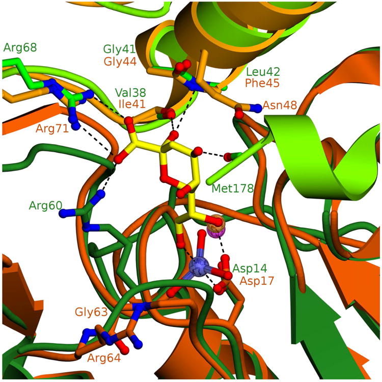
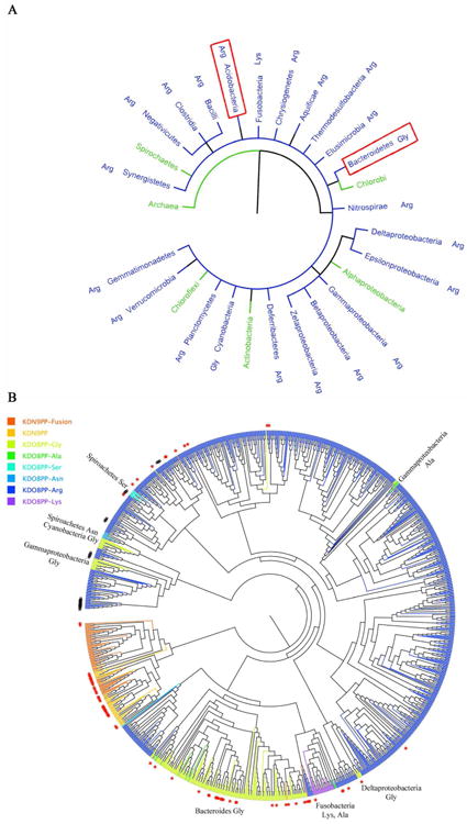
The haloacid dehalogenase enzyme superfamily (HADSF) is largely composed of phosphatases that have been particularly successful at adaptating to novel biological functions relative to members of other phosphatase families. Herein, we examine the structural basis for the divergence of function in two bacterial homologues: 2-keto-3-deoxy-d-manno-octulosonate 8-phosphate phosphohydrolase (KDO8P phosphatase, KDO8PP) and 2-keto-3-deoxy-9-O-phosphonononic acid phosphohydrolase (KDN9P phosphatase, KDN9PP). KDO8PP and KDN9PP catalyze the final step in KDO and KDN synthesis, respectively, prior to transfer to CMP to form the activated sugar nucleotide. KDO8PP and KDN9PP orthologs derived from an evolutionarily diverse collection of bacterial species were subjected to steady-state kinetic analysis to determine their specificities toward catalyzed KDO8P and KDN9P hydrolysis. Although each enzyme was more active with its biological substrate, the degree of selectivity (as defined by the ratio of kcat/Km for KDO8P vs KDN9P) varied significantly. High-resolution X-ray structure determination of Haemophilus influenzae KDO8PP bound to KDO/VO3(-) and Bacteriodes thetaiotaomicron KDN9PP bound to KDN/VO3(-) revealed the substrate-binding residues. The structures of the KDO8PP and KDN9PP orthologs were also determined to reveal the differences in their active-site structures that underlie the variation in substrate preference. Bioinformatic analysis was carried out to define the sequence divergence among KDN9PP and KDO8PP orthologs. The KDN9PP orthologs were found to exist as single-domain proteins or fused with the pathway nucleotidyl transferases; the fusion of KDO8PP with the transferase is rare. The KDO8PP and KDN9PP orthologs share a stringently conserved Arg residue that forms a salt bridge with the substrate carboxylate group. The split of the KDN9PP lineage from the KDO8PP orthologs is easily tracked by the acquisition of a Glu/Lys pair that supports KDN9P binding. Moreover, independently evolved lineages of KDO8PP orthologs exist, and are separated by diffuse active-site sequence boundaries. We infer a high tolerance of the KDO8PP catalytic platform to amino acid replacements that in turn influence substrate specificity changes and thereby facilitate the divergence in biological function.


Figures

Figure 1
Figure 1. KD9PP monomer (A) and tetramer (B) structures (PDB ID 3E8M)
HADSF Rossmann fold domain colored gray or light blue with the tetramerization flap colored yellow. HADSF catalytic motifs colored: motif 1, red; motif 2, green; motif 3, cyan; motif 4, orange; magnesium cofactor is shown in magenta. Figure (and all others unless indicated) generated with MOLSCRIPT and POVscript+.
Figure 2
Figure 2. Structure of BT-KDN9PP-Mg2+−VO3-KDN
(A) Dimer from tetrameric BT-KDN9PP with “catalytic” subunit colored dark blue and “cap” subunit colored light blue, HAD catalytic motifs are shown as dark blue sticks, KDN is shown as yellow sticks, vanadium is colored slate blue and magnesium is a magenta sphere. (B) HAD catalytic residues and (C) KDN binding residues. (B) and (C) are colored as in (A).
Figure 3
Figure 3. Structure of HI-KDO8PP-Mg2+-VO3-KDO
(A) Dimer of HI-KDO8PP (generated from symmetry mates comprising tetramer) with “catalytic” subunit colored forest green and “cap” subunit colored light green, HAD catalytic motifs are shown as forest green sticks, KDN is shown as yellow sticks, vanadium is colored slate blue and magnesium is a magenta sphere. (B) HAD catalytic residues and (C) KDN binding residues. (B) and (C) are colored as in (A).
Figure 4
Figure 4. Overlay of HI-KDO8PP and BT-KDO8PP active sites
Dimer of HI-KDO8PP (generated from symmetry mates comprising tetramer) with “catalytic” subunit colored forest green and “cap” subunit colored light green, KDN is shown as yellow sticks, vanadium is colored slate blue and magnesium is a magenta sphere. Dimer of BT-KDO8PP with “catalytic” subunit colored dark orange and “cap” subunit colored light orange, magnesium is an orange sphere. Putative substrate binding residues are shown as sticks and labeled in their corresponding enzyme color. Hydrogen bonds and coordination bonds are shown as dashed lines.
Figure 5
Figure 5
Schematics of Bioinformatic Analyses. (A) The tree of life generated from iTOL (itol.embl.de), displaying phyla (and class in the case of proteobacteria) that contain KDO8PP and KDN9PP sequences. Phyla are color-coded (KDO8PP blue, KDN9PP green) based on the majority presence of enzymes. Red boxes indicate the two phyla that contain species with both KDN9PP and KDO8PP. The type of KDO8PP enzyme (Arg, Lys, or Gly) is indicated next to the phyla name. (B) An unrooted phylogenetic tree generated using a multiple sequence alignment of all KDO8PP and KDN9PP enzymes identified herein. KDO8PP-Arg enzymes are colored blue, KDO8PP-Gly green, KDN9PP light orange and KDN9PP-KDN:CMP transferase fusions deep orange. Red stars indicate enzymes that were identified where an additional KDN9PP or KDO8PP gene exists within the species. Black number signs (#) indicate KDO8PP-Arg-KDO:CMP transferase fusion genes. The phyla of some KDO8PP-Gly enzymes have been labeled. Figure generated with FigTree (http://tree.bio.ed.ac.uk/software/figtree/).
Chart I
Chart I
Scheme 1
Scheme 1
The biosynthetic pathways leading to the cytidine monophosphate (CMP) substituted β-anomers of KDO and KDN.
Scheme 2
Scheme 2. Chemical pathway catalyzed by the HADSF phosphatase

References

    1. Nguyen HH, Wang L, Huang H, Peisach E, Dunaway-Mariano D, Allen KN. Structural determinants of substrate recognition in the HAD superfamily member D-glycero-D-manno-heptose-1,7-bisphosphate phosphatase (GmhB) Biochemistry. 2010;49:1082–1092. - PMC - PubMed
    1. Huang H, Patskovsky Y, Toro R, Farelli JD, Pandya C, Almo SC, Allen KN, Dunaway-Mariano D. Divergence of structure and function in the haloacid dehalogenase enzyme superfamily: Bacteroides thetaiotaomicron BT2127 is an inorganic pyrophosphatase. Biochemistry. 2011;50:8937–8949. - PMC - PubMed
    1. Rangarajan ES, Proteau A, Wagner J, Hung MN, Matte A, Cygler M. Structural snapshots of Escherichia coli histidinol phosphate phosphatase along the reaction pathway. J Biol Chem. 2006;281:37930–37941. - PubMed
    1. Burroughs AM, Allen KN, Dunaway-Mariano D, Aravind L. Evolutionary genomics of the HAD superfamily: understanding the structural adaptations and catalytic diversity in a superfamily of phosphoesterases and allied enzymes. J Mol Biol. 2006;361:1003–1034. - PubMed
    1. Allen KN, Dunaway-Mariano D. Markers of fitness in a successful enzyme superfamily. Curr Opin Struct Biol. 2009;19:658–665. - PMC - PubMed

Publication types

MeSH terms

LinkOut - more resources