Review
. 2013 Jul 10;79(1):16-29.
doi: 10.1016/j.neuron.2013.06.028.

The brain on stress: vulnerability and plasticity of the prefrontal cortex over the life course

Affiliations
Review

The brain on stress: vulnerability and plasticity of the prefrontal cortex over the life course

Bruce S McEwen et al. Neuron. .

Abstract

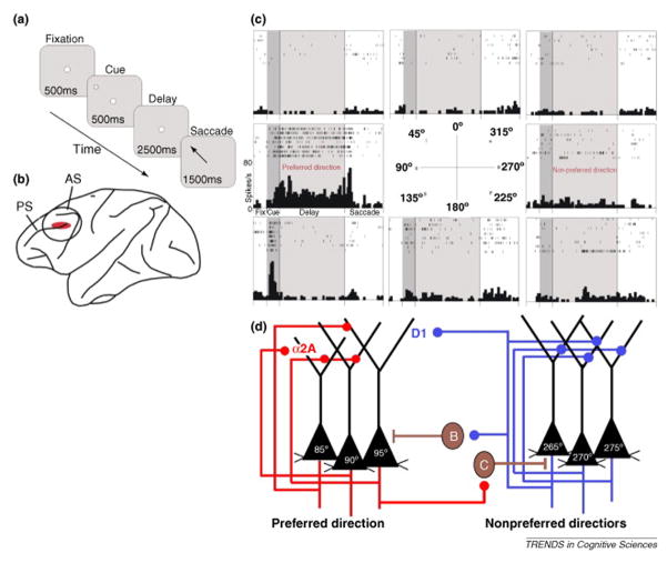
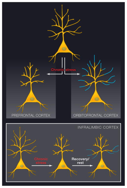
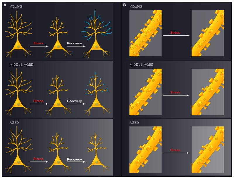
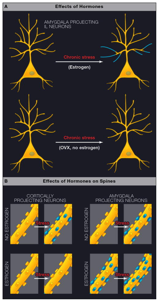
The prefrontal cortex (PFC) is involved in working memory and self-regulatory and goal-directed behaviors and displays remarkable structural and functional plasticity over the life course. Neural circuitry, molecular profiles, and neurochemistry can be changed by experiences, which influence behavior as well as neuroendocrine and autonomic function. Such effects have a particular impact during infancy and in adolescence. Behavioral stress affects both the structure and function of PFC, though such effects are not necessarily permanent, as young animals show remarkable neuronal resilience if the stress is discontinued. During aging, neurons within the PFC become less resilient to stress. There are also sex differences in the PFC response to stressors. While such stress and sex hormone-related alterations occur in regions mediating the highest levels of cognitive function and self-regulatory control, the fact that they are not necessarily permanent has implications for future behavior-based therapies that harness neural plasticity for recovery.


Figures

Figure 1
Figure 1
The neuronal response properties and neural circuitry underlying spatial working memory task as envisioned by Goldman-Rakic and colleagues (Arnsten et al, 2010; Goldman-Rakic, 1995) (a) The oculomotor delayed response (ODR) task, which is a test of spatial working memory as mediated by Brodmann area 46 in the dlPFC of the monkey. (b) Area 46, delineated in red surrounding the principal sulcus. The electrophysiological response properties depicted in c are generated from recordings of pyramidal neurons within this region. PS=principal sulcus; AS=arcuate sulcus. (c) Recordings from a representative neuron in area 46 with spatially tuned firing during the delay period of the ODR task. For details, see (Wang et al 2007). (d) The PFC microcircuits subserving spatially tuned firing during the delay period in a spatial working memory task. Brown neurons designated by B and C represent GABAergic neurons innervating pyramidal neurons mediating working memory. The red circuit on the left represents the noradrenergic inputs modulating α2A receptors on the pyramidal neurons, and the blue circuit on the right depicts the dopaminergic inputs acting through D1 receptors. From Arnsten (Arnsten et al 2010) with permission.
Figure 2
Figure 2
Schematic diagrams depicting dendritic shrinkage and expansion in response to chronic stress and recovery. A) Chronic stress leads to dendritic shrinkage in layer 3 pyramidal neurons in the prelimbic and anterior cingulate cortex, whereas it causes dendritic expansion in the corresponding neurons within orbitofrontal cortex. Both effects are seen primarily in the distal apical dendritic tree. B) While shrinkage and recovery both affect distal dendrites in neurons depicted in A, layer 5 neurons in infralimbic cortex lose distal dendritic branches in response to stress, yet recovery occurs primarily in proximal dendrites, shifting the dendritic architecture (see text for details).
Figure 3
Figure 3
Schematic diagrams depicting the interactive effects between stress and aging on layer 3 pyramidal neurons in the prelimbic area of mPFC. 3A (left) shows the effects on dendritic arbor and 3B (right), shows the effects on spines. In both cases the upper panel represents young male rats, middle panel represents middle-aged rats, and the bottom panel represents aged rats. A) In young animals, chronic stress leads to shrinkage of distal apical dendrites. After cessation of chronic stress, dendritic trees regrow. Such recovery after stress cessation is blunted by middle age and gone in the aged animals. B) Spines are also lost in young animals exposed to chronic stress, and it is primarily the thin spines that are affected. No further spine loss is induced by stress in middle-aged or aged rats, and this is likely due to the fact that age on its own leads to a loss of the thin spine class (See text for details)
Figure 4
Figure 4
Interactive effects of stress and estrogen on neurons within layer 3 of infralimbic cortex. - Schematic depicting a three-way interaction between stress, estrogen, and circuit-specificity in female rats. A) Chronic stress increased dendritic arbor in layer 3 neurons in IL neurons that project to amygdala, whereas this is not the case with the general population of layer 3 pyramidal neurons (not shown). Furthermore, in OVX females, this effect is only seen if the rat receives estrogen treatment. B) Schematic representation of the different effects of estrogen on stress-induced spine formation in the female rat PFC in cortically-projecting and amygdala-projecting neurons. On left, for cortically-projecting neurons, chronic stress induces spine formation in OVX females but fails to do so in OVX females treated with estrogen; on right, for amygdala-projecting neurons, chronic stress induces spine formation in OVX females and estrogen treatment increases spine density in non-stressed animals and promotes further spinogenesis in chronically stressed estrogen-treated OVX females. These dendritic and spine effects differ from those seen in males.

Comment in

  • An Overly Permissive Extension.
    Kagan J. Kagan J. Perspect Psychol Sci. 2016 Jul;11(4):442-50. doi: 10.1177/1745691616635593. Perspect Psychol Sci. 2016. PMID: 27474132

References

    1. Ahima R, Krozowski Z, Harlan R. Type I corticosteroid receptor-like immunoreactivity in the rat CNS: distribution and regulation by corticosteroids. J Comp Neurol. 1991;313:522–538. - PubMed
    1. Ahima RS, Harlan RE. Charting of type II glucocorticoid receptor-like immunoreactivity in the rat central nervous system. Neuroscience. 1990;39:579–604. - PubMed
    1. Akers KG, Yang Z, DelVecchio DP, Reeb BC, Romeo RD, McEwen BS, Tang AC. Social competitiveness and plasticity of neuroendocrine function in old age: influence of neonatal novelty exposure and maternal care reliability. PLoS ONE. 2008;3(7):e2840. - PMC - PubMed
    1. Akirav I, Richter-Levin G. Biphasic modulation of hippocampal plasticity by behavioral stress and basolateral amygdala stimulation in the rat. J Neurosci. 1999;19:10530–10535. - PMC - PubMed
    1. Anda RF, Butchart A, Felitti VJ, Brown DW. Building a framework for global surveillance of the public health implications of adverse childhood experiences. Am J Prev Med. 2010;39:93–98. - PubMed

MeSH terms