. 2013 Jul 11;51(1):80-91.
doi: 10.1016/j.molcel.2013.06.013.

Division of labor between the chromodomains of HP1 and Suv39 methylase enables coordination of heterochromatin spread

Affiliations

Division of labor between the chromodomains of HP1 and Suv39 methylase enables coordination of heterochromatin spread

Bassem Al-Sady et al. Mol Cell. .

Abstract

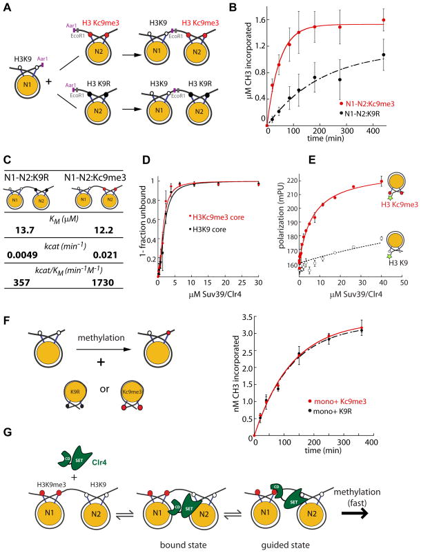
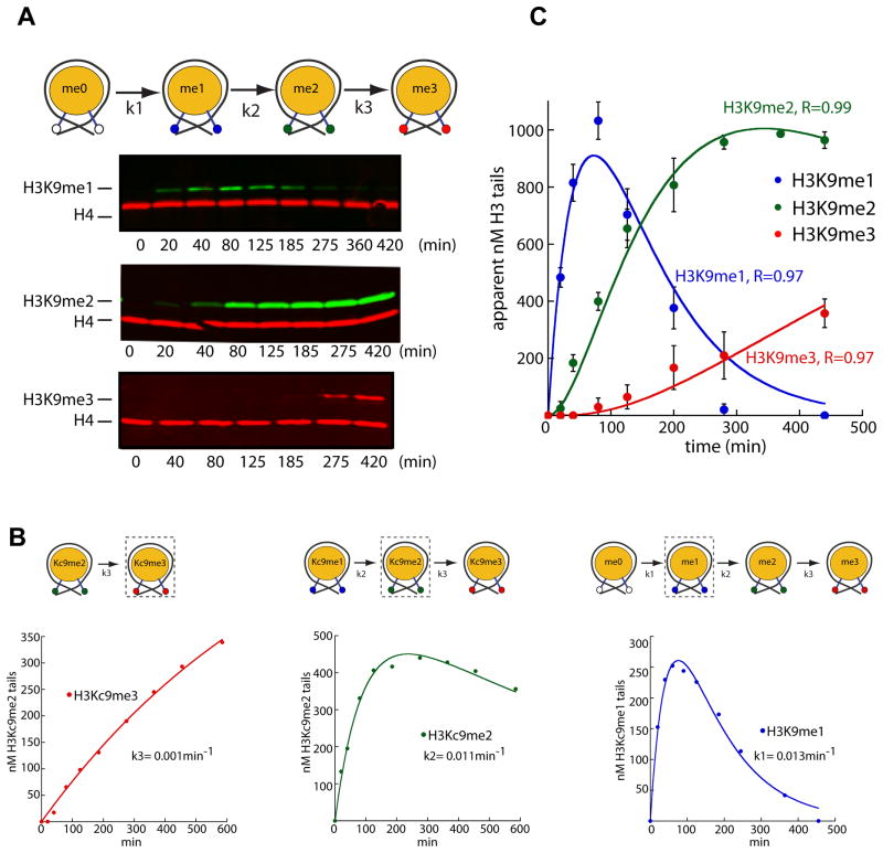
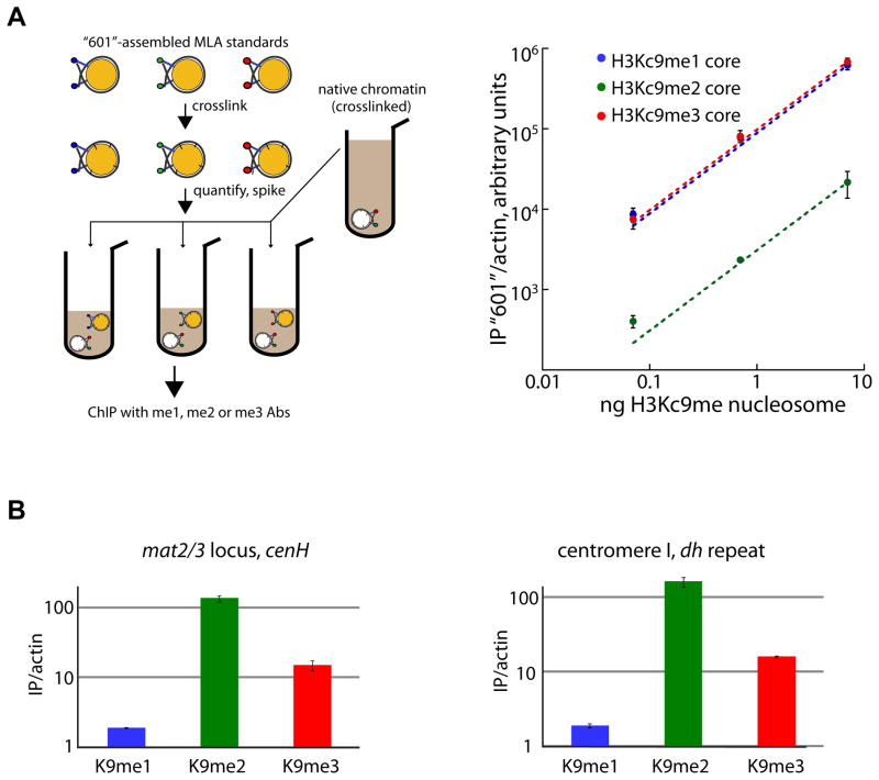
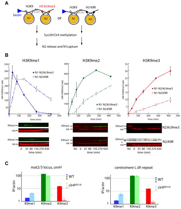
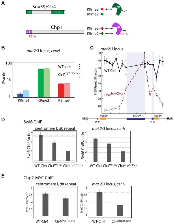
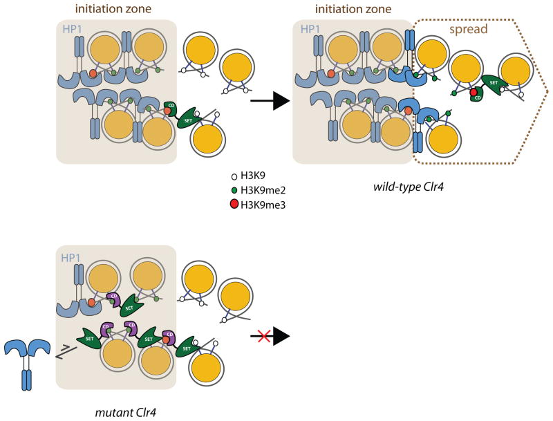
In Schizosaccharomyces pombe, heterochromatin spread, which is marked by histone 3 lysine 9 methylation (H3K9me), requires the chromodomains (CDs) of the H3K9 methylase Suv39/Clr4 and the HP1/Swi6 protein. It is unclear how the actions of these two H3K9me-recognizing CDs are coordinated. We find that the intrinsic preference of Suv39/Clr4 is to generate dimethylated H3K9 product. The recognition of pre-existing H3K9me marks by the CD of Suv39/Clr4 stimulates overall catalysis, enabling the accumulation of small amounts of trimethylated product in vivo. Coincidentally, the Suv39/Clr4 CD, unlike the HP1/Swi6 CD, has been shown to prefer the trimethyl state over the dimethyl state. We show that this preference enables efficient heterochromatin spread in vivo by reducing competition with HP1 proteins for the more prevalent dimethyl state. Our results reveal a strategy by which "writers" and "readers" of a chromatin mark exploit different methylation states on the same residue in order to facilitate collaboration and avoid competition.


Figures

Figure 1
Figure 1. H3K9me3 raises the kcat of Suv39/Clr4 only when present in cis in a dinucleosome context
A. Scheme for the production of asymmetric dinucleosomes. B. Dinucleosome methylation time courses at saturating (20μM) Suv39/Clr4. 100nM N1-N2:K9R (black) or N1-N2:Kc9me3 dinucleosomes (red) were used as substrates. The kcat values for Suv39/Clr4 for N1-N2:K9R and N1-N2:Kc9me3 were 0.0049min−1 and 0.021min−1, respectively. The kcat was derived from normalized data points from 4 separate experiments. Error bars denote standard error of five repeats. C. Enzymological parameters for Suv39/Clr4 activity on N1-N2:K9R or N1-N2:Kc9me3 dinucleosomes. D. Electrophoretic mobility shift experiments with H3K9 (black circles) and H3Kc9me3 (red circles) core mononucleosomes to determine the overall binding K1/2. The K1/2 of Suv39/Clr4 for H3K9 and H3Kc9me3 core mononucleosomes was 1.8μM and 1.5μM, respectively. Error bars denote standard error of three repeats. E. Fluorescence polarization (FP) on nucleosomes. H3K9 (open black circles) and H3Kc9me3 (red circles) core mononucleosomes were fluorescently labeled at the 5′ end of the 147-bp positioning sequence. FP detected by this label is sensitive to local binding events, such as at the nearby H3 tail, as previously described (Canzio et al.,2011). The Suv39/Clr4 dependent FP increase at this position is at least 25- fold more sensitive to the presence of the H3Kc9me3 mark over the H3K9 control. In this panel the bars shown do not denote standard error, but instead show the variation between two independent repeats. F. Methylation of mononucleosomes in presence of effector nucleosomes in trans. LEFT: Experimental design. RIGHT: Methylation time courses at 2μM Suv39/Clr4 (below the KM for di- and mononucleosomes). 300nM wild-type core mononucleosomes were used as substrates. K9R (black) or Kc9me3 core mononucleosomes (red) were added as effector in trans at 10μM. In this panel the bars shown do not denote standard error, but instead show the variation between two independent repeats. G. Model for role of the Suv39/Clr4 CD in catalytic enhancement of H3K9 methylation on chromatin. Formation of the enzyme-substrate ground state complex (bound state) is independent of pre-existing H3K9 methyl marks. Catalysis requires formation of a guided state, which optimally positions the active site relative to the H3K9 substrate. A pre-existing H3K9 methyl mark stabilizes the guided state. The absence of a pre-existing H3K9 methyl mark, slows catalysis as formation of the guided state is destabilized.
Figure 2
Figure 2. Suv39/Clr4 is primarily a H3K9 dimethylase on mononucleosomes
A. TOP: H3K9 methylation by Suv39/Clr4 proceeds in a series of consecutive steps: monomethylation (k1), dimethylation (k2) and trimethylation (k3). BOTTOM: Quantitative western blots of H3K9me1, me2 and me3. Methylation reactions were performed at 20μM Suv39/Clr4 and 125nM core mononucleosomes. Reactions were stopped and separated by SDS-PAGE and probed for H4 (red) and H3K9me1 (green, top panel), H3K9me2 (green, middle panel) or H3K9me3 (red, bottom panel). B. Determination of k1, k2 and k3. Methyl-Lysine Analog (MLA) core mononucleosomes representing intermediate steps of the consecutive reaction depicted in (A) were used as methylation substrates as in (A). The concentration of methylated tails in nanomolar (nM) is derived from the H4-adjusted amount of H3K9me1, H3Kc9me2 or H3Kc9me3 at each time point as determined by H3Kc9me1, me2 or me3 standards. C. Fitting the consecutive methylation reaction. k1, k2 and k3 as determined from (B) were used to model the formation of H3K9me1, me2 and me3 starting from unmethylated core mononucleosomes. The lines describing the model fit well to the data obtained from unmethylated H3K9 nucleosomes. H3K9me1, me2 and me3 quantities determined as in (B). Error bars denote standard error of three timecourses.
Figure 3
Figure 3. The dimethyl state is the predominant H3K9 methylation state in vivo alongside a small trimethyl pool
A. The Q-ChIP method. LEFT: overview of the H3Kc9me nucleosome ChIP spiking scheme. RIGHT: Double log plot of amount of crosslinked H3Kc9me1, me2, or me3 core mononucleosomes spiked into ChIP reactions (x-axis), vs. amount of precipitated, actin-normalized 601 nucleosome DNA (y-axis) as quantified by RT-qPCR. Error bars denote standard error of three immunoprecipitations (IPs). B. ChIP with anti-H3K9me1 (blue), anti-H3K9me2 (green) or anti-H3K9me3 (red) antisera at the mating type locus cenH element (LEFT) and the centromeric dh repeat (RIGHT). Enrichment is represented as the ratio of the actin-normalized signal in Suv39/Clr4 WT over the clr4Δ (no methylation) mutant. Relative H3K9me1, me2 and me3 signals are normalized by a parallel Q-ChIP reaction and shown on a log scale. Error bars denote standard error of three IPs.
Figure 4
Figure 4. Impact of product guidance on H3K9me3 accumulation in vitro and in vivo.
A. Scheme for measuring mono-, di- and trimethylation on Biotin-N1-N2K9R and Biotin-N1-N2MLA dinucleosomes. Methylation reactions were performed as in Figure 2A, reactions were stopped with 2mM SAH. Following reactions, N2 effector nucleosomes were released by EcoRI restriction digestion and N1 nucleosomes separated by SDS-PAGE and probed with indicated antisera. B. TOP: Quantification of Western blot data as in Figure 2B. BOTTOM: Representative western blots. N2 represents effector nucleosomes from t=0 min time point. The N1-N2:K9R anti-H3K9me3 blot is enhanced to show the very small pool of detectable H3K9me3. Error bars denote standard error of three timecourses. C. The Suv39/Clr4 W31A mutation leads to loss of H3K9me3 at the mat2/3 locus cenH element or centromere dh repeat. Q-ChIP was performed as in Figure 3B. The Suv39/Clr4W31A is denoted in light colors. Wild-type data are the same as in Figure 3B and shown for comparison. Error bars denote standard error of three IPs.
Figure 5
Figure 5. Impact of changing H3K9me3 selectivity of the Suv39/Clr4 CD on methylation spread HP1 assembly in vivo.
A. The CD of Suv39/Clr4 (residue 1–64) was swapped for the F61A mutant CD of Chp1 (1–77). The F61A Chp1 CD has a similar preference for H3K9me2 and H3K9me3, while the Clr4 CD is more H3K9me3 specific. B. H3K9me1, me2 or me3 signals in Suv39/Clr4 WT or Suv39/Clr4chp1CDF61A at the cenH initiation region probe of the mat2/3 locus were quantified as in Figure 3B. Error bars represent standard error of three IPs. C. H3K9me2 signals across the mat2/3 locus in Suv39/Clr4 WT or Suv39/Clr4chp1CDF61A backgrounds. Error bars represent standard error of three IPs. D. The Suv39/Clr4chp1CDF61A CD domain swap decreases HP1/Swi6 at initiation sites. Swi6 enrichment over a clr4Δ mutant, normalized to an actin control is shown. Error bars represent standard error of three of three IPs. E. The Suv39/Clr4chp1CDF61A CD domain swap decreases Chp2:7xMYC at the mat2/3 locus and to a lesser degree at the centromere. Chp2:7xMYC enrichment over a clr4Δ mutant normalized to an actin control is shown. Error bars represent standard error of three IPs.
Figure 6
Figure 6. Model for co-ordination of Suv39/Clr4 and HP1 protein chromodomains in H3K9 methylation spread across nucleosomes
In the initiation zone, a pool of H3K9me2 and H3K9me3 marks is formed by directly recruited Suv39/Clr4 molecules. The CDs of HP1 proteins such as Swi6 and Chp2 cover most of the central H3K9me2 and H3K9me3 marks. Specific H3K9me2/3 recognition of some HP1 proteins like Swi6 requires oligomerization mediated by the chromatin template (Canzio et al., 2011), exposing H3K9me2/3 at the edge of the heterochromatic domain. In the context of wild-type enzyme, exposed H3K9me3 marks act as a guidance mark for Suv39/Clr4. Any nearby unmethylated nucleosome transiently available for the guided state of Suv39/Clr4 will be methylated, resulting mostly in H3K9 dimethylation, and a small H3K9 trimethylated pool. These new sites are covered by the HP1 proteins, preferentially exposing H3K9 methylated tails at the edge of the growing heterochromatin structure. Suv39/Clr4 does not significantly compete for HP1 proteins in the central domain due its high preference for H3K9me3, lacking in HP1 proteins, which efficiently recognize H3K9me2 marks and are also present in higher concentration. In the context of Suv39/Clr4chp1CDF61A (“mutant Clr4”), the increased affinity for H3K9me2 now allows Suv39/Clr4 to engage the guided state at a majority of methylated nucleosomes, competing non-productively with HP1 proteins.

References

    1. Bhalla KN. Epigenetic and Chromatin Modifiers As Targeted Therapy of Hematologic Malignancies. J Clin Oncol. 2005;23:3971–3993. - PubMed
    1. Buhler M, Haas W, Gygi SP, Moazed D. RNAi-dependent and -independent RNA turnover mechanisms contribute to heterochromatic gene silencing. Cell. 2007;129:707–721. - PubMed
    1. Canzio D, Chang EY, Shankar S, Kuchenbecker KM, Simon MD, Madhani HD, Narlikar GJ, Al-Sady B. Chromodomain-mediated oligomerization of HP1 suggests a nucleosome-bridging mechanism for heterochromatin assembly. Mol Cell. 2011;41:67–81. - PMC - PubMed
    1. Carbone R, Botrugno OA, Ronzoni S, Insinga A, Di Croce L, Pelicci PG, Minucci S. Recruitment of the Histone Methyltransferase SUV39H1 and Its Role in the Oncogenic Properties of the Leukemia-Associated PML-Retinoic Acid Receptor Fusion Protein. Mol Cell Biol. 2006;26:1288–1296. - PMC - PubMed
    1. Ceol CJ, Houvras Y, Jane-Valbuena J, Bilodeau S, Orlando DA, Battisti V, Fritsch L, Lin WM, Hollmann TJ, Ferre F, et al. The histone methyltransferase SETDB1 is recurrently amplified in melanoma and accelerates its onset. Nature. 2011;471:513–517. - PMC - PubMed

Publication types

MeSH terms