

Review
doi: 10.1016/j.virol.2013.06.006.
Epub 2013 Jul 10.
The papillomavirus E2 proteins
Affiliations
- PMID: 23849793
- PMCID: PMC3783563
- DOI: 10.1016/j.virol.2013.06.006
Item in Clipboard
Review
The papillomavirus E2 proteins
Virology.
2013 Oct.
Abstract
The papillomavirus E2 proteins are pivotal to the viral life cycle and have well characterized functions in transcriptional regulation, initiation of DNA replication and partitioning the viral genome. The E2 proteins also function in vegetative DNA replication, post-transcriptional processes and possibly packaging. This review describes structural and functional aspects of the E2 proteins and their binding sites on the viral genome. It is intended to be a reference guide to this viral protein.
Keywords: E2; Genomics; HPV; Mutation; Papillomavirus; Replication; Structure; Tethering; Transcription.
Published by Elsevier Inc.
Figures
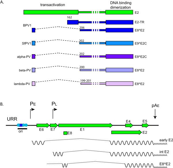
A. Isoforms The two conserved domains of the full-length E2 protein, encoded by all PVs, are
shown in green at the top. The region between the domains is of variable length
and is named the Hinge. Many PVs have been shown to encode an E8^E2 (or
equivalent) repressor form as shown and all PVs encode homologous sequences and
splice donor/acceptor sites. BPV1 encodes two repressor forms. Some examples of
E2 repressor proteins are shown. B. E2 transcripts The URR and early region of an alpha-PV genome is shown. In the lower layers of a
papilloma, mRNAs are transcribed from the early promoter, PE, and terminated at
the early polyadenylation signal, pAE. At the next stage of differentiation, the
late promoter is activated, but messages are still truncated at pAE. Transcripts
encoding the full-length E2 protein are transcribed from the early and late
promoters (reviewed in (Johansson and Schwartz,
2013)). It is not clear whether both promoters can transcribe the
E8^E2 mRNAs, though it seems most likely that they would be transcribed from the
early promoter in undifferentiated cells. The origin of replication (ori) is
indicates with the E1 binding site shown as a blue square and E2 binding sites
as cyan circles.
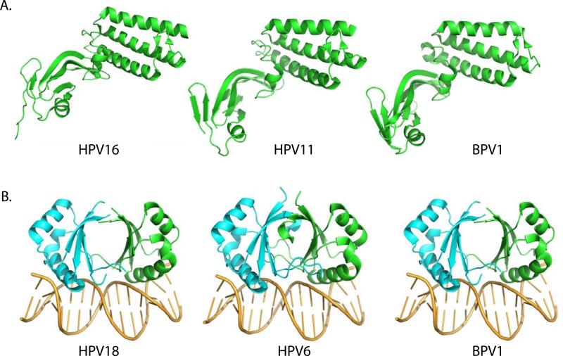
A. Transactivation Domain The transactivation domain structures for three E2 proteins are shown. Structures
PDB: 2JEU (BPV1), PDB: 1R6K (HPV11) and PDB: 1DTO (HPV16) were rendered in
Pymol. B. DNA binding Domain The DNA binding domain structures for three E2 proteins are shown. Structures
PDB: 1JJ4 (HPV18), PDB: 2AYB (HPV6) and PDB: 2BOP (BPV1) were rendered in
Pymol.
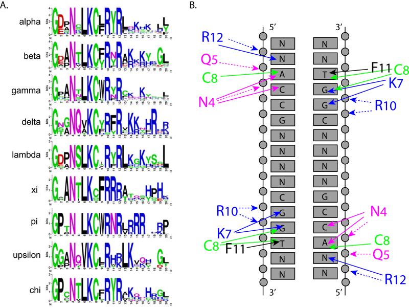
A. Recognition Helix Sequence Logos Sequence logos were created for the recognition helix of all genera of PVs with
more than six members. The structure of the recognition helix shown at the top
is derived from PDB: 2AYB HPV6 E2 bound to DNA (Hooley et al., 2006). Logos were created using Weblogo (http://weblogo.berkeley.edu/logo.cgi ) (Crooks et al., 2004). B. Contacts between the Recognition Helix and DNA Binding Site This figure was adapted from (Hegde, 2002;
Hegde et al., 1998). It shows the
contacts between residues in the recognition helix of BPV1 E2 (with residue
numbers converted to match those shown in A) and the consensus DNA binding site.
Solid arrows signify contacts with specific bases. Dotted arrows represent
contacts with the phosphate backbone.
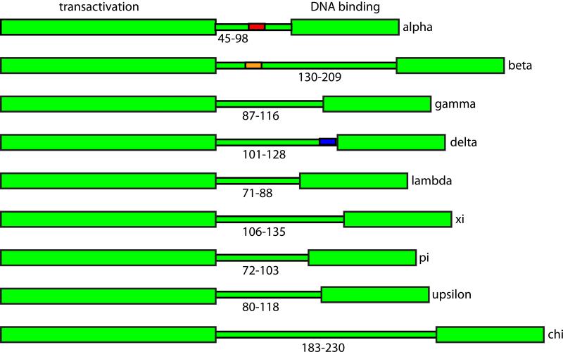
The variable lengths of the hinge regions from nine genera of papillomaviruses
are shown. Functional, conserved regions that have been mapped to the hinge
region are shown: Alpha E2s, nuclear localization and nuclear matrix attachment
(HPV11, in red); Beta E2s, chromatin attachment and PKA phosphorylation site
(HPV8, orange); Delta E2s, conformational switch and CK2 phosphorylation site
(BPV1 blue).
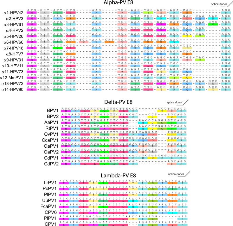
The E8 moieties from a series of Alpha PVs, all Delta PVs and all Lambda PVs are
shown. Sequences were extracted from the PaVE database
and aligned using the ClustalW module of Geneious v6 created by Biomatters.
Available from http://www.geneious.com/ .

A. E2 Binding Site Sequence Logos E2 binding sites conforming to ACCN6GGT were extracted from the URR
sequences in the PaVE database from genera
of PVs with more than six members. This amounted to 281 Alpha, 130 Beta, 127
Gamma, 68 Delta, 21 Lambda, 19 Xi, 13 Pi, 15 Epsilon and 48 Chi E2 consensus
motifs. Sequence logos were created for the recognition helix of sites from each
genera using Weblogo (http://weblogo.berkeley.edu/logo.cgi ) (Crooks et al., 2004). B. E2 Binding Site Sequence Logos for Individual Alpha E2 Sites Individual E2 binding sites sequences conforming to ACCN6GGT and
corresponding to the positions shown (#1, 2, 3, 4) were extracted from the 54
alpha URR sequences in the PaVE database that have the
traditional four sites shown. URRs containing non-consensus E2 binding sites
were not included. Sequence logos were created for the E2 site at each position
using Weblogo (http://weblogo.berkeley.edu/logo.cgi ) (Crooks et al., 2004). C. Number of E2 Binding Sites in the URRs of PV types in Different Genera The number of consensus (ACCN6GGT) E2 binding sites in the URR of PV
types from genera of PVs with more than six members.
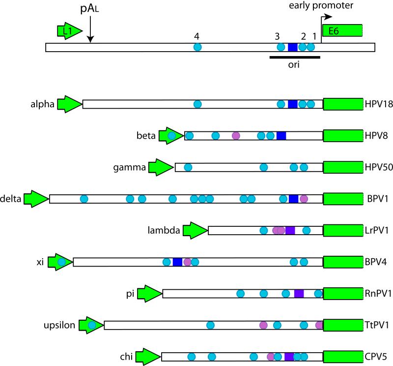
The URRS of PVs selected from nine different genera are shown. The ends of the L1
and E6 ORFs are indicated. The E1 binding site, when mapped, is indicated by a
blue box, while putative sites are indicated by purple boxes. E2 binding sites
that match the consensus ACCN6GGT are shown as cyan circles. E2 sites
that deviate from this consensus are shown as light purple circles.
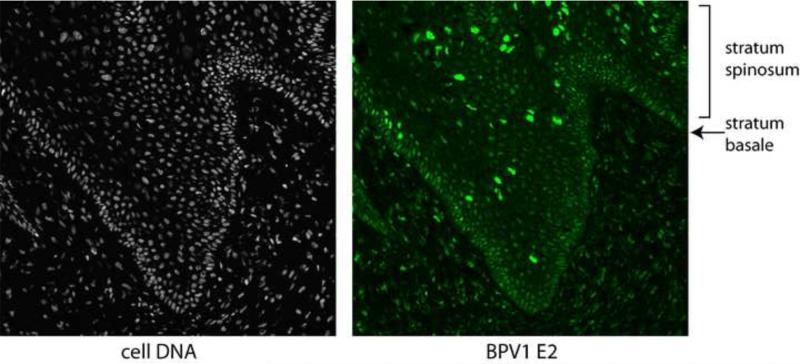
Shown is a section of tissue from a BPV1 infected fibropapilloma. Host cells are
visualized in the left panel using DAPI to stain nuclei. The E2 protein is
detected in the right panel using a BPV1 E2 specific monoclonal antibody (B201)
with an epitope between residues 160 and 200 in the E2 protein. B201 antiserum
was a gift from Elliot Androphy (Yao et al.,
1998).
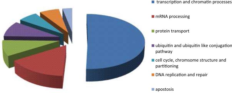
The E2 associated proteins shown in Table
2 were categorized according to Gene Ontology functions using the
DAVID v6.7 bioinformatics resource (Vempati,
2012) with some manual curation.
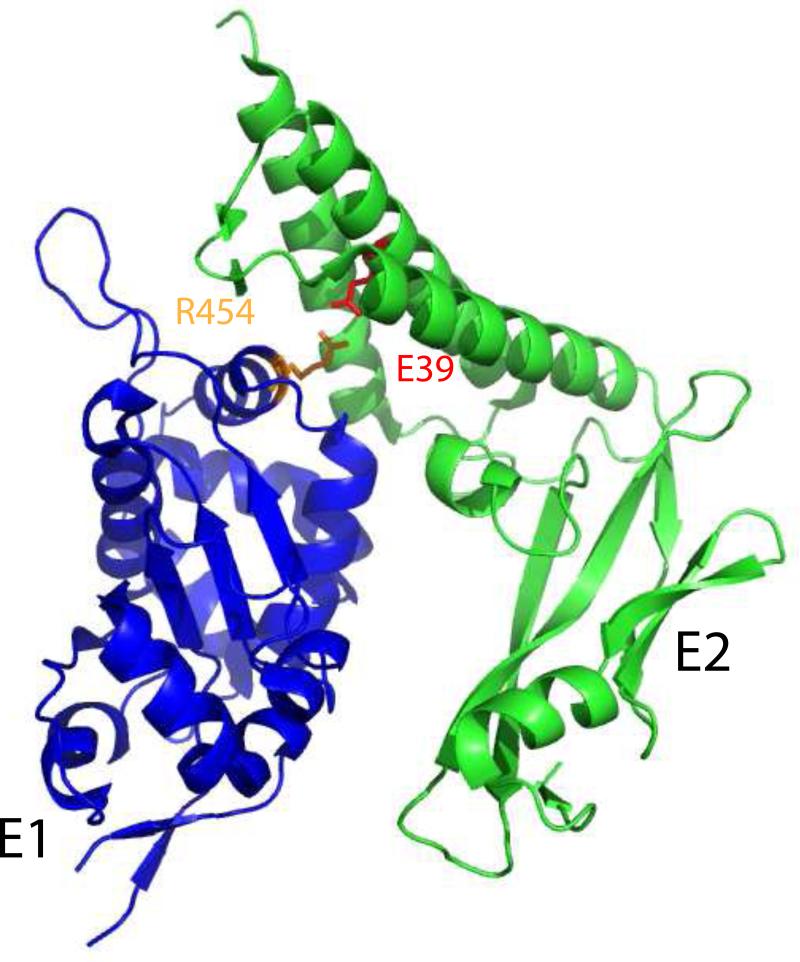
The structure of the HPV18 E2 transactivation domain in complex with a C-terminal
fragment of E1 (residues 428-631) was rendered in Pymol (PDB: 1TUE). Highlighted
on the structure in red is E39, which forms a buried salt bridge with the highly
conserved residue arginine 454 in E1 (shown in orange).
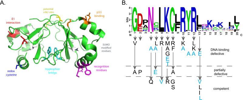
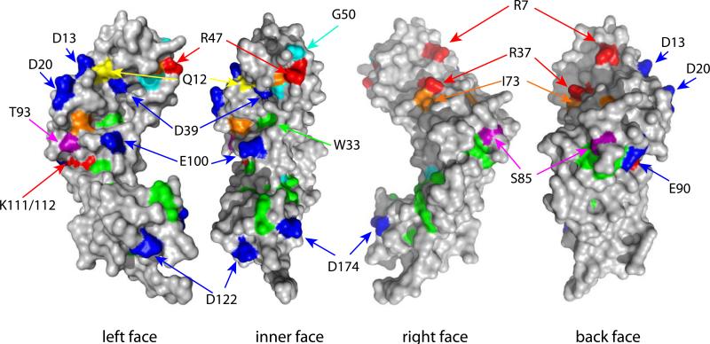
A. Key residues on the DNA Binding Domain The BPV1 E2 DNA binding domain structure PDB: 1DBD was rendered in Pymol.
Highlighted on the structure are regions of interest. In red is the region in
BPV1 E2 that interacts with E1 when both proteins are bound to adjacent sites
(Gillitzer et al., 2000). The same
helix interacts with p53, and shown in orange on the other monomer are residues
that when mutated (HPV16 D338, E340, W341, D344) reduce p53 binding (Brown et al., 2008). Also indicated are
potential SUMO modification sites (grey) and cdk2 phosphorylation sites (yellow)
(Johansson et al., 2009; Wu et al., 2008). In cyan are two
tryptophan residues that are readily crosslinked in the dimer by UV radiation
(Corina et al., 1993). These sites
have been mapped in different E2 proteins and are shown on the BPV1 domain for
illustrative purposes only. B. Mutational analysis of the E2 Recognition Helix The phenotype of the amino acid substitutions shown are divided into three
categories according to their ability to specifically bind DNA. Mutations shown
in black are from BPV1 (Carruth and McBride,
2001; Grossel et al., 1996;
McBride et al., 1992; Prakash et al., 1992; Ustav et al., 1993). Mutations shown in
light blue are from HPV11 (Matsumoto et al.,
1997).
The transactivation domain structure PDB: 1DTO (HPV16) was rendered in Pymol.
Key, conserved residues on the surface of the domain are indicated.
References
-
- Abbate EA, Voitenleitner C, Botchan MR. Structure of the papillomavirus DNA-tethering complex E2:Brd4 and a peptide that ablates HPV chromosomal association. Mol.Cell. 2006;24:877–889. - PubMed
-
- Akgul B, Pfefferle R, Marcuzzi GP, Zigrino P, Krieg T, Pfister H, Mauch C. Expression of matrix metalloproteinase (MMP)-2, MMP-9, MMP-13, and MT1-MMP in skin tumors of human papillomavirus type 8 transgenic mice. Exp.Dermatol. 2006;15:35–42. - PubMed
Publication types
MeSH terms
Substances
Grants and funding
LinkOut - more resources
Full Text Sources
Other Literature Sources
