Review
. 2013 Oct;3(5):546-57.
doi: 10.1016/j.coviro.2013.06.008. Epub 2013 Jul 12.

A virocentric perspective on the evolution of life

Affiliations
Review

A virocentric perspective on the evolution of life

Eugene V Koonin et al. Curr Opin Virol. 2013 Oct.

Abstract

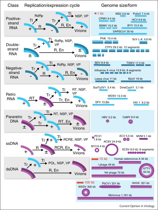
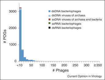
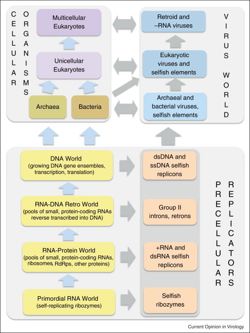
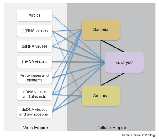
Viruses and/or virus-like selfish elements are associated with all cellular life forms and are the most abundant biological entities on Earth, with the number of virus particles in many environments exceeding the number of cells by one to two orders of magnitude. The genetic diversity of viruses is commensurately enormous and might substantially exceed the diversity of cellular organisms. Unlike cellular organisms with their uniform replication-expression scheme, viruses possess either RNA or DNA genomes and exploit all conceivable replication-expression strategies. Although viruses extensively exchange genes with their hosts, there exists a set of viral hallmark genes that are shared by extremely diverse groups of viruses to the exclusion of cellular life forms. Coevolution of viruses and host defense systems is a key aspect in the evolution of both viruses and cells, and viral genes are often recruited for cellular functions. Together with the fundamental inevitability of the emergence of genomic parasites in any evolving replicator system, these multiple lines of evidence reveal the central role of viruses in the entire evolution of life.


Figures

Figure 1
Figure 1
The diversity of the virus world. Seven major classes of viruses with distinct genome replication/expression cycles are shown at the left; representative examples of the corresponding genome sizes and architectures are shown at the right approximately to scale indicated in red. Abbreviations: Tl, translation; Tr, transcription; R, replication; RT, reverse transcription; RCR, rolling-circle replication; kb, kilobase. En, encapsidation; RdRp, RNA-dependent RNA polymerase; NSP, non-structural proteins; VP, virion proteins; RCRE, rolling-circle replication endonuclease. Designations: (+), (−), (±), positive-strand, negative-strand, and double-stranded nucleic acids, respectively. Virus acronyms: MS2, bacteriophage MS2; PolioV, Poliomyelitis virus; CPMV, Cowpea mosaic virus; HCV, Hepatitis C virus; BSMV, Barley stripe mosaic virus; BYV, Beet yellows virus; SARS CoV, SARS Coronavirus; Phi6, bacteriophage phi 5; ScV L-A, Saccharomyces cerevisiae virus L-A; CTFV, Colorado tick fever virus; BDV, Borna disease virus; TSWV, Tomato spotted wilt virus; EboV, Ebola virus; SceTy3V1, Saccharomyces cerevisiae Ty virus 1; DmeCopV1, Drosophilla melanogaster Copia virus 1; WDSV, Walleye dermal sarcoma virus; SFV, Simian foamy virus; HIV1, Human immunodeficiency virus 1; HBV, Hepatitis B virus; CaMV, Cauliflower mosaic virus; PCV1, Porcine circovirus 1; ACV, African cassava mosaic virus; MVM, Minute virus of mice; M13, bacteriophage M13; ACSV, Aeropyrum coil-shaped virus; SCSV, Subterranean clover stunt virus; ATV, Acidianus two-tailed virus; WSSV, White spot syndrome virus; PbCV1, Paramecium bursaria Chlorella virus 1; HHV6, Human herpesvirus 6.
Figure 2
Figure 2
The size distribution of clusters of orthologous genes from prokaryotic viruses: the lack of a conserved core. POGs, Phage Orthologous Groups. Altogether, 487 genomes of bacteriophages and viruses of archaea were analyzed.
Figure 3
Figure 3
The network of hallmark genes connecting different classes of viruses and capsid-less selfish elements. The colored circles show classes of viruses and related capsid-less selfish elements (see Figure 1) and other shapes show distinct classes of non-viral selfish elements. The size of each shape roughly reflects the abundance and diversity of the respective class. The color-coded edges connecting the shapes denote shared hallmark genes; the thickness of each line roughly reflects the prevalence of the respective gene in the corresponding classes of viruses and selfish elements (in most cases, any given hallmark gene is present only in a subset of the class members). The dashed line reflects the tentative link between RNA-dependent RNA polymerases of positive-strand and negative-strand RNA viruses. JRC, Jelly Roll Capsid protein; RdRp, RNA-dependent RNA polymerase; RT, reverse transcriptase; S3H, superfamily 3 helicase; Int, integrase; ATPase, packaging ATPase of the FtsK family; Pro, C5-family thiol protease; RCRE, rolling circle replication (initiation) endonuclease. Compared with the original list of viral hallmark genes [12], integrase and thiol proteases were addionally included whereas several genes that are widespread among diverse dsDNA viruses but not found in other classes of selfish elements are not shown. The Int gene is present in numerous but not all prokaryotic and eukaryotic DNA transposons. Helitrons are eukaryotic transposons that replicate via the RCR mechanism. Polintons are large, self-replicating eukaryotic transposons.
Figure 4
Figure 4
The model of the evolution of all extant life forms (top) from a virus-like primordial state (bottom).
Figure 5
Figure 5
Classification of life forms: the viral and cellular empires. The lines denote parasite-host associations between selfish elements and cellular life forms as well as fluxes of genetic information. The thickness of the lines roughly reflects the prevalence of these associations and the intensity of the fluxes. The dashed line shows a putative RNA virus infecting a hyperthermophilic archaeon [88].

References

    1. Bergh O., Borsheim K.Y., Bratbak G., Heldal M. High abundance of viruses found in aquatic environments. Nature. 1989;340:467–468. - PubMed
    1. Rosario K., Breitbart M. Exploring the viral world through metagenomics. Curr Opin Virol. 2011;1:289–297. - PubMed
    2. An overview of the discovery of new viruses and virus–host associations through analysis of metagenomic data.

    1. Culley A.I. Virophages to viromes: a report from the frontier of viral oceanography. Curr Opin Virol. 2011;1:52–57. - PubMed
    2. An overview of the discovery of novel marine viruses, including giant viruses and virophages, and their roles in the ecology of the oceans.

    1. Rohwer F. Global phage diversity. Cell. 2003;113:141. - PubMed
    1. Colson P., de Lamballerie X., Fournous G., Raoult D. Reclassification of giant viruses composing a fourth domain of life in the new order Megavirales. Intervirology. 2012;55:321–332. - PubMed

Publication types