. 2013 Jul 25;4(2):302-315.
doi: 10.1016/j.celrep.2013.06.022. Epub 2013 Jul 11.

Polyester modification of the mammalian TRPM8 channel protein: implications for structure and function

Affiliations

Polyester modification of the mammalian TRPM8 channel protein: implications for structure and function

Chike Cao et al. Cell Rep. .

Abstract

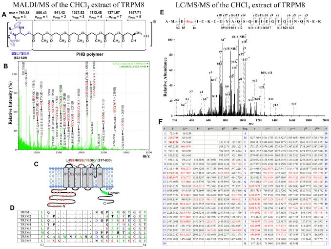
The TRPM8 ion channel is expressed in sensory neurons and is responsible for sensing environmental cues, such as cold temperatures and chemical compounds, including menthol and icilin. The channel functional activity is regulated by various physical and chemical factors and is likely to be preconditioned by its molecular composition. Our studies indicate that the TRPM8 channel forms a structural-functional complex with the polyester poly-(R)-3-hydroxybutyrate (PHB). We identified by mass spectrometry a number of PHB-modified peptides in the N terminus of the TRPM8 protein and in its extracellular S3-S4 linker. Removal of PHB by enzymatic hydrolysis and site-directed mutagenesis of both the serine residues that serve as covalent anchors for PHB and adjacent hydrophobic residues that interact with the methyl groups of the polymer resulted in significant inhibition of TRPM8 channel activity. We conclude that the TRPM8 channel undergoes posttranslational modification by PHB and that this modification is required for its normal function.


Figures

Figure 1
Figure 1
Mass spectrometric analysis of the chloroform-extracted peptide LHSSNKSSLYSGR (817–829) of the TRPM8 protein derived from MALDI/MS experiments: Panel A: Molecular composition of PHBylated serine residue on the SSLYSGR (823–829) peptide with a number of PHB units attached via an ester bond, numbers indicate the PHB modification with a shift in the monoisotopic masses. Panel B: Intensity peaks of the LHSSNKSSLYSGR (817–829) peptide with indicated PHB modifications detected in mass spectrum of the chloroform-extracted peptides derived from MALDI/MS. Masses were analyzed with the ExPASy FindMod Tool (Swiss Proteomics Bioinformatics Resources) run against the TRPM8 sequence with possible PHB modifications up to 30 units, with 0 or 1 missed cleavage cites for trypsin-digested protein (error window ±50 ppm). The variability of different length of PHB might be due to the breakage of labile ester bonds under the MS laser beam (Xian et al., 2007). Panel C: Cartoon of the putative PHB-modification sites on the TRPM8 protein with a sequence indication for the extracellular PHBylated peptides. The red spheres indicate putative PHBylated peptides on the N-terminus of TRPM8, derived from MALDI-MS experiments. D: The amino-acid sequence of the S3–S4 linker of TRPM family ion channels is not conserved. Panel E: MS/MS spectrum of the quadruply charged ion (m/z 775.354) corresponding to the peptide 63AMESICKCGYAQSQHIEGTQINQNEK88 showing the methionine oxidation and 2 units of PHB modification on serine residues. The observed y- and b-ion series confirmed the peptide sequence. The b4, b9, b10, b11, b12, b14, b15, b16, b17, b19, b20, and b21 ions confirmed PHB localization on the serine residue. Panel F: A table showing the identified peptide fragment ions from the spectrum (red) versus theoretical fragment ions not found in the spectrum (black). The bold italic red ions are the relatively abundant y- or b-ions that contributed to the scoring of the peptide and PTM identification. Additional matched ions, including bold red ions, were not used for the calculation of the identification score.
Figure 2
Figure 2
Screening of the serine mutants within the peptide LHSSNKSSLYSGR (817–829) for TRPM8 activity in intracellular Ca2+-measurements. Fluorescence measurements of intracellular Ca2+ concentration were performed on HEK-293 cells transiently transfected with the wild type or mutants TRPM8 (0.7 μg) and GFP (0.2 μg) constructs. Panels A–I are representatives from single cover slips, the total number of measurements (n) and total number of cells for each variety are indicated in parentheses A: TRPM8 wild type (n = 5, ncells = 71); B: double serine mutant S823G/S824G (n = 8, ncells = 99); C: quadruple serine mutant S819G/S820G/S823G/S824G or GGNKGG (n = 4, ncells = 76); D: quintuple serine mutant 5S-G: S819G/S820G/S823G/S824G/S827G or GGNKGGLYG (n = 8, ncells = 61); E: double serine mutant S819P/S820P (n = 4, ncells = 53); F: single serine mutant S827G (n = 7, ncells = 171); G: triple serine mutant S823G/S824G/S827P or GGLYP (n = 5, ncells = 67); H: quintuple serine mutant S823G/S824G/L825G/Y826G/S827P or GGGGP (n = 7, ncells = 88); I: single serine mutant S827P (n = 6, ncells = 83); J: the summary of cold, menthol, icilin, and second cold applications (error bars stand for ±s.e.m.). Alternatively, these measurements were carried with a single application of icilin, which resulted in the similar observations (Figure S6). We have also performed measurements with subsequent ligand washout, but those resulted in a slow Ca2+ run down and prolonged experiment time that might have an effect on cells (data not shown). K: Cartoon of the putative PHB-modification sites on the TRPM8 protein with a sequence indication for the extracellular PHBylated peptides targeted for the mutagenesis.
Figure 3
Figure 3
Inhibition of TRPM8 activity by PHB-depolymerase, PhaZ7, in intracellular Ca2+-measurements Panels A–C: Fluorescence measurements of intracellular Ca2+ concentration were performed on HEK-293 TRPM8 stable cell lines with transiently transfected either with GFP (0.2 μg) alone (panel A) (n = 7, ncells= 209) or together with the PhaZ7 clone (0.7 μg) (n = 15, ncells= 277) and the S136A-PhaZ7 mutant – inactive enzyme (0.7 μg) (n = 3, ncells= 61) (panel B). The summaries of averaged cold and menthol responses with GFP-positive cells are represented in panel C (p < 0.0005; error bars stand for ±s.e.m.). Panels D–K: Fluorescence measurements of intracellular Ca2+ concentration were performed on HEK-293 transiently transfected with TRPM8 (0.7 μg) and GFP (0.2 μg) panel D (n = 7, ncells=71). Further, TRPM8-expressing cells were treated for 1 h with an inactive form of PHB-depolymerase – S136A PhaZ7 (panel E) (n = 3, ncells= 26) and wild-type PhaZ7 (panel F) (n = 3, ncells= 40). Ca2+ signals obtained from the HEK cells transiently expressing TRPM8 mutants: L825G (panel G) (n = 5, ncells= 83), V830G (panel H) (n = 6, ncells= 90), R829E (panel I) (n = 5, ncells= 39), and Y826G (panel J) (n = 5, ncells= 37). Panels A–J are representative of single cover slips; the total number of measurements and the total number of cells for each variety are indicated in parentheses. The summaries of averaged cold, menthol, and icilin responses are represented in panel K (error bars stand for ±s.e.m.).
Figure 4
Figure 4. Inhibition of TRPM8 activity by PHB-depolymerase PhaZ7 in DRG neurons
Fluorescence measurements of intracellular Ca2+ concentration were performed on DRG neurons with transiently transfected TRPM8 (3 μg) and GFP (0.5 μg) (panel A) (n = 4, cells= 7), TRPM8 together with the PhaZ7 clone (3 μg) (panel B) (n = 5, ncells= 10), and mutant S827P (3 μg) (panel C) (n = 3, ncells= 6). Panels A–C are representatives of single cover slips; total number of experiments and number of neurons are indicated in parentheses. The summaries of averaged cold and menthol responses are represented in panel D (error bars stand for ±s.e.m.). Details of the neuronal transfection are given in supplementary information. Panel E: PHB signals enhanced in DRG neurons expressing TRPM8. DRG neurons expressing TRPM8/GFP or TRPM8/PhaZ7/GFP were immuno-probed with anti-PHB-IgG and Alexa-546 (red) as secondary antibodies. The transfection conditions are the same as described for the panels A and B. PHB-depolymerase hydrolyzes PHB, which results in reduced signals from plasma membrane and neurites. The upper panel shows PHB staining in DRG neurons transiently expressing TRPM8 and GFP; the two middle panels demonstrate PHB in neurons expressing TRPM8, PhaZ7, and GFP; and the lower panel shows PHB in un-transfected neurons. For immunocytochemical analysis cells were observed with a Zeiss (Oberkochen, Germany) LSM-510 confocal microscope (60X objective), equipped with an Argon laser (488 nm), a Red Diode laser (637 nm), and a Green HeNe laser (543 nm), in the Confocal Imaging Facility of the New Jersey Medical School.
Figure 5
Figure 5. Electrophysiological characterization of TRPM8 and other mutants' sensitivity to menthol and cold stimulus
Whole-cell current-voltage relationships from voltage ramps (−100 +100mV) were obtained for HEK-293 cells expressing the wild type TRPM8 (n = 7) and the mutant channels: 5S-G (n = 5), S827G (n = 4), S827P (n = 4), L825G (n = 4) Y826G (n = 3), V830G (n = 4); measurements were performed in the presence of 2 mM Ca2+. Panel A demonstrates averaged ramp-recordings for menthol-induced currents (500 μM) and panel B shows cold-induced currents (10 °C). Control values for background current for the wild type TRPM8 and mutants are very close, and only background wild-type TRPM8 current was demonstrated. Summary of the menthol-induced whole-cell currents at −100 and +100 mV potentials are shown in panel C, and those for cold-induced currents, in panel D. For each group background current marked in gray color. Panel E. Representative current traces for −100 +100 mV potentials of the cold-induced and subsequent menthol-induced currents obtained for the wild type TRPM8 and single mutants, S827G and S827P. In comparison to the wild type TRPM8, the menthol-induced activity of the PHB-mutants showed a reduction in current density in the following sequence S827G (p = 0.212 at +100 mV; p=0.0465 at −100 mV) > L825G (p = 0.044 at 100 mV; p = 0.0097 at −100 mV) > 5S-G (p = 0.023 at +100 mV; p = 0.0088 at −100 mV) > S827P (p = 0.012 at +100 mV; p = 0.024 at −100 mV) > V830G (p = 0.0024 at +100 mV; p = 0.0041 at −100 mV) > Y826G (p = 0.0072 at +100 mV; p = 0.0085 at −100 mV) (panels A and C). The cold-induced activity of the mutants was inhibited to a greater degree and exhibited a similar pattern of inhibition: S827G (p = 0.105 at +100 mV; p = 0.141 at −100 mV) > L825G (p = 0.0106 at 100 mV; p = 0.0559 at −100 mV) > 5S-G (p = 0.0054 at +100 mV; p = 0.096 at −100 mV) > S827P (p = 0.0016 at +100 mV; p = 0.186 at −100 mV) > V830G (p = 0.00005 at +100 mV; p = 0.0569 at −100 mV) > Y826G (p = 0.002 at +100 mV; p = 0.092 at −100 mV) (panels B and D). The number of asterisks in the graphs indicates the significance level: one star (*) for p ≤ 0.05, two (**) for ≤ 0.01, and three (***) for ≤ 0.001. Kinetics of cold activation of TRPM8 and mutants 5S-G, S827G, and L825G and their menthol-induced desensitization: Panel F: Summary plot of experiments showing cold ramp (drop to 10 °C in 15 sec) and menthol-induced (panel G) whole-cell currents for the wild type TRPM8 (n = 10) and mutants 5G (n = 5), S827G (n = 9), L825G (n = 8) at +100 mV potentials. To minimize current desensitization this set of experiments was performed in Ca2+-free extracellular solution. Panel G demonstrates slight but non-significant current desensitization for all 3 experimental replications of menthol application.
Figure 6
Figure 6. Menthol-induced activity of wild type TRPM8, and 5S-G, S827P, and Y826G mutant channels in the Planar Lipid Bilayers
Representative single-channel current recordings of TRPM8 and the mutant channels incorporated in planar lipid bilayers formed from POPC/POPE (3:1) in n-decane, between symmetric bathing solutions of 150 mM KCl, 0.2 mM MgCl2 in 20mM HEPES buffer, pH 7.4 at 22 °C. Between 0.2 and 0.5 μl of 0.2 μg/ml the protein was incorporated into POPC/POPE micelles, and added to the cis compartment (ground). Clamping potential was +100 mV. The closed state is delineated by a horizontal line under the traces on the left. Panel A: representative currents traces in the presence of 4 μM of DiC8 PtdIns(4,5)P2 and 500 μM of menthol. Panel B: Open probabilities of the wt TRPM8 and mutants obtained at +100 mV. Cold activation of 5S-G mutant channels in Planar Lipid Bilayer and Po change for the wild type TRPM8 and mutants: Panel C: Representative single-channel current recordings of 5S-G mutant channels activated by cold in the presence of 4 μM diC8PIP2. Clamping potential was +100 mV. Panel D: Open probability for the wild-type TRPM8 and 5S-G mutant obtained during cooling the planar lipid bilayers. Panel E: Two-phase TRPM8 temperature dependence and single-phase of 5S-G mutant in log(Po) versus T plot. Panel F: Van't Hoff plot of the equilibrium constant Keq demonstrates two-phase channel transitions for wild type TRPM8 and a single transition obtained for 5S-G mutant. Changes in enthalpy and entropy of TRPM8 and 5S-G activation are indicated on the graph.
Figure 7
Figure 7
Extracellular PHB levels of cells expressing the 5S-G and Y826G mutants are significantly reduced. Panel A: PHB signals on the cell surface obtained by immunocytochemical analyses of the polymer with anti-PHB-IgG on non-permeabilized cells detected with confocal microscopy (for details see supplementary methods section). The images were obtained with a Zeiss (Oberkochen, Germany) LSM-510 confocal microscope (60X objective). HEK-293 cells were transiently transfected with the wild type TRPM8 (1 μg), the 5S-G (1 μg), and the Y826G (1 μg) mutants along with GFP (0.2 μg) for detection purposes. Panel B: Summary of intensities of the PHB signals on PM measured for the wild type TRPM8 and the mutants 5S-G and Y826G. Panel C: The surface expression of the protein is not altered. The wild type TRPM8 and the mutants (5S-G, Y826G) were biotinylated and captured with streptavidin beads as described under the supplementary methods section. Panel D: Arithmetic means of the relative density of the proteins in the membrane fraction of wtTRPM8 and the 5S-G and Y826G mutants, n = 3. Panel E: Cartoon of a model for the PHB-TRPM8 complex with PHB attachment to the extracellular peptide LHSSNHSSLYSGR (817–829) and supported by a covalent bond to S827 and by hydrophobic interactions with Y826 and other hydrophobic residues in the region; red spheres show putative intracellular PHBylation sites of TRPM8.

References

    1. Bandell M, Dubin AE, Petrus MJ, Orth A, Mathur J, Hwang SW, Patapoutian A. High-throughput random mutagenesis screen reveals TRPM8 residues specifically required for activation by menthol. Nature neuroscience. 2006;9:493–500. - PubMed
    1. Bautista DM, Siemens J, Glazer JM, Tsuruda PR, Basbaum AI, Stucky CL, Jordt S-E, Julius D. The menthol receptor TRPM8 is the principal detector of environmental cold. Nature. 2007;448:204–208. - PubMed
    1. Behrendt HJ, Germann T, Gillen C, Hatt H, Jostock R. Characterization of the mouse cold-menthol receptor TRPM8 and vanilloid receptor type-1 VR1 using a fluorometric imaging plate reader (FLIPR) assay. Br J Pharmacol. 2004;141:737–745. - PMC - PubMed
    1. Braaz R, Handrick R, Jendrossek D. Identification and characterisation of the catalytic triad of the alkaliphilic thermotolerant PHA depolymerase PhaZ7 of Paucimonas lemoignei. FEMS Microbiology Letters. 2003;224:107–112. - PubMed
    1. Brauchi S, Orio P, Latorre R. Clues to understanding cold sensation: thermodynamics and electrophysiological analysis of the cold receptor TRPM8. Proceedings of the National Academy of Sciences of the United States of America. 2004;101:15494–15499. - PMC - PubMed

Publication types

LinkOut - more resources