. 2013 Jul 25;4(2):287-301.
doi: 10.1016/j.celrep.2013.06.019. Epub 2013 Jul 11.

Sympathetic innervation during development is necessary for pancreatic islet architecture and functional maturation

Affiliations

Sympathetic innervation during development is necessary for pancreatic islet architecture and functional maturation

Philip Borden et al. Cell Rep. .

Abstract

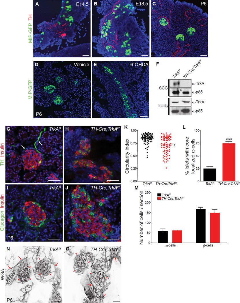
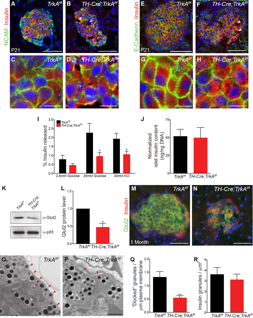
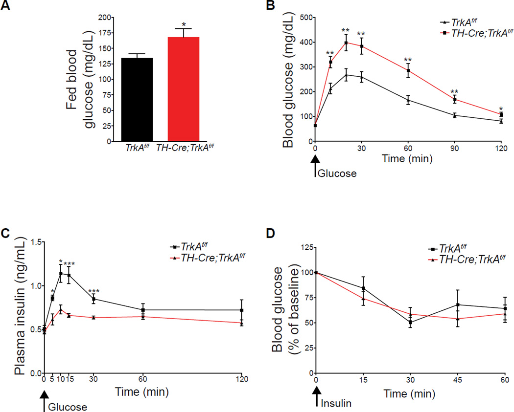
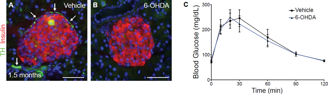
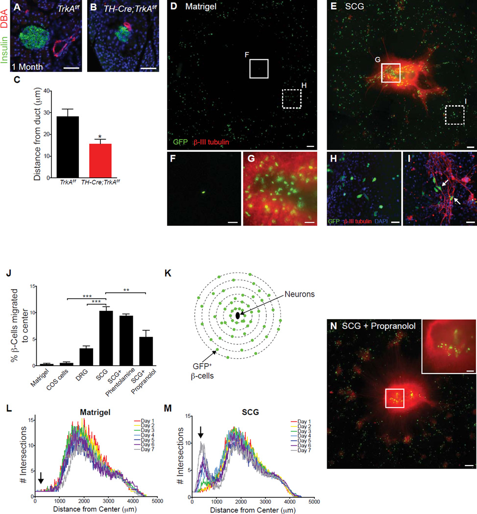
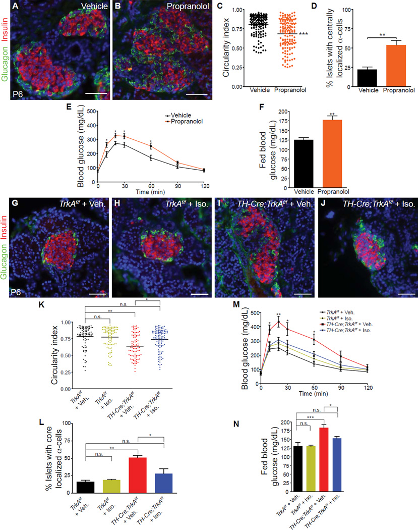
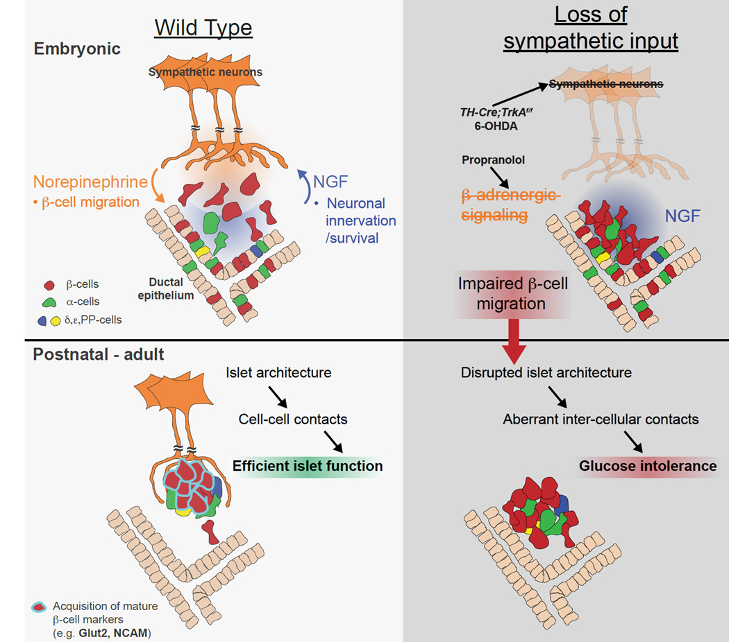
Sympathetic neurons depend on target-derived neurotrophic cues to control their survival and growth. However, whether sympathetic innervation contributes reciprocally to the development of target tissues is less clear. Here, we report that sympathetic innervation is necessary for the formation of the pancreatic islets of Langerhans and for their functional maturation. Genetic or pharmacological ablation of sympathetic innervation during development resulted in altered islet architecture, reduced insulin secretion, and impaired glucose tolerance in mice. Similar defects were observed with pharmacological blockade of β-adrenergic signaling. Conversely, the administration of a β-adrenergic agonist restored islet morphology and glucose tolerance in deinnervated animals. Furthermore, in neuron-islet cocultures, sympathetic neurons promoted islet cell migration in a β-adrenergic-dependent manner. This study reveals that islet architecture requires extrinsic inductive cues from neighboring tissues such as sympathetic nerves and suggests that early perturbations in sympathetic innervation might underlie metabolic disorders.


Figures

Figure 1
Figure 1. Sympathetic innervation establishes islet architecture and the spatial distribution of endocrine cells
(A–C) TH immunohistochemistry and GFP fluorescence reveals the organization of β-cells into discrete clusters near sympathetic fibers during development in MIP-GFP mice. (D,E) Chemical sympathectomy with 6-OHDA administration disrupts islet clustering in neonatal MIP-GFP mice. GFP-positive β-cells (green) and DAPI (nuclei) staining are shown. (F) Genetic ablation of sympathetic innervation in TH-Cre;TrkAf/f mice. Immunoblotting shows depleted TrkA protein levels in superior cervical ganglia (SCGs) from TH-Cre;TrkAf/f mice at postnatal day 2 (P2), and intact TrkA expression in islets. The p85 subunit of phosphatidylinositol-3-kinase (PI-3K) was used to normalize for protein amounts. (G,H) Immunohistochemistry for TH and insulin shows that sympathetic axons (arrows) surrounding control TrkAf/f islets (G) are lost in TH-Cre;TrkAf/f islets (H) by postnatal day 6 (P6). (I,J) Immunostaining for islet hormones shows disrupted arrangement of endocrine cells within islets in neonatal (P6) TH-Cre;TrkAf/f mice. (K) Islet shape is quantified by circularity index. (L) Percentage of islets with mis-localized α-cells. (M) Normal endocrine cell numbers in P6 TH-Cre;TrkAf/f mutants. For all quantifications, n = 3 mice per genotype; mean ± SEM; *p < 0.05, ***p < 0.001, t test. (N,O) Intact islet vasculature in sympathectomized animals. The density and morphology of the pancreatic blood vessels (arrows) and islet capillaries (arrowheads) are not compromised, despite the alterations in islet architecture in TH-Cre;TrkAf/f pups. Blood vessels were fluorescently painted by perfusing Alexa-633-conjugated wheat germ agglutinin (WGA) into TH-Cre;TrkAf/f and control mice at P6. Scale bars, 50µm.
Figure 2
Figure 2. Defects in islet cell-cell contacts and glucose-stimulated insulin secretion in sympathectomized mice
(A, B) Reduced N-CAM expression in islets from postnatal (P21) TH-Cre;TrkAf/f mice (P21). (C, D) Higher magnification images of regions from (A, B). (E, F) Reduced expression of E-cadherin in islets from P21 TH-Cre;TrkAf/f animals compared to control mice. (G, H) Higher magnification images of regions in (E, F) show an uneven distribution of E-cadherin at junctions between adjacent β-cells in mutant islets. Mutant islets also exhibit abnormalities in cellular spacing and gaps devoid of any cells (arrowheads in B and F). Scale bars 50µm for (A, B, E, F) and 10 µm for (C, D, G, H). (I) Isolated islets from one month-old TH-Cre;TrkAf/f mice secrete less insulin when challenged with high glucose or with potassium chloride treatment. Insulin released is expressed as a percentage of the total insulin content. n = 5 mice per genotype; mean ± SEM; *p<0.05, t test. (J) Total insulin content is normal in isolated islets from TH-Cre ;TrkAf/f mice. Insulin content is normalized to total islet DNA content. n=4 mice per genotype; mean ± SEM. (K–N) Reduced expression and surface localization of the glucose transporter, Glut2, in mutant β-cells as revealed by immunostaining and immunoblotting. n=3 mice per genotype; mean ± SEM; *p<0.05, t test. (O–Q) Ultrastructural analysis of β-cells reveals fewer insulin granules within 50nm of the plasma membrane (arrowheads), in mutant islets. Numbers in (Q) represent insulin granules docked per µm of the plasma membrane n=3 mice per genotype, mean ± SEM; **p < 0.01, t test. (R) The total number of insulin granules (per µm2) are not different between mutant and control islets. n=3 mice per genotype, mean ± SEM; t test.
Figure 3
Figure 3. Sympathectomized mice exhibit impaired glucose tolerance but normal insulin sensitivity
(A) Elevated blood glucose levels in one month-old TH-Cre;TrkAf/f compared to control mice when fed ad libitum. n=9 mice per genotype; mean ± SEM; *p < 0.05, t test. (B) Glucose intolerance in TH-Cre;TrkAf/f mice. Mice, at one month of age, were fasted overnight, then injected i.p with a bolus of 2g/kg glucose at t=0. Blood glucose measurements were made from tail vein at the indicated time intervals after glucose injection. n=10 mice per genotype; mean ± SEM; *p < 0.05, **p < 0.01, t test. (C) Reduced plasma insulin levels in response to a glucose injection (3g/kg glucose in TH-Cre;TrkAf/f mice compared to control mice. n=8 control, 10 mutant mice, mean ± SEM; *p<0.05, ***p<0.001, t test. (D) Normal insulin sensitivity in TH-Cre;TrkAf/f mice. A significant decrease (p<0.001, t test) in blood glucose levels was seen in both TH-Cre;TrkAf/f and control mice at 30 minutes after insulin injection compared to t=0). Mice were treated with 0.75U/kg of insulin, and blood glucose measurements made from tail blood at the indicated times post-injection. n=5 mice per genotype, t test, mean ± SEM,
Figure 4
Figure 4. Sympathetic de-innervation in mature animals does not affect islet architecture and glucose tolerance
(A–C) Normal islet morphology and glucose tolerance with 6-OHDA administration at a late postnatal stage (postnatal day 21). P21-old mice were serially injected with 6-OHDA or vehicle for 7 days, and then analyzed at 1.5 months of age. TH-positive sympathetic fibers (arrows in A) are sparser in mature animals, and are completely lost with 6-OHDA administration (B). n=6 mice for each treatment; t test, mean ± SEM.
Figure 5
Figure 5. Sympathetic neurons promote β-cell migratory behavior through norepinephrine signaling
(A–C) Ductal proximity of islets in one-month old TH-Cre;TrkAf/f mice. n=4 control and 3 mutant animals; mean ± SEM; *p < 0.05, t test. (D,E) Isolated β-cells exhibit directed migration towards sympathetic (SCG) tissue explants plated in the center of a Matrigel culture dish. Scale bars, 200µm. (F,G) Higher magnification images of the insets shown in D,E. Scale bars, 50 µm. (H,I) β-cells align along sympathetic axons and adopt polarized morphologies when cultured with neurons (arrows, I). Scale bars, 50 µm. (J) Percentage of β-cells that have migrated toward the center of Matrigel cultures containing COS cell aggregates, DRG or SCG explants with or without adrenergic antagonists after 7 days. n= at least 3 independent experiments; mean ± SEM; **p < 0.01, ***p < 0.001 by one-way ANOVA and Tukey’s post-hoc test. (K–M) Sholl analysis to quantify the distribution of GFP+ β-cells over 7 days in culture. The distribution of β-cells cultured alone does not change significantly over 7 days (L), while there is a gradual shift in the distribution of β-cells towards the center of the culture containing SCGs (M, arrow). Averaged profiles of four independent experiments are plotted. (N) β-cells exhibit decreased migration towards sympathetic explants in the presence of the β-adrenergic antagonist, propranolol (see J for quantification).
Figure 6
Figure 6. β-adrenergic signaling is necessary during development to regulate islet architecture and glucose metabolism
(A,B) In vivo administration of propranolol (20 mg/kg body weight) during development (E18-P6) results in defects in islet architecture in neonatal (P6) mice, compared to saline-injected controls. (C,D) Quantification of islet shape and endocrine cell compartmentalization in mice injected with saline or propranolol. n=5 animals per genotype; mean ± SEM; **p < 0.01, ***p < 0.001, t test. (E) Glucose intolerance in one month-old animals injected with propranolol during development (E18.5-P6), compared to vehicle-treated controls. n=7,9 mice for vehicle and propranolol respectively;, mean ± SEM; *p < 0.05, t test. (F) Developmental administration of propranolol results in elevated fed blood glucose levels in mature animals at one month. n=7,9 mice for vehicle and propranolol respectively; mean ± SEM; **p < 0.01, t test. (G–L) Restoration of islet morphologies in neonatal TH-Cre;TrkAf/f mice treated with a β-adrenergic receptor agonist, isoproterenol, from E18-P6. Isoproterenol administration (Iso, 30 mg/kg body weight) has no effect on islet architecture in control TrkAf/f mice (G,H), but normalizes islet structure and cytoarchitecture in TH-Cre;TrkAf/f mice (J) compared to saline-injected TH-Cre;TrkAf/f mice (I). (K,L) Quantification of islet shape and endocrine cell compartmentalization in TH-Cre;TrkAf/f and control mice injected with saline or isoproterenol. n = 3 mice for all treatments except for TH-Cre;TrkAf/f + Iso where n=4 mice; mean ± SEM; *p < 0.05, **p<0.01 one-way ANOVA with Tukey’s post-hoc test. (M) Developmental administration of isoproterenol administration (E18-P6) rescues the glucose intolerance in one-month old TH-Cre;TrkAf/f mice, compared to vehicle-injected TH-Cre;TrkAf/f mice. Isoproterenol had no effect on glucose metabolism in control TrkAf/f mice. n=5 mice for vehicle- and Iso-injections in TrkAf/f mice; n=6 for vehicle-injected TH-Cre;TrkAf/f mice and n=7 for Iso-injected TH-Cre;TrkAf/f mice; mean ± SEM; *p < 0.05, **p<0.01 as determined by one-way ANOVA and Tukey’s post-hoc test. (N) Normalization of fed blood glucose levels in in one-month old TH-Cre;TrkAf/f mice treated with isoproterenol between E18-P6, compared to saline-treated TH-Cre;TrkAf/f mice. n=5 mice for vehicle- and Iso-injections in TrkAf/f mice; n=6 for vehicle-injected TH-Cre;TrkAf/f mice and n=7 for Iso-injected TH-Cre;TrkAf/f mice; mean ± SEM; *p < 0.05, ***p<0.001 one-way ANOVA with Tukey’s post-hoc test.
Figure 7
Figure 7. Developmental interactions between sympathetic neurons and pancreatic endocrine cells
Sympathetic innervation of the embryonic pancreas occurs in response to target-derived NGF, which then supports survival of innervating neurons (Glebova and Ginty, 2005). Sympathetic nerves release norepinephrine, which acts through β-adrenergic receptors on β-cells to contribute to endocrine cell migration and formation of discrete islet clusters. Nerve-dependent establishment of islet architecture is essential for intercellular contacts within islets, β-cell maturation (i.e. Glut2 levels) and optimal glucose-stimulated insulin secretion.

References

    1. Ahren B. Autonomic regulation of islet hormone secretion--implications for health and disease. Diabetologia. 2000;43:393–410. - PubMed
    1. Asensio C, Jimenez M, Kuhne F, Rohner-Jeanrenaud F, Muzzin P. The lack of beta-adrenoceptors results in enhanced insulin sensitivity in mice exhibiting increased adiposity and glucose intolerance. Diabetes. 2005;54:3490–3495. - PubMed
    1. Bonner-Weir S. Morphological evidence for pancreatic polarity of beta-cell within islets of Langerhans. Diabetes. 1988;37:616–621. - PubMed
    1. Bosco D, Orci L, Meda P. Homologous but not heterologous contact increases the insulin secretion of individual pancreatic B-cells. Experimental cell research. 1989;184:72–80. - PubMed
    1. Brissova M, Fowler MJ, Nicholson WE, Chu A, Hirshberg B, Harlan DM, Powers AC. Assessment of human pancreatic islet architecture and composition by laser scanning confocal microscopy. The journal of histochemistry and cytochemistry : official journal of the Histochemistry Society. 2005;53:1087–1097. - PubMed

Publication types

MeSH terms