. 2013 Aug 15;500(7462):354-8.
doi: 10.1038/nature12329. Epub 2013 Jul 14.

An Sp1 transcription factor coordinates caspase-dependent and -independent apoptotic pathways

Affiliations

An Sp1 transcription factor coordinates caspase-dependent and -independent apoptotic pathways

Takashi Hirose et al. Nature. .

Abstract

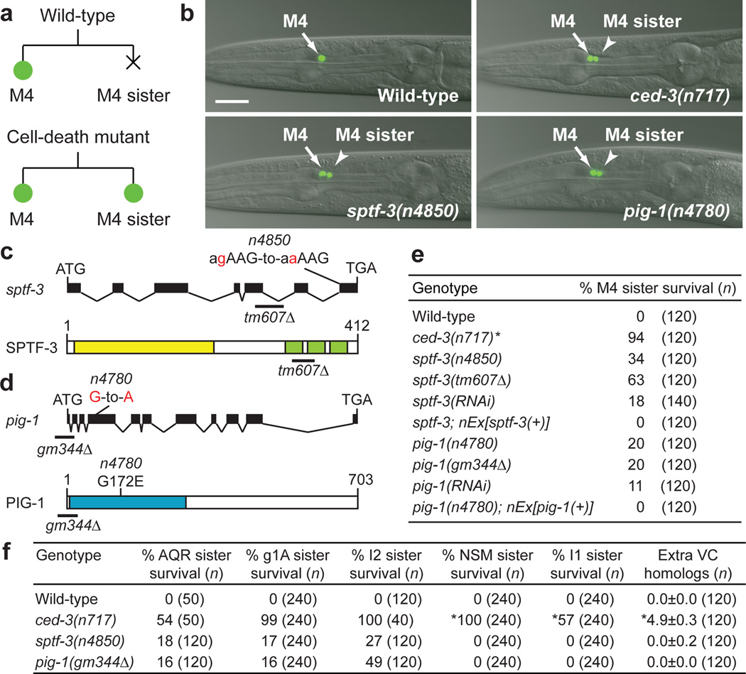
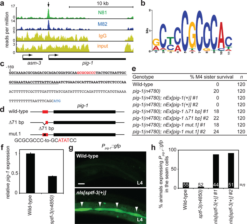
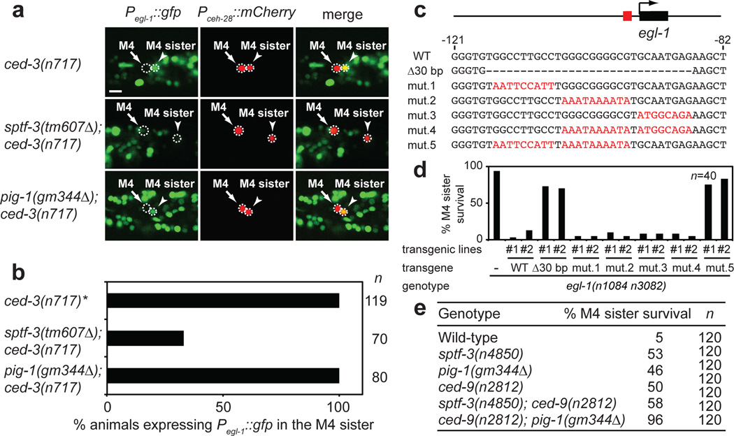
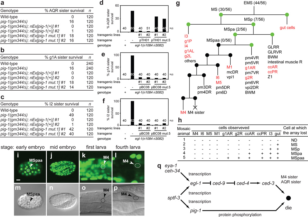
During animal development, the proper regulation of apoptosis requires the precise spatial and temporal execution of cell-death programs, which can include both caspase-dependent and caspase-independent pathways. Although the mechanisms of caspase-dependent and -independent cell killing have been examined extensively, how these pathways are coordinated within a single cell that is fated to die is unknown. Here we show that the Caenorhabditis elegans Sp1 transcription factor SPTF-3 specifies the programmed cell deaths of at least two cells-the sisters of the pharyngeal M4 motor neuron and the AQR sensory neuron-by transcriptionally activating both caspase-dependent and -independent apoptotic pathways. SPTF-3 directly drives the transcription of the gene egl-1, which encodes a BH3-only protein that promotes apoptosis through the activation of the CED-3 caspase. In addition, SPTF-3 directly drives the transcription of the AMP-activated protein kinase-related gene pig-1, which encodes a protein kinase and functions in apoptosis of the M4 sister and AQR sister independently of the pathway that activates CED-3 (refs 4, 5). Thus, a single transcription factor controls two distinct cell-killing programs that act in parallel to drive apoptosis. Our findings reveal a bivalent regulatory node for caspase-dependent and -independent pathways in the regulation of cell-type-specific apoptosis. We propose that such nodes might act as features of a general mechanism for regulating cell-type-specific apoptosis and could be therapeutic targets for diseases involving the dysregulation of apoptosis through multiple cell-killing mechanisms.


Figures

Figure 1
Figure 1. sptf-3 and pig-1 promote the death of the M4 sister cell
a, Schematic representation of the M4 cell lineage in the wild type and mutants defective in M4 sister cell death. b, Merged epifluorescence and Nomarski images of the pharynx in wild-type, ced-3(n717),sptf-3(n4850) and pig-1(n4780) animals expressing Pceh-28∷gfp. Scale bar, 20 µm. c, d, Genomic organizations and protein structures of sptf-3 and pig-1, including the mutations n4850 and n4780. The yellow and green boxes represent the Q-rich and C2H2-type zinc finger domains of SPTF-3, respectively. The blue box indicates the kinase domain of PIG-1. e, The percentages of M4 sister survival in animals of the indicated genotypes. f, The percentages of survival of the indicated cells and the number of extra VC homologs are shown. Error, s.d. *Data from ref. .
Figure 2
Figure 2. pig-1 is a direct transcriptional target of SPTF-3 in the regulation of the death of the M4 sister cell
a, ChIP-seq results obtained by immunoprecipitation with the antibodies anti-SPTF-3 N81, anti-SPTF-3 M82 or normal IgG and input chromatin are shown. Arrow, an enriched SPTF-3-bound region. b, The consensus SPTF-3 binding motif determined by ChIP-seq is represented. c, DNA sequence of the pig-1 promoter. Bold letters, SPTF-3-bound regions identified by ChIP-seq. Red letters, the GC-rich sequence that contains the consensus SPTF-3 binding motif. Blue letters, the ATG start site of the pig-1 coding region. Underline, the deleted pig-1 sequences in the pig-1 Δ71 bp transgene. d, Representations of the pig-1 transgenes used in rescue experiments. Wild-type, a wild-type pig-1 transgene; Δ71 bp lacks 71 base pairs of the SPTF-3-bound region; mut.1, mutated as shown for the consensus SPTF-3 binding motif. Red box, SPTF-3-bound regions identified by ChIP-seq. e, The percentages of M4 sister survival in animals of the indicated genotypes. f,pig-1 mRNA levels of wild-type and sptf-3(n4850) embryos were measured by quantitative RT-PCR. pig-1 expression levels of sptf-3(n4850) mutants relative to that of the wild type are represented. Error bars, s.d. of three technical replicates. g, Expression of Ppig-1∷gfp in animals of the indicated genotypes at the fourth larval stage (L4). Arrowheads, seam cells. Scale bar, 20 µm. h, The percentages of L4 larvae expressing Ppig-1∷gfp in the seam cells of animals of the indicated genotypes.
Figure 3
Figure 3. sptf-3 directly drives egl-1 expression in the M4 sister
a, egl-1 expression in the M4 sister cell of animals of the indicated genotypes. b, The percentages of animals expressing Pegl-1∷gfp in the M4 sister of the indicated genotypes. *Data from ref. . c, Representations of the egl-1 transgenes used in rescue experiments. WT, a wild-type egl-1 transgene; Δ30 bp lacks 30 base pairs of the SPTF-3-bound region; mut.1 to mut.5, mutated as shown. Black box, egl-1 coding region. Red box, SPTF-3-bound region identified by ChIP-seq. Red letters, mutated bases. d, The percentages of M4 sister survival in animals of the indicated genotypes. e, The percentages of M4 sister survival in animals of the indicated genotypes. All strains were homozygous for ced-3(n2446).
Figure 4
Figure 4. SPTF-3 functions cell autonomously to promote apoptosis of the M4 sister and the AQR sister
a–f, The percentages of survival of the (a, d) AQR sister, (b, e) g1A sister and (c, f) I2 sister in animals of the indicated genotypes. The pig-1(+) transgene carries the wild-type pig-1 genomic locus, and the pig-1 mut.1 transgene has mutations in the consensus SPTF-3 binding motif in the pig-1 promoter region, as described in Fig. 2d. The egl-1 transgene pTH01 contains 6.5 kb of the 5’ promoter of egl-1, the coding region and 2.2 kb of the 3’ of the stop codon of egl-1. The egl-1 transgene pBC08 contains 1.1 kb of the 5’ promoter of egl-1, the coding region and 5.7 kb of the 3’ of the stop codon of egl-1. pTH01 mut.5 and pBC08 mut.5 have mutations in the consensus SPTF-3 binding motif of the egl-1 promoter region, as described in Fig. 3c. g, Diagram of the M4 cell lineage. The cells in red were scored to determine the cell division at which the sptf-3-containing extrachromosomal array was lost in a mosaic animal. Shown are numbers of animals carrying the array in the indicated cell of 56 mosaic animals observed. Green circles, blastomeres in which the array was present. Black circles, blastomeres in which the array was lost. h, Examples of mosaic animals carrying an extrachromosomal array in the indicated cells. +, extrachromosomal array present. −, extrachromosomal array absent. i-p, Nomarski and epifluorescence images of a strain expressing a gfp∷sptf-3 transgene. Scale bar, 10 µm. q, A model for the pathways that regulate M4 sister and AQR sister cell-specific programmed cell death.

Comment in

References

    1. Fuchs Y, Steller H. Programmed cell death in animal development and disease. Cell. 2011;147:742–758. - PMC - PubMed
    1. Yuan J, Kroemer G. Alternative cell death mechanisms in development and beyond. Genes Dev. 2010;24:2592–2602. - PMC - PubMed
    1. Metzstein MM, Stanfield GM, Horvitz HR. Genetics of programmed cell death in C. elegans: past, present and future. Trends Genet. 1998;14:410–416. - PubMed
    1. Cordes S, Frank CA, Garriga G. The C. elegans MELK ortholog PIG-1 regulates cell size asymmetry and daughter cell fate in asymmetric neuroblast divisions. Development. 2006;133:2747–2756. - PubMed
    1. Denning DP, Hatch V, Horvitz HR. Programmed elimination of cells by caspase-independent cell extrusion in C. elegans . Nature. 2012;488:226–230. - PMC - PubMed

Methods References

    1. Marson A, et al. Connecting microRNA genes to the core transcriptional regulatory circuitry of embryonic stem cells. Cell. 2008;134:521–533. - PMC - PubMed
    1. Bailey TL. MEME SUITE: tools for motif discovery and searching. Nucleic Acids Res. 2009;37:W202–W208. - PMC - PubMed
    1. Bailey TL, Gribskov M. Combining evidence using p-values: application to sequence homology searches. Bioinformatics. 1998;14:48–54. - PubMed
    1. Beissbarth T, Speed TP. GOstat: find statistically overrepresented Gene Ontologies within a group of genes. Bioinformatics. 2004;20:1464–1465. - PubMed
    1. Conradt B, Horvitz HR. The C. elegans protein EGL-1 is required for programmed cell death and interacts with the Bcl-2-like protein CED-9. Cell. 1998;93:519–529. - PubMed

Publication types

MeSH terms