

Structural basis for molecular recognition of folic acid by folate receptors
- PMID: 23851396
- PMCID: PMC5797940
- DOI: 10.1038/nature12327
Structural basis for molecular recognition of folic acid by folate receptors
Abstract
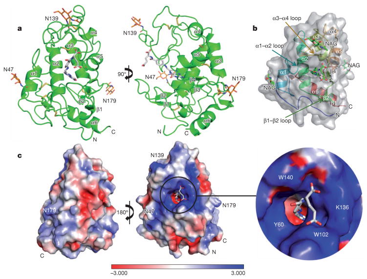
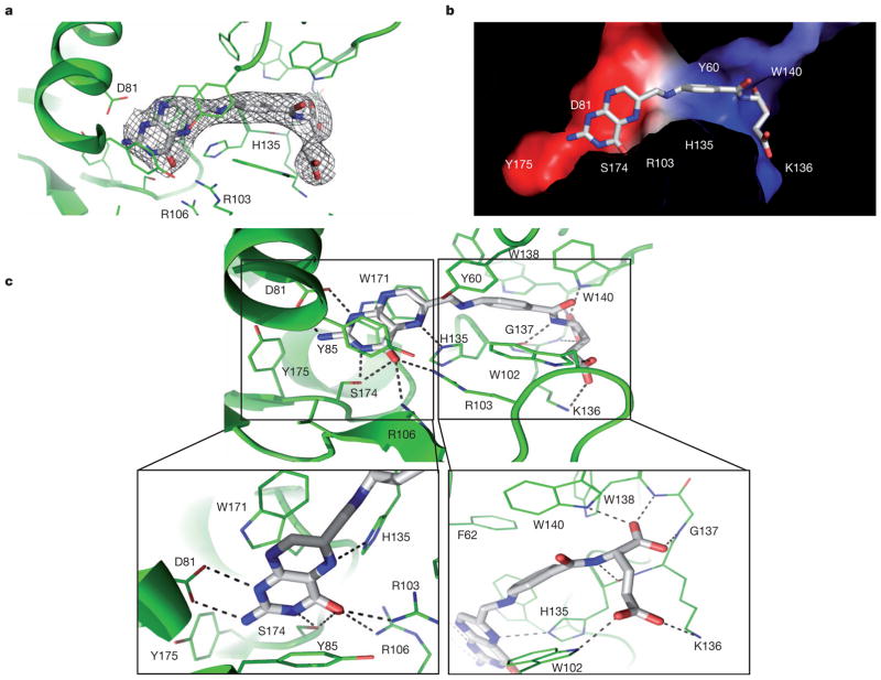
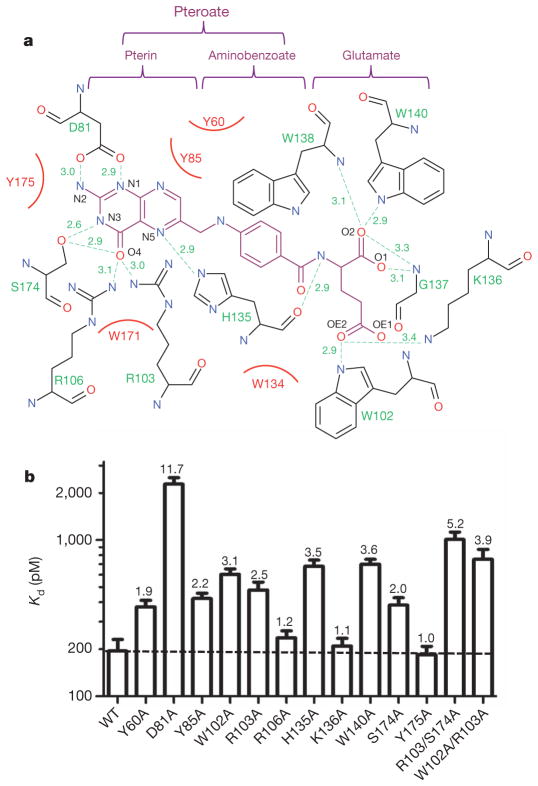
Folate receptors (FRα, FRβ and FRγ) are cysteine-rich cell-surface glycoproteins that bind folate with high affinity to mediate cellular uptake of folate. Although expressed at very low levels in most tissues, folate receptors, especially FRα, are expressed at high levels in numerous cancers to meet the folate demand of rapidly dividing cells under low folate conditions. The folate dependency of many tumours has been therapeutically and diagnostically exploited by administration of anti-FRα antibodies, high-affinity antifolates, folate-based imaging agents and folate-conjugated drugs and toxins. To understand how folate binds its receptors, we determined the crystal structure of human FRα in complex with folic acid at 2.8 Å resolution. FRα has a globular structure stabilized by eight disulphide bonds and contains a deep open folate-binding pocket comprised of residues that are conserved in all receptor subtypes. The folate pteroate moiety is buried inside the receptor, whereas its glutamate moiety is solvent-exposed and sticks out of the pocket entrance, allowing it to be conjugated to drugs without adversely affecting FRα binding. The extensive interactions between the receptor and ligand readily explain the high folate-binding affinity of folate receptors and provide a template for designing more specific drugs targeting the folate receptor system.
Conflict of interest statement
The authors declare no competing financial interests.
Figures
References
-
- Kelemen LE. The role of folate receptor α in cancer development, progression and treatment: cause, consequence or innocent bystander? Int J Cancer. 2006;119:243–250. - PubMed
-
- McGuire JJ. Anticancer antifolates: current status and future directions. Curr Pharm Des. 2003;9:2593–2613. - PubMed
-
- Deng YJ, et al. Synthesis and biological activity of a novel series of 6-substituted thieno 2,3-d pyrimidine antifolate inhibitors of purine biosynthesis with selectivity for high affinity folate receptors over the reduced folate carrier and proton-coupled folate transporter for cellular entry. J Med Chem. 2009;52:2940–2951. - PMC - PubMed
Publication types
MeSH terms
Substances
Associated data
- Actions
Grants and funding
LinkOut - more resources
Full Text Sources
Other Literature Sources
Medical
Molecular Biology Databases
Research Materials
