. 2013 Oct;11(10):1203-14.
doi: 10.1158/1541-7786.MCR-13-0072. Epub 2013 Jul 12.

Angiogenin mediates androgen-stimulated prostate cancer growth and enables castration resistance

Affiliations

Angiogenin mediates androgen-stimulated prostate cancer growth and enables castration resistance

Shuping Li et al. Mol Cancer Res. 2013 Oct.

Abstract

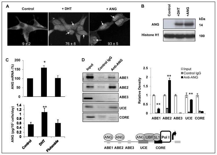
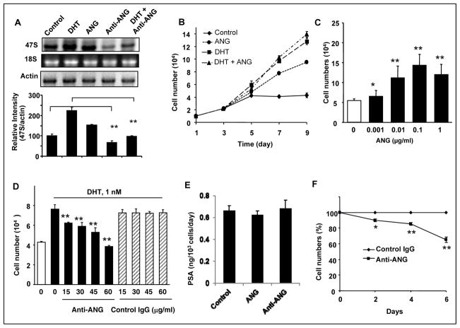
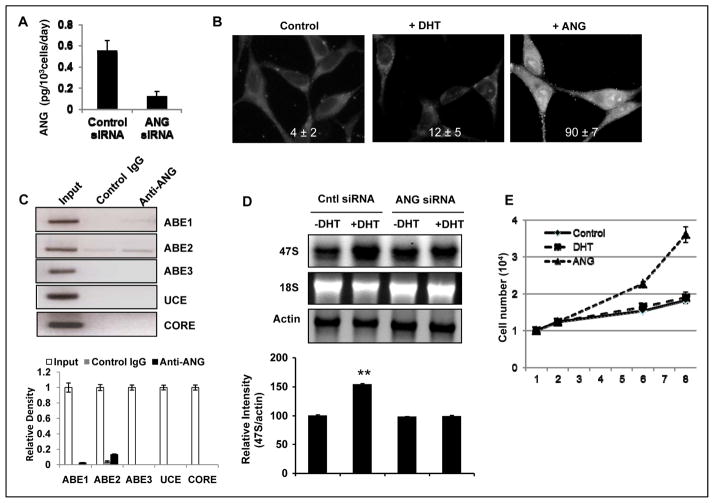
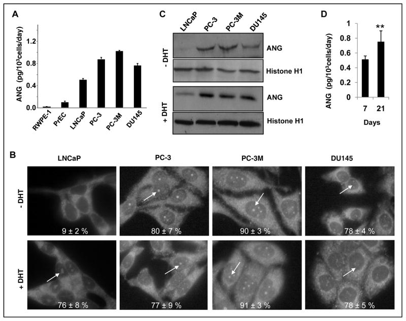
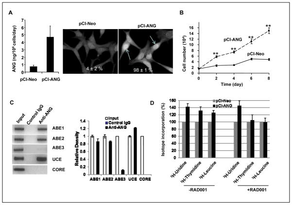
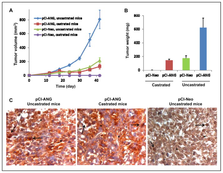
The androgen receptor (AR) is a critical effector of prostate cancer development and progression. Androgen-dependent prostate cancer is reliant on the function of AR for growth and progression. Most castration-resistant prostate cancer (CRPC) remains dependent on AR signaling for survival and growth. Ribosomal RNA (rRNA) is essential for both androgen-dependent and castration-resistant growth of prostate cancer cells. During androgen-dependent growth of prostate cells, androgen-AR signaling leads to the accumulation of rRNA. However, the mechanism by which AR regulates rRNA transcription is unknown. Here, investigation revealed that angiogenin (ANG), a member of the secreted ribonuclease superfamily, is upregulated in prostate cancer and mediates androgen-stimulated rRNA transcription in prostate cancer cells. Upon androgen stimulation, ANG undergoes nuclear translocation in androgen-dependent prostate cancer cells, where it binds to the rDNA promoter and stimulates rRNA transcription. ANG antagonists inhibit androgen-induced rRNA transcription and cell proliferation in androgen-dependent prostate cancer cells. Interestingly, ANG also mediates androgen-independent rRNA transcription through a mechanism that involves its constitutive nuclear translocation in androgen-insensitive prostate cancer cells, resulting in a constant rRNA overproduction and thereby stimulating cell proliferation. Critically, ANG overexpression in androgen-dependent prostate cancer cells enables castration-resistant growth of otherwise androgen-dependent cells. Thus, ANG-stimulated rRNA transcription is not only an essential component for androgen-dependent growth of prostate cancer but also contributes to the transition of prostate cancer from androgen-dependent to castration-resistant growth status.

Implications: The ability of angiogenin to regulate rRNA transcription and prostate cancer growth makes it a viable target for therapy.


Conflict of interest statement

Conflicts of Interest: The authors declare no conflict of interest

Figures

Figure 1
Figure 1
Effect of DHT on ANG. A, DHT stimulates nuclear translocation of ANG in LNCaP cells. Cells were cultured in phenol red-free RPMI 1640 medium + 10% charcoal/dextran-stripped FBS, and incubated in the absence (left panel) or presence of 1 nM DHT (middle panel) or 0.1 μg/ml ANG (right panel) for 30 min. Cells were fixed and ANG localization was determined by IF staining with ANG mAb 26-2F and Alexa 555-labeled goat F(ab′)2 anti-mouse IgG. ANG positive cells were counted in a total of 200 cells in five randomly selected areas. B, Western blotting analysis of nuclear ANG. Nuclear proteins were extracted and analyzed by Western blotting (150 μg per lane) with ANG pAb R112. Histone H1 was used as a loading control. C, Effect of androgen and anti-androgen on ANG expression. Cells were treated with DHT (1 nM) or Flutamide (1 μM) for 2 days. ANG mRNA and secreted ANG protein were determined by qRT-PCR and ELISA, respectively. D, ANG binds to the promoter region of rDNA. Left panels, ChIP analyses of ANG binding to ABE1, ABE2, ABE3, UCE, and CORE regions of the rDNA promoter. PCR primers were designed using MacVector software. The input control in each panel contains 1% of the total DNA. Left panel, ImageJ analysis of the ChIP bands. *, p<0.01; **, p<0.001. Bottom panel, a schematic view of ANG occupancy in the rDNA promoter.
Figure 2
Figure 2
ANG mediates DHT-stimulated rRNA transcription and cell proliferation. A, ANG stimulates, and ANG mAb inhibits, rRNA transcription in LNCaP cells. Top panel, Northern blotting analysis of 47S rRNA. Cells were cultured in phenol red-free medium and charcoal/dextran-stripped FBS and incubated with DHT (1 nM), ANG (0.1 μg/ml), ANG mAb 26-2F (60 μg/ml) or a mixture of DHT and 26-2F for 2 h. The level of 47S rRNA was determined by Northern blotting with a probe specific to the initiation site sequence of the rRNA precursor. EB staining of 18S rRNA and Northern blotting of actin mRNA were used as the loading controls. The bar graph at the bottom shows the relative intensity of 47S rRNA to actin mRNA determined by ImageJ. B, ANG stimulates LNCaP cell proliferation in the absence of androgens. Cells were culture in phenol red-free and charcoal/dextran-stripped FBS for 2 day and stimulated with DHT (1 nM), ANG (0.1 μg/ml), or a mixture of the two for the time indicated. Cell numbers were determined with a Coulter counter and with confirmed with MTS assay. C, Dose dependence. ANG was added to the cells and cultured for 4 days. D, ANG mAb 26-2F inhibits DHT-induced cell proliferation. LNCaP cells were stimulated with 1 nM DHT in the presence of 26-2F or a control non-immune IgG at the concentrations indicated for 3 days. E, PSA expression is not dependent on ANG level. LNCaP cells were seeded at 1×10⁵ cells per 35 mm2 dish in RPMI 1640 + 10% FBS and cultured for 2 days. After 2 days, the medium was switched to phenol red-free RPMI 1640 + 10% Charcoal/dextran-treated FBS. The cells were treated with 0.1 μg/ml of ANG or 60 μg/ml of 26-2F for another 2 days. The culture media were collected and PSA level was determined by ELISA. F, ANG mAb inhibits PC-3 cell proliferation. PC-3 cells were cultured in DMEM plus 10% FBS in the presence of 30 μg/ml 26-2F or a control non-immune IgG for the time indicated. *, p<0.01; **, p<0.001.
Figure 3
Figure 3
ANG siRNA abolishes DHT-stimulated rRNA transcription and cell proliferation. A, ELISA analysis of secreted ANG level in control and ANG siRNA-transfected cells. B, DHT-stimulated nuclear translocation of ANG is decreased in ANG knockdown cells. Cells were cultured in steroid-free medium for 24 h and stimulated with 1 nM DHT (middle) or 0.1 μg/ml ANG for 30 min. IF detection of ANG was carried out with 26-2F and Alexa 488-labeled goat F(ab′)2 anti-mouse IgG as described in the legend to Fig. 1A. ANG positive cells were counted in a total of 200 cells in five randomly selected areas. C, ChIP analysis of ANG occupancy in rDNA promoter. ANG knockdown cells were cultured in steroid-free medium for 24 h and stimulated with 1 nM DHT for 1 h. ChIP were carried out as described in the legend to Fig. 1D. Top panel, ChIP bands; bottom panel, ImageJ analysis. D, ANG siRNA abolishes DHT-stimulated rRNA transcription. Top panel, Northern blotting analysis of 47S rRNA in control and ANG siRNA-transfected cells with actin mRNA as loading controls. Bottom panel, relative intensity of 47S rRNA to actin mRNA determined by densitometry. **, p<0.01. E, ANG siRNA abolishes DHT-stimulated cell proliferation. ANG knockdown cells were cultured in steroid-free medium and stimulated with 1 nM DHT or 0.1 μg/ml ANG. Cell numbers were determined with a Coulter counter. Data shown are means ± SD of triplicates in a representative experiment.
Figure 4
Figure 4
Constant nuclear translocation of ANG in androgen-insensitive PCa cells. A, Upregulation of ANG in human PCa cells. Secreted ANG proteins in normal human prostate epithelial cells and PCa cells were determined by ELISA. B, Nuclear translocation of ANG in PCa cells. LNCaP, PC-3, PC-3M, and DU145 cells were cultured in their respective media supplemented with 10% FBS for two days. FBS was then replaced with charcoal/dextran-stripped serum and the cells were cultured in the absence or presence of DHT (1 nM) for 2 days. IF of ANG was carried out with ANG mAb 26-2F (50 μg/ml) and Alexa 488-labeled goat F(ab′)2 anti-mouse IgG (1:100 dilution) as described in the legend to Fig. 1A. Nucleolar ANG was indicated by arrows. C, Western blotting analysis of nuclear ANG. Nuclear proteins were extracted from the cells cultured in the absence (top two panels) and presence (bottom two panels) of DHT and analyzed by Western blotting (150 μg per lane) with ANG pAb R112. Histone H1 was used as a loading control. D, ANG expression is upregulated in ANG knockdown LNCaP cells after prolonged culture in steroid-free medium. Control and ANG siRNA transfectants were continuously cultured in steroid-free medium for one or three weeks without subculture. Medium was changed every two days. ANG secreted into the medium between day 6 and day 7, and between day 20 and day 21 was determined by ELISA. Cell number was determined from parallel dishes by a Coulter counter.
Figure 5
Figure 5
ANG over-expression promotes androgen-independent growth of LNCaP cells. A, Endogenous ANG in stable vector control (pCI-Neo) and ANG (pCI-ANG) transfectants. Left panel, ELISA analysis of secreted ANG protein in vector control and ANG transfectants. Right panels, IF detection of cellular ANG in control and ANG transfectants. Cells positive for nuclear ANG were counted in 200 cells in 5 randomly selected areas. B, ANG overexpression promotes LNCaP proliferation in vitro in the absence of androgen. Cells were cultured in phenol red-free medium and charcoal/dextran-stripped FBS. Cell numbers were determined with a Coulter counter. Data shown are means ± SD of triplicates in a representative experiment. C, ChIP analysis of ANG occupancy in rDNA promoter. ANG over-expression cells were cultured in steroid-free medium for 24 h and ChIP analyses were carried out as described in the legend to Fig. 1D. Right panel, ChIP bands; right panel, ImageJ analysis. D, RAD001 inhibits ANG-stimulated DNA synthesis and protein translation but not RNA transcription. Vector (pCI-Neo) and ANG (pCI-ANG) transfectants were cultured in steroid-free medium and were pulsed with 1 μCi 3H-uridine, 3H-thymidine, or 3H-leucine for 6 h. Radioisotope incorporated into TCA-insoluble fraction was normalized to cell number and the value from the vector control transfectants was set as 100. Data shown are means ± SD of triplicates.
Figure 6
Figure 6
ANG overexpression is correlated with castration-resistant growth of LNCaP cells in vivo. A, Xenograft growth of vector control and ANG transfectants in castrated SCID mice. A mixture of 70 μl cell suspension (1×10⁶ cells) and 30 μl Matrigel was injected s.c. per mouse (6 per group). The mice were castrated or sham-operated at the same time. Tumors sizes were measured with a caliper and recoded in mm3 (length × width2). B, Wet weight of dissected tumors. C, Expression and localization of human ANG detected by IHC with mAb 26-2F. Representative nuclear and extracellular localizations of ANG were indicated with black arrows and stars, respectively. **, p<0.001.

Similar articles

Cited by

References

    1. Taplin ME. Androgen receptor: role and novel therapeutic prospects in prostate cancer. Expert Rev Anticancer Ther. 2008;8:1495–508. - PubMed
    1. Chen Y, Sawyers CL, Scher HI. Targeting the androgen receptor pathway in prostate cancer. Curr Opin Pharmacol. 2008;8:440–8. - PMC - PubMed
    1. Feldman BJ, Feldman D. The development of androgen-independent prostate cancer. Nat Rev Cancer. 2001;1:34–45. - PubMed
    1. Balk SP, Knudsen KE. AR, the cell cycle, and prostate cancer. Nucl Recept Signal. 2008;6:e001. - PMC - PubMed
    1. Wang Q, Li W, Liu XS, Carroll JS, Janne OA, Keeton EK, et al. A hierarchical network of transcription factors governs androgen receptor-dependent prostate cancer growth. Mol Cell. 2007;27:380–92. - PMC - PubMed

Publication types

MeSH terms