. 2014 Jun 12;33(24):3172-82.
doi: 10.1038/onc.2013.279. Epub 2013 Jul 15.

GSK3β controls epithelial-mesenchymal transition and tumor metastasis by CHIP-mediated degradation of Slug

Affiliations

GSK3β controls epithelial-mesenchymal transition and tumor metastasis by CHIP-mediated degradation of Slug

S-H Kao et al. Oncogene. .

Erratum in

Abstract

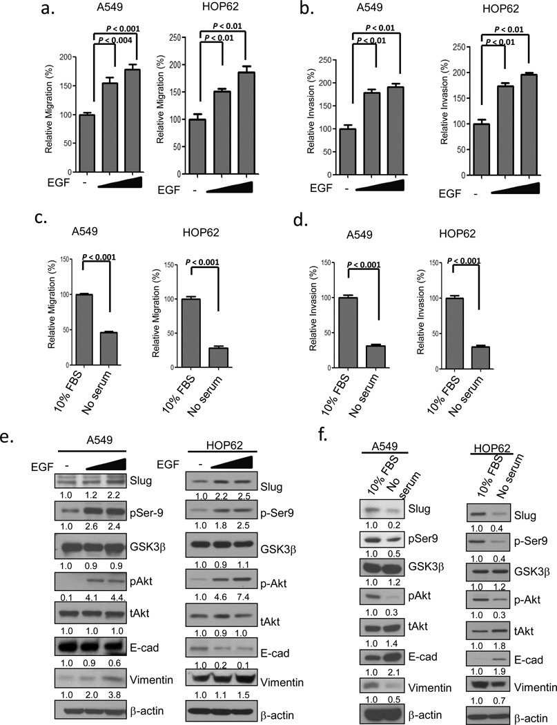
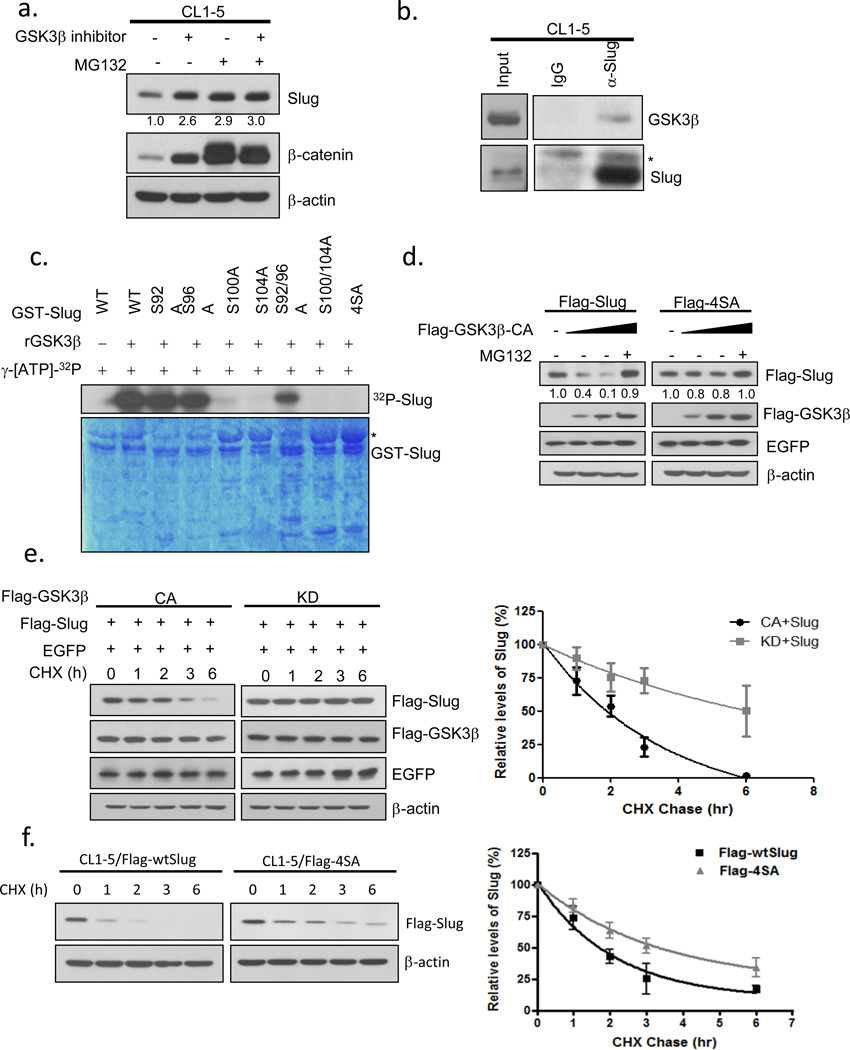
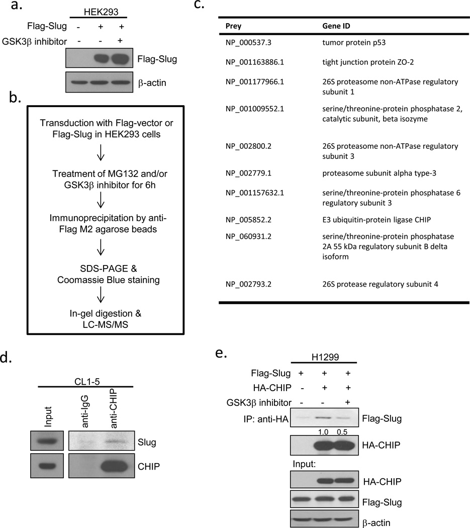
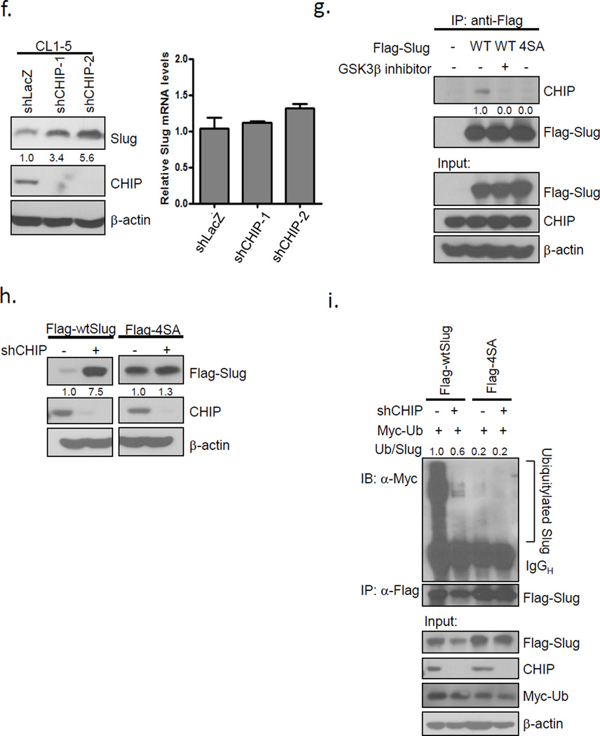
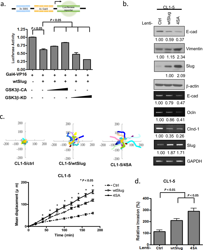
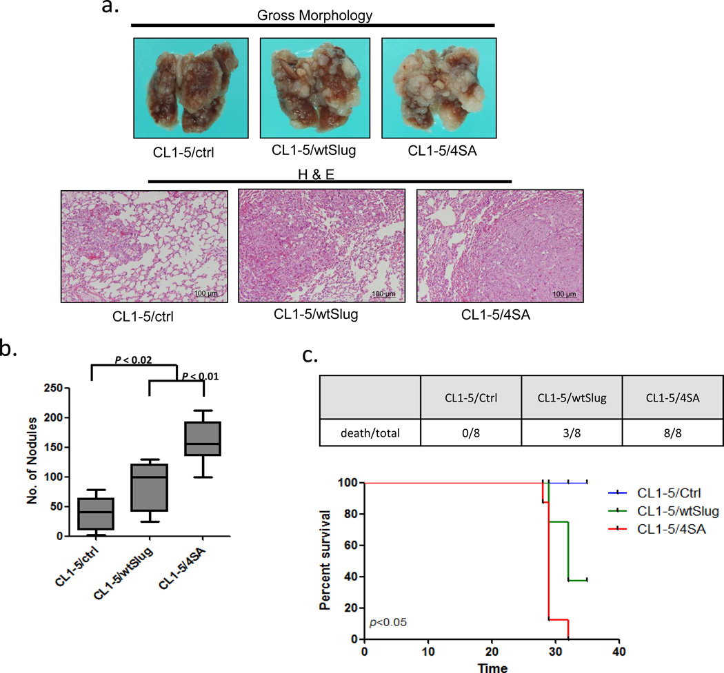
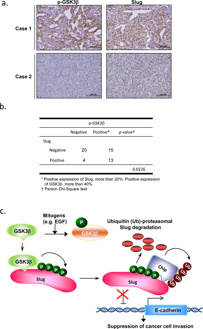
Glycogen synthase kinase 3 beta (GSK3β) is highly inactivated in epithelial cancers and is known to inhibit tumor migration and invasion. The zinc-finger-containing transcriptional repressor, Slug, represses E-cadherin transcription and enhances epithelial-mesenchymal transition (EMT). In this study, we find that the GSK3β-pSer9 level is associated with the expression of Slug in non-small cell lung cancer. GSK3β-mediated phosphorylation of Slug facilitates Slug protein turnover. Proteomic analysis reveals that the carboxyl terminus of Hsc70-interacting protein (CHIP) interacts with wild-type Slug (wtSlug). Knockdown of CHIP stabilizes the wtSlug protein and reduces Slug ubiquitylation and degradation. In contrast, nonphosphorylatable Slug-4SA is not degraded by CHIP. The accumulation of nondegradable Slug may further lead to the repression of E-cadherin expression and promote cancer cell migration, invasion and metastasis. Our findings provide evidence of a de novo GSK3β-CHIP-Slug pathway that may be involved in the progression of metastasis in lung cancer.


Conflict of interest statement

CONFLICTS OF INTEREST

The authors declare that they have no competing financial interests.

Figures

Figure 1
Figure 1
The activity of GSK3β inversely correlates with Slug protein levels. (a-b) EGF induces cell migration and invasion. A549 and HOP62 cells were cultured in serum-free medium for 12 h with or without EGF treatment for 6 h. Cell migration (a) and invasion (b) were determined by the Transwell assay at 6 h or 12 h after treatment. (c-d) Serum starvation reduces cell migration and invasion. A549 and HOP62 cells were cultured in DMEM or RPMI medium supplemented with 10% FBS or in serum-free medium for 6 h. Cell migration (c) and invasion (d) were determined by the Transwell assay at 6 h or 12 h after treatment. Data are shown as mean ± s.e.m. in three different experiments (n=3). (e-f) Correlation between the Slug protein level and the activity of GSK3β. Cell lysates of the experiments in (a-d) were analyzed by Western blotting.
Figure 2
Figure 2
GSK3β phosphorylates Slug and modulates its protein stability. (a) Inhibition of GSK3β and proteasome pathways stabilizes Slug. CL1-5 cells were treated with or without 10 μM MG132 or 25 μM GSK3β inhibitor VIII, or both, for 6 h and analyzed by Western blotting. (b) Interaction of endogenous GSK3β and Slug. CL1-5 cells incubated with MG132 for 6 h and crosslinked were lysed and either analyzed directly by Western blotting or subjected to immunoprecipitation with control and anti-Slug antibodies. The asterisk indicates a non-specific band. (c) GSK3β phosphorylates Slug at serine-92, -96, -100, and -104. Recombinant GSK3β (50U) was incubated with glutathione S-transferase (GST)-Slug fusion proteins (GST-WT, -92A, -96A, -100A, -104A, -92/96A, -100/104A, -4SA) in the presence of γ-[32P]-ATP. The protein kinase reaction products were resolved by SDS-PAGE, and phosphorylation was detected by autoradiography. The asterisk indicates a non-specific band. (d) Lysates were prepared from CL1-5 cells transfected with plasmids expressing Flag-wtSlug or Flag-4SA in the presence or absence of increasing amounts of Flag-tagged constitutively-active GSK3β for 24 h. A plasmid encoding EGFP was used as a negative control for transfection efficiency. Proteins were analyzed by Western blotting. (e) GSK3β modulates Slug protein stability. H1299 cells were transfected with the Flag-Slug construct together with the indicated Flag-GSK3β-expressing plasmids. A plasmid encoding EGFP was used as a negative control for transfection efficiency. Twenty-four hours after transfection, the cells were treated with 300 μgml−1 cycloheximide (CHX). At the indicated time points, lysates were prepared and Western blotting analysis was performed with antibodies specific for the indicated proteins (left panel). The Slug band intensity was normalized to EGFP and then normalized to the t=0 controls (right panel). (f) CL1-5 cells were stably transduced with viruses expressing Flag-Slug-WT or Flag-Slug-4SA for 24 h. Cycloheximide treatment and Western blotting (left panel) were conducted as in (e). The Slug band intensity was normalized to actin and then normalized to the t=0 controls (right panel). Data are shown as mean ± s.e.m. in three different experiments (n=3).
Figure 3
Figure 3
The E3 ligase CHIP is involved in GSK3β-mediated phosphorylation-dependent Slug degradation. (a) HEK293 cells were infected with control or Flag-Slug-expressing viruses in the presence or absence of GSK3β inhibitor for 8 h. The Slug expression was assessed by immunoblotting. (b) Schematic representation of the experimental procedure of immunoprecipitation. (c) List of the associated genes of Flag-Slug. The probability of an interaction partner of the wild-type Slug immunoprecipitate was computed and compared against that of the vector control and GSK3β inhibitor using the SAINT method. (d) Slug interacts with CHIP. CL1-5 cells were treated with 10 μM MG132 for 6 h before cell collection. The lysates were immunoprecipitated with anti-CHIP antibodies and resolved by SDS-PAGE. (e) H1299 cells were transfected with the plasmids expressing the indicated proteins, HA-CHIP and Flag-Slug (or the control). Twenty-four hours after transfection, the cells were treated with 10 μM MG132 with or without 25 μM GSK3β inhibitor VIII for 8 h before cell collection. The lysates were immunoprecipitated with anti-HA antibodies and resolved by SDS-PAGE. (f) CHIP knockdown leads to the accumulation of Slug protein levels. CL1-5 cells were infected with control shLacZ or two different shCHIP viruses. The Slug expression was assessed by real-time PCR (right) and immunoblotting (left). GAPDH and β-actin were used as the internal controls, respectively. Data are shown as mean±s.e.m. in three different experiments (n=3). (g) HEK293 cells were transfected with the vectors expressing Flag-Slug-WT or -4SA. Twenty-four hours after transfection, the cells were treated with 10 μM MG132 and 25 μM GSK3β inhibitor VIII (where indicated) for 8 h before cell collection. The lysates were subjected to immunoprecipitation using anti-Flag antibodies. Western blot analysis was conducted with the indicated antibodies to determine the protein interactions. (h) A549 cells were infected with control shLacZ or shCHIP viruses. Seventy-two hours after infection, the cells were re-seeded for the transduction with Flag-Slug-WT or -4SA lentiviruses. Forty-eight hours after transduction, the lysates were prepared and Western analysis was performed. (i) A549 cells infected in (h) were transfected with the plasmid expressing Myc-ubiquitin. Twenty-four hours after transfection, the cells were treated with 10 μM MG132 for 6 h before cell collection. The lysates were subjected to immunoprecipitation using anti-Flag antibodies. Western blot analysis was conducted with the indicated antibodies to determine the protein interactions. Ub, ubiquitin.
Figure 3
Figure 3
The E3 ligase CHIP is involved in GSK3β-mediated phosphorylation-dependent Slug degradation. (a) HEK293 cells were infected with control or Flag-Slug-expressing viruses in the presence or absence of GSK3β inhibitor for 8 h. The Slug expression was assessed by immunoblotting. (b) Schematic representation of the experimental procedure of immunoprecipitation. (c) List of the associated genes of Flag-Slug. The probability of an interaction partner of the wild-type Slug immunoprecipitate was computed and compared against that of the vector control and GSK3β inhibitor using the SAINT method. (d) Slug interacts with CHIP. CL1-5 cells were treated with 10 μM MG132 for 6 h before cell collection. The lysates were immunoprecipitated with anti-CHIP antibodies and resolved by SDS-PAGE. (e) H1299 cells were transfected with the plasmids expressing the indicated proteins, HA-CHIP and Flag-Slug (or the control). Twenty-four hours after transfection, the cells were treated with 10 μM MG132 with or without 25 μM GSK3β inhibitor VIII for 8 h before cell collection. The lysates were immunoprecipitated with anti-HA antibodies and resolved by SDS-PAGE. (f) CHIP knockdown leads to the accumulation of Slug protein levels. CL1-5 cells were infected with control shLacZ or two different shCHIP viruses. The Slug expression was assessed by real-time PCR (right) and immunoblotting (left). GAPDH and β-actin were used as the internal controls, respectively. Data are shown as mean±s.e.m. in three different experiments (n=3). (g) HEK293 cells were transfected with the vectors expressing Flag-Slug-WT or -4SA. Twenty-four hours after transfection, the cells were treated with 10 μM MG132 and 25 μM GSK3β inhibitor VIII (where indicated) for 8 h before cell collection. The lysates were subjected to immunoprecipitation using anti-Flag antibodies. Western blot analysis was conducted with the indicated antibodies to determine the protein interactions. (h) A549 cells were infected with control shLacZ or shCHIP viruses. Seventy-two hours after infection, the cells were re-seeded for the transduction with Flag-Slug-WT or -4SA lentiviruses. Forty-eight hours after transduction, the lysates were prepared and Western analysis was performed. (i) A549 cells infected in (h) were transfected with the plasmid expressing Myc-ubiquitin. Twenty-four hours after transfection, the cells were treated with 10 μM MG132 for 6 h before cell collection. The lysates were subjected to immunoprecipitation using anti-Flag antibodies. Western blot analysis was conducted with the indicated antibodies to determine the protein interactions. Ub, ubiquitin.
Figure 4
Figure 4
Non-degradable Slug promotes cell migration and invasion. (a) CL1-5 cells transfected with the luciferase reporter plasmid 3xSBS-Luc were co-transfected with plasmids expressing the indicated proteins. The luciferase activity was assayed 24 h later and normalized to the β-galactosidase activity, with pSV-β-gal used as an internal control plasmid. Each data point represents the mean ± s.d. The experiments were performed twice, in triplicate. (b) CL1-5 cells were stably transduced with the control, Slug-WT, or -4SA lentiviruses. The indicated RNA and protein expressions were assessed by RT-PCR and immunoblotting. GAPDH and β-actin were used as the internal controls, respectively. (c) Single-cell tracking migration analysis of control, wtSlug, and 4SA-overexpressing CL1-5 cells by time-lapse video microscopy. The movement of individual cells was followed using cell-tracking software and representative trajectories are shown (upper panel). Total displacement of the indicated cell lines was quantified from the track plots (mean ± s.e.m. of 20 cells analyzed in two independent experiments.) (d) Cell invasion was determined by the Transwell assay. Data are shown as mean ± s.e.m. in three different experiments (n=3).
Figure 5
Figure 5
Non-degradable Slug increases in vivo cancer metastasis of lung adenocarcinoma cells. (a) Effects of Slug overexpression on metastasis in vivo. Upper panel: representative lungs of mice i.v. injected with CL1-5/control, CL1-5/wtSlug, or CL1-5/4SA cells. Lower panel: Histological examination of the lungs by hematoxylin-eosin staining. (b) Quantitative evaluation of lung metastatic nodules 4 weeks after tail-vein injection. Data are expressed as the mean±s.e.m. (n=8 per group). (c) The survival curve (lower panel) of the mice i.v. injected with CL1-5/control (n=8), CL1-5/wtSlug (n=8), or CL1-5/4SA cells (n=8). The number of death/total in each group at 35 days post i.v. injection is presented in the upper panel. Data are expressed as the mean±s.d. *P < 0.05 by the log-rank (Mantel-Cox) test.
Figure 6
Figure 6
The GSK3β-pSer9 level is associated with the expression of Slug in NSCLC tumor specimens. (a-b) Immunohistochemistry of GSK3β-pSer9 and Slug in serial sections of NSCLC tumor specimens. Representative specimens of p-GSK3β-Ser9 and Slug staining of the same case are shown in (a). Scale bar represents 100 μm. (c) Schematic representation depicting that Slug is phosphorylated by GSK3β at four consecutive serine sites and is bound by CHIP for ubiquitylation. GSK3β suppression by mitogens, i.e. EGF, releases Slug from this negative regulation, thus enhancing Slug protein stability and resulting in EMT of cancer cells, their migration/invasion, and metastasis.

References

    1. Herbst RS, Heymach JV, Lippman SM. Lung cancer. N Engl J Med. 2008;359(13):1367–1380. - PMC - PubMed
    1. Valastyan S, Weinberg RA. Tumor metastasis: molecular insights and evolving paradigms. Cell. 2011;147(2):275–292. - PMC - PubMed
    1. Steeg PS. Tumor metastasis: mechanistic insights and clinical challenges. Nat Med. 2006;12(8):895–904. - PubMed
    1. Gupta GP, Massague J. Cancer metastasis: building a framework. Cell. 2006;127(4):679–695. - PubMed
    1. Peinado H, Olmeda D, Cano A. Snail, Zeb and bHLH factors in tumour progression: an alliance against the epithelial phenotype? Nat Rev Cancer. 2007;7(6):415–428. - PubMed

Publication types

MeSH terms