. 2013:3:2184.
doi: 10.1038/srep02184.

Mimicking cellular transport mechanism in stem cells through endosomal escape of new peptide-coated quantum dots

Affiliations

Mimicking cellular transport mechanism in stem cells through endosomal escape of new peptide-coated quantum dots

Karthikeyan Narayanan et al. Sci Rep. 2013.

Abstract

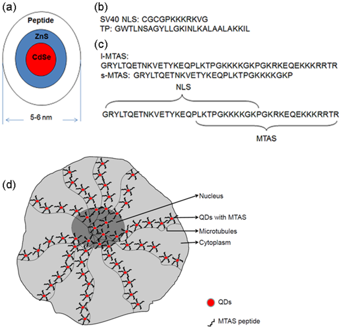
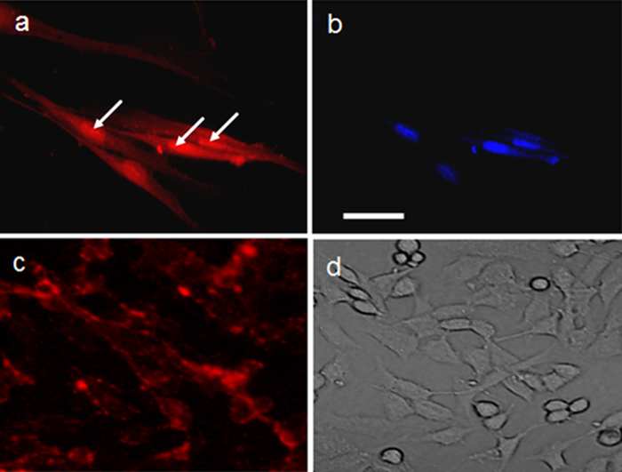
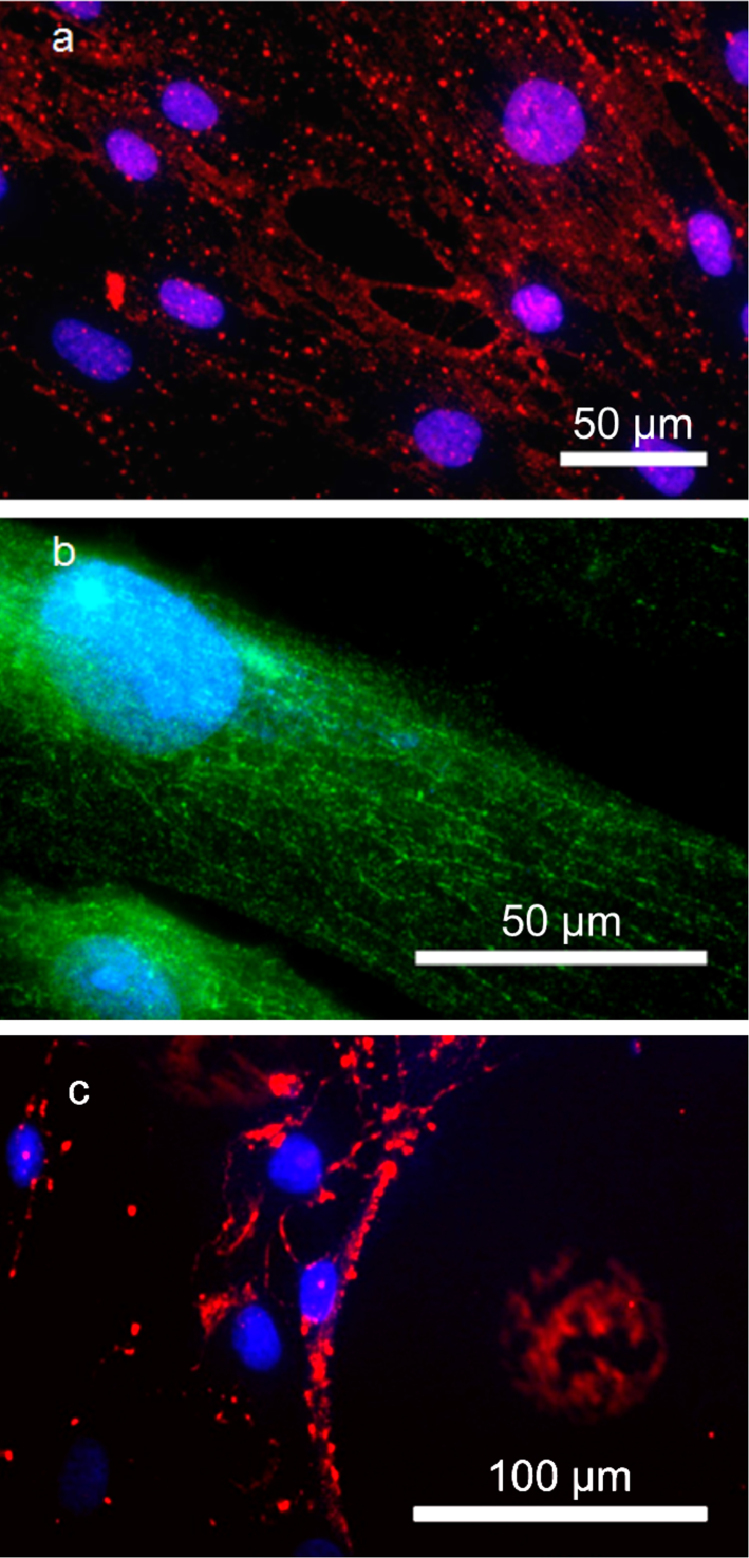
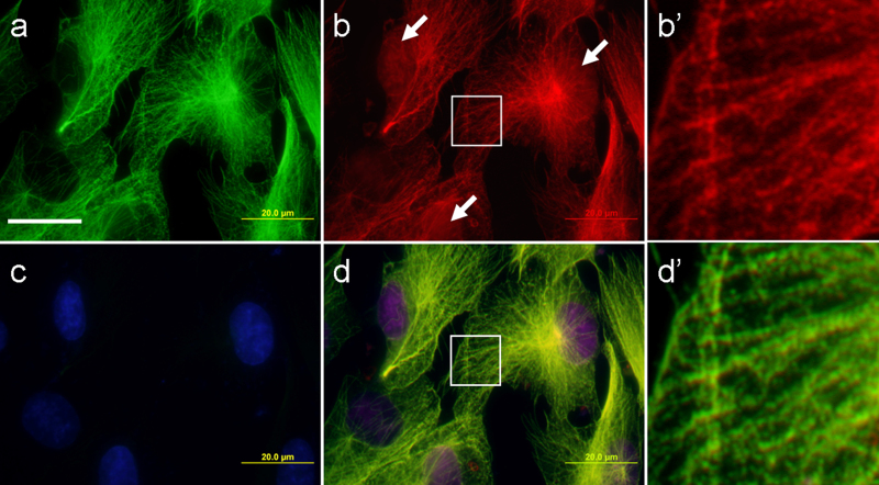
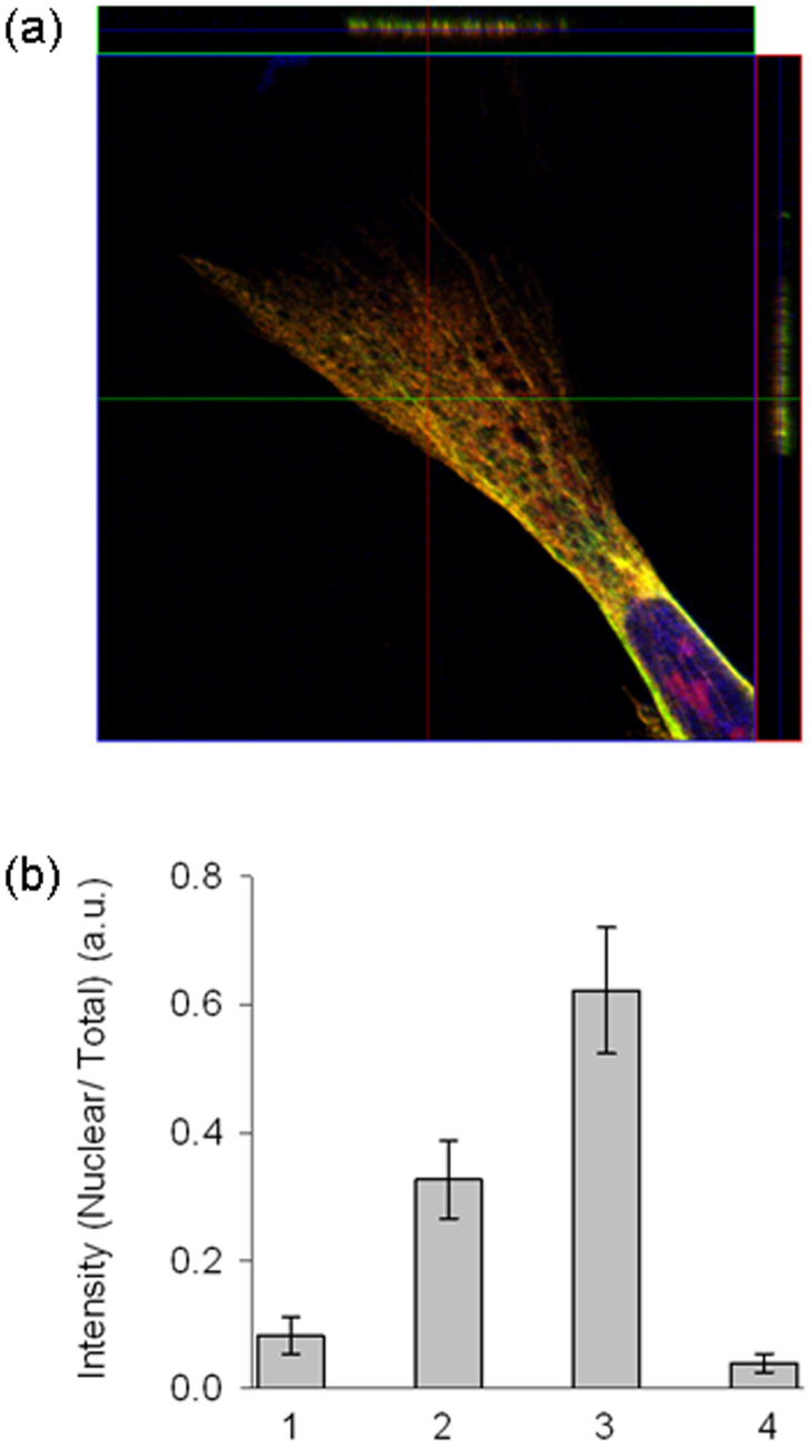
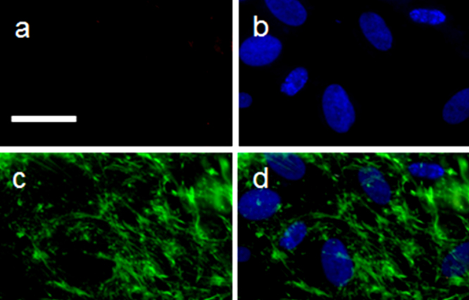
Protein transport is an important phenomenon in biological systems. Proteins are transported via several mechanisms to reach their destined compartment of cell for its complete function. One such mechanism is the microtubule mediated protein transport. Up to now, there are no reports on synthetic systems mimicking the biological protein transport mechanism. Here we report a highly efficient method of mimicking the microtubule mediated protein transport using newly designed biotinylated peptides encompassing a microtubule-associated sequence (MTAS) and a nuclear localization signaling (NLS) sequence, and their final conjugation with streptavidin-coated CdSe/ZnS quantum dots (QDs). Our results demonstrate that these novel bio-conjugated QDs enhance the endosomal escape and promote targeted delivery into the nucleus of human mesenchymal stem cells via microtubules. Mimicking the cellular transport mechanism in stem cells is highly desirable for diagnostics, targeting and therapeutic applications, opening up new avenues in the area of drug delivery.


Figures

Figure 1
Figure 1. Design of different peptide sequences for making water-soluble core/shell CdSe/ZnS quantum dots (QDs).
(a) A cartoon showing the mean diameter of peptide-coated QDs. (b) Peptides used in this study: SV40 NLS – nuclear localization signalling sequence, TP – transportan protein. (c) Peptide sequence deduced from parathyroid hormone related protein (PTHrP). The microtubule associated sequence (MTAS) domain spans 17–44 amino acids, while NLS spans 1–30 amino acids. Long (l-) MTAS represents 1–44 amino acids while short (s-) MTAS represents 1–30 amino acids. The s-MTAS lacks the complete sequence required for the association with microtubules. (d) A schematic illustration of the transportation of QDs to the nucleus of hMSCs via microtubules.
Figure 2
Figure 2. Labeling of hMSCs.
(a,b) SV40-NLS-TP and (c,d) SV40-NLS peptide-coated red-emitting CdSe/ZnS QDs. See Figure 1 for the anatomy of peptides. (a, c) Cells under fluorescence microscope. (b) DAPI staining indicating the nucleus of the cell. White arrows in (a) point out the accumulation of QDs in the nucleus. (d) Light microscope image. It is clear from (c) that SV40-NLS peptide-coated QDs are present on the cell membrane and endosomes without nuclear targeting. Scale bar: 100 μm.
Figure 3
Figure 3. Microtubule assisted transport of QDs.
Nuclear transport of streptavidin-QDs conjugated with biotinylated l-MTAS of (a) red emission and (b) green emission in hMSCs. The nuclear compartment is stained with DAPI (blue). The transport via microtubules and nuclear accumulation of QDs can be clearly seen. (c) Transport of QDs (red) conjugated with s-MTAS peptide. The s-MTAS peptide lacks the complete MTAS domain and thus is not able to transport QDs via microtubules. The cell nucleus is stained with DAPI (blue).
Figure 4
Figure 4. Co-localization of l-MTAS QDs, anti-tubulin antibody and DAPI in hMSC.
(a) Anti-tubulin antibody (green), (b) QDs conjugated to l-MTAS (red), (c) DAPI (blue) and (d) Composite image. The alignment of QDs on the microtubules and nucleus labelling (d) are clearly seen. The white arrows mark the presence of QDs in the nucleus (b). The regions corresponding to the white boxes in (b) and (d) were enlarged and shown in (b') and (d') respectively. Scale bar = 20 μm.
Figure 5
Figure 5. Z-stack imaging for the co-localization of l-MTAS–QDs on the tubulin filaments observed by confocal laser scanning microscope.
(a) Z-stack imaging and orthogonal analysis of the stained cells. Scanning was done along the lines drawn on adjacent extensions of the image. (b) The efficiency of nuclear targeting for the four different peptides reported here. The total cellular fluorescence and nuclear compartment fluorescence were measured for five cells of each group. The efficiency of the nuclear targeting was measured as the ratio of fluorescence intensity between nucleus and total hMSC labeling (represented as nuclear/total). X-axis represents the different peptide groups: 1, 2, 3 and 4 corresponding to SV40-NLS, SV40-NLS-TP, l-MTAS and s-MTAS, respectively.
Figure 6
Figure 6. Microtubule disruption results in no uptake of QDs.
hMSCs were treated with 5 μg/ml of nocodazole for 4 h prior to the addition of l-MTAS-conjugated QDs. The cells were fixed and stained with QDs (a) and DAPI (blue, (b)). The disrupted microtubules can be seen with anti-tubulin antibody staining (green, (c)) and in the composite image (d). It is clear that the uptake of QDs conjugated with l-MTAS peptide is inhibited completely in the absence of organized microtubules (a). Scale bar: 50 μm.

References

    1. Gao X., Cui Y., Levenson R. M., Chung L. W. & Nie S. In vivo cancer targeting and imaging with semiconductor quantum dots. Nat. Biotechnol. 22, 969–976 (2004). - PubMed
    1. Medintz I. L., Uyeda H. T., Goldman E. R. & Mattoussi H. Quantum dot bioconjugates for imaging, labelling and sensing. Nat. Mater. 4, 435–446 (2005). - PubMed
    1. Michalet X. et al. Quantum dots for live cells, in vivo imaging, and diagnostics. Science 307, 538–544 (2005). - PMC - PubMed
    1. Shen R. et al. Multifunctional conjugates to prepare nucleolar-targeting CdS quantum dots. J. Am. Chem. Soc. 132, 8627–8634 (2010). - PubMed
    1. Jiang W., Kim B. Y., Rutka J. T. & Chan W. C. Nanoparticle-mediated cellular response is size-dependent. Nat. Nanotechnol. 3, 145–150 (2008). - PubMed

Publication types