. 2013 Aug 15;191(4):1818-26.
doi: 10.4049/jimmunol.1300379. Epub 2013 Jul 12.

Critical role of p38 and GATA3 in natural helper cell function

Affiliations

Critical role of p38 and GATA3 in natural helper cell function

Jun-ichi Furusawa et al. J Immunol. .

Erratum in

  • J Immunol. 2014 Aug 1;193(3):1512

Abstract

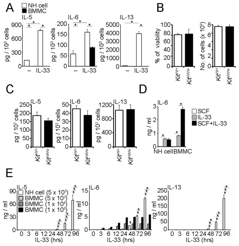
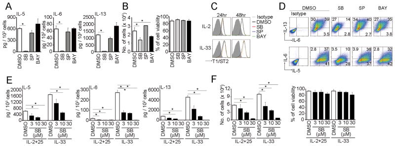
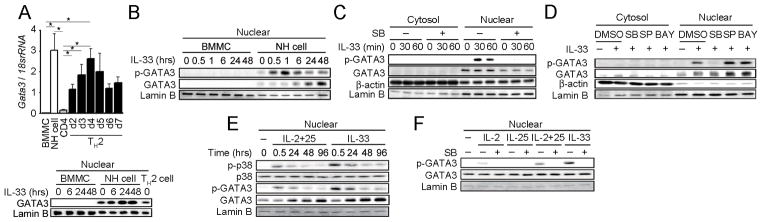
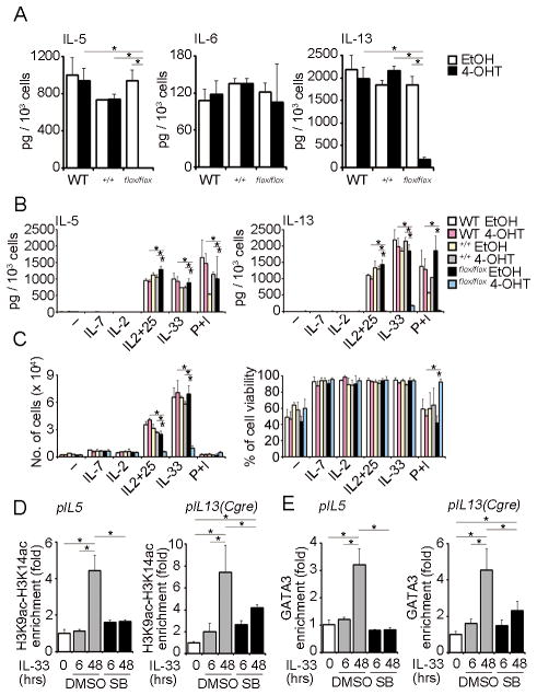
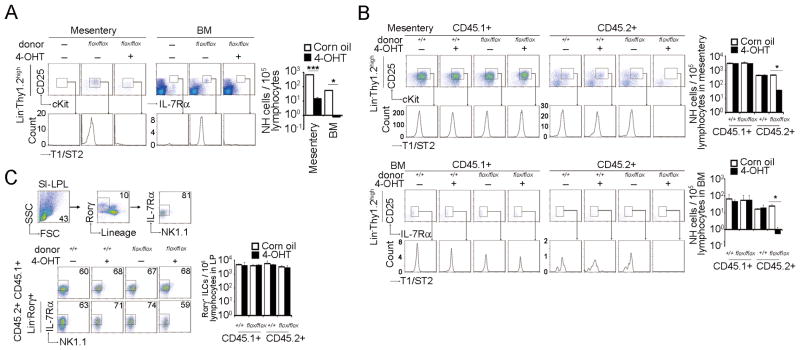
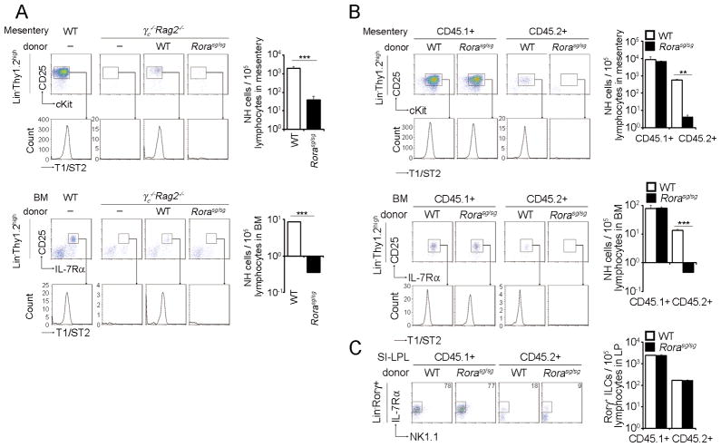
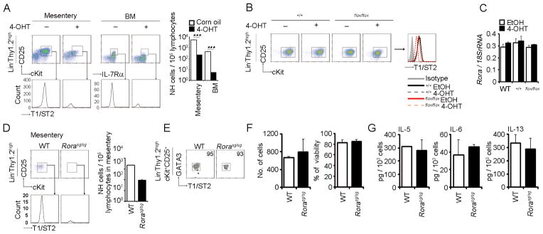
Natural helper (NH) cells, a member of Lin(-)IL-2R(+)IL-7R(+)IL-25R(+)IL-33R(+)GATA3(+) group 2 innate lymphoid cell subset, are characterized by the expression of transcription factors GATA3 and RORα and production of large amounts of Th2 cytokines such as IL-5, IL-6, and IL-13 upon IL-33 stimulation or a combination of IL-2 and IL-25. We have studied the signal transduction pathways critical for the cytokine expression and development of NH cell. Either stimulation with IL-33 or a combination of IL-2 and IL-25 induced p38 activation and phosphorylation of GATA3 in NH cells, and the phosphorylated form of GATA3 bound to the IL-5 and IL-13 promoters. All these events were blocked by SB203580, a p38 inhibitor. Inhibition of p38 also blocked IL-6 production. The mature NH cells lacking Gata3 were impaired in the proliferation and production of IL-5 and IL-13, but not IL-6, indicating that both p38 and GATA3 are critical for the proliferation and production of IL-5 and IL-13 and that the mechanisms downstream of p38 differ between IL-6 and IL-5/IL-13. In contrast, the NH cells lacking RORα showed no impairment in the proliferation and cytokine production, indicating that GATA3 but not RORα plays a pivotal role in the effector functions of mature NH cell. However, deletion of either GATA3 or RORα in hematopoietic stem cells severely blocked the development into NH cells. Our results demonstrate the important roles of p38 and GATA3 in NH cell functions.


Conflict of interest statement

Disclosure

S.K. is a consultant for Medical and Biological Laboratories, Co. Ltd. The authors otherwise have no financial conflicts of interest.

Figures

FIGURE 1
FIGURE 1. NH cells produce IL-5, IL-6 and IL-13 upon stimulation by IL-33 in a different manner from BMMCS
(A) Five thousand mesenteric NH cells or BMMCS were stimulated with IL-33 (10 ng/ml) for 96 hrs. The amounts of IL-5, IL-6 and IL-13 in the supernatants were detected by ELISA and are presented as pg production per 1 × 10³ cells. Error bars show s.e.m. (n = 2–3). *, P < 0.05. (B, C) Five thousand mesenteric NH cells from KitW/+ or KitW/Wv mice were stimulated with IL-33 (10 ng/ml) for 96 hrs. Amounts of IL-5, IL-6 and IL-13 in the supernatants were detected by ELISA and are presented as pg production per 1 × 10³ cells. Cell numbers and viability at the end of culture are also shown. Error bars show s.e.m. (n = 3). (D) Five thousand mesenteric NH cells or a one hundred thousand BMMCS were stimulated with SCF (50 ng/ml), IL-33 (10 ng/ml) or SCF and plus IL-33 for 96 hrs. The amounts of IL-6 in the supernatants were detected by ELISA. Error bars show s.e.m. (n = 3). *, P < 0.05. (E) Mesenteric NH cells or BMMCS were stimulated by IL-33 (10 ng/ml) and supernatants harvested at the indicated times. The amounts of IL-5, IL-6 and IL-13 were detected by ELISA. Error bars show s.e.m. (n = 3). *, P < 0.05. All results are representatives of two or three independent experiments with similar results.
FIGURE 2
FIGURE 2. p38 MAPK is important for cytokine production in NH cells
(A) Five thousand mesenteric NH cells were pretreated with DMSO (0.1%), SB203580 (10 μM), SP600125 (3 μM) or BAY11-7082 (100 nM) for 1 hr prior to IL-33 (10 ng/ml) stimulation for 96 hrs. Amounts of IL-5, IL-6 and IL-13 in the supernatants were detected by ELISA and are presented as pg production per 1 × 10³ cells. (B) Cell numbers and viability at the end of culture are also shown. Error bars show s.e.m. (n = 3). *, P < 0.05. (C) Mesenteric NH cells were pretreated as in (A) and cultured with IL-2 (10 ng/ml) or IL-33 (10 ng/ml) for the indicated time periods and examined the expression levels of T1/ST2. (D) Mesenteric NH cells were pretreated as in (A) and stimulated IL-33 (10 ng/ml) for 24 hrs. Cells were incubated with Brefeldin A for the last 3 hrs of IL-33 treatment. IL-5, IL-6 and IL-13 expression levels were detected by intracellular cytokine staining. Numbers indicate the percentage of each population within the gate. (E) Five thousand mesenteric NH cells were pretreated with DMSO (0.1%) or SB203580 (3–30 μM) for 1 hr prior to IL-2+25 (10 ng/ml each) or IL-33 (10 ng/ml) stimulation for 96 hrs. Amounts of IL-5, IL-6 and IL-13 in the supernatants were detected by ELISA and are presented as pg production per 1 × 10³ cells. (F) Cell numbers and viability at the end of culture are also shown. Error bars show s.e.m. (n = 3). *, P < 0.05. All results are representatives of two or three independent experiments with similar results.
FIGURE 3
FIGURE 3. IL-33-induced GATA3 phosphorylation by p38 MAPK in NH cells
(A) Total RNA was extracted from the indicated cells and Gata3 mRNA levels were detected by real-time PCR (upper panel). Error bars show s.e.m. (n = 3). *, P < 0.05. Nuclear fractions were prepared from IL-33 stimulated BMMCS and mesenteric NH cells as well as Th2 cells and GATA3 levels examined by western blotting (lower panel). (B) Purified mesenteric NH cells and BMMCS were stimulated with IL-33 (10 ng/ml) for the indicated periods. Phosphorylation and expression levels of GATA3 in nuclear extracts were detected by immunoblot analysis. NH cells were pretreated with DMSO (0.1%) or SB203580 (10 μM) (C), or DMSO (0.1%), SB203580 (10 μM), SP600125 (3 μM) or BAY11-7082 (100 nM) (D) prior to IL-33 (10 ng/ml) stimulation for 1 hr. Phosphorylation and expression levels of GATA3 in cytoplasmic and nuclear extracts were detected by immunoblot analysis. (E) NH cells were stimulated with IL-2+25 (10 ng/ml each) or IL-33 (10 ng/ml) for the indicated periods. Phosphorylation and expression levels of p38 and GATA3 in nuclear extracts were detected by immunoblot analysis. We used anti-pGATA3 (phosphor-S308) antibody purchased from Abcam (Cat No.ab61052). (F) NH cells were pretreated with DMSO (0.1%) or SB203580 (10 μM) prior to IL-2 (10 ng/ml), IL-25 (10 ng/ml), IL-2+25 (10 ng/ml each) or IL-33 (10 ng/ml) stimulation for 30 min. Phosphorylation and expression levels of GATA3 in nuclear extracts were detected by immunoblot analysis. All results are representative of two or three independent experiments with similar results.
FIGURE 4
FIGURE 4. IL-33 induced p38-mediated GATA3 phosphorylation is critical for IL-5 and IL-13 but not IL-6 production in NH cells
(A) Mesenteric NH cells (5 × 10³ cells) isolated from WT, Cre-Ert2:Gata3+/+ or Cre-Ert2:Gata3flox/flox mice were cultured in media containing IL-2 (10 ng/ml) with EtOH (0.01%) or 4-OHT (100 nM) for 48hr. After deleting Gata3, cells were stimulated with IL-33 (10 ng/ml) for 96 hrs. Amounts of IL-5, IL-6 and IL-13 in the supernatants were detected by ELISA and are presented as pg production by 1 × 10³ cells. Error bars show s.e.m. (n = 3). *, P < 0.05. (B, C) After deleting Gata3 as in (A), cells were cultured with IL-7 (10 ng/ml), IL-2 (10 ng/ml), IL-2 and 25 (10 ng/ml each), IL-33 (10 ng/ml) or PMA (30 ng/ml) plus ionomycin (500 ng/ml) for 96 hrs. (B) Amounts of IL-5, IL-6 and IL-13 in the supernatants were detected by ELISA and are presented as pg production by 1 × 10³ cells. (C) Cell numbers and viability at the end of culture are shown. Error bars show s.e.m. (n = 2–3). *, P < 0.05. (D, E). WT mesenteric NH cells were pretreated with DMSO (0.1%) or SB203580 (10 μM) for 1 hr prior to IL-33 (10 ng/ml) stimulation for the indicated time periods. ChIP analysis for the acetylation of histone H3 at Lys9 and Lys14 (H3K9ac-H3K14ac) (D) or GATA3 (E) binding to the Il5 and Il13 loci were detected by quantitative real-time PCR. Results are presented as relative enrichment compared to input DNA prepared from untreated cells. Error bars show s.e.m. (n = 3–4). *, P < 0.05. All results are representative of two or three independent experiments with similar results.
FIGURE 5
FIGURE 5. GATA3 is critical for the differentiation of NH cells
BMCs from Cre-Ert2:Gata3+/+ or Cre-Ert2:Gata3flox/flox mice (2.0 × 10⁷ cells) were transferred intravenously into γc−/−Rag2−/− mice (A) or B6.SJL-Rag2−/− mice (B, C). One day after transfer, corn oil or 4-OHT (1 mg/25 g mouse body weight) was injected intraperitoneally for 3 consecutive days and mice were analyzed on day 30. (A) Mesenteric LinThy1.2highc-Kit+CD25+ and BM LinIL-7Rα+CD25+ NH cells were analyzed by flow cytometry. Histograms in the lower panels show the expression levels of T1/ST2 on NH cells. Graphs indicate the numbers of NH cells per 1 × 10⁵ lymphocytes in mesentery or BM. (B) Mesenteric (upper panels) and BM (lower panels) NH cells from CD45.1+ recipient (left panels) and CD45.2+ donor (right panels) mice were analyzed by flow cytometry as in (A). (C) LinIL-7Rα+RORγ+ LTi-like cells in the small intestine were analyzed by flow cytometry and cell numbers examined per 1 × 10⁵ lymphocytes. Error bars show s.e.m. (n = 2). * and ***, P < 0.05 and 0.005, respectively. Results are representative of two or three independent experiments with similar results.
FIGURE 6
FIGURE 6. RORα is important for the differentiation of NH cells
(A–C) BMCs from WT or Rora sg/sg mice (6 × 10⁶ (A) or 5 × 10⁶ (B) cells) were transferred intravenously into γc−/−Rag2−/− mice (A) or B6.SJL-Rag2−/− mice (B, C) and analyzed 30 days after transfer. Mesenteric (upper panels) and BM (lower panels) NH cells (A, B) and LinIL-7Rα+RORγ+ LTi-like cells in the small intestine (C) were analyzed by flow cytometry. Graphs indicate the numbers of NH cell per 1 × 10⁵ lymphocytes in mesentery, bone marrow or small intestine. Error bars show s.e.m. (n = 2–4). ** and ***, P < 0.01 and 0.005, respectively. Results are representative of two or three independent experiments with similar results.
FIGURE 7
FIGURE 7. RORα is dispensable for Th2 cytokine production in NH cells
(A) Corn oil or 4-OHT (1 mg/25 g mouse body weight) was injected intraperitoneally for 3 consecutive days to Cre-Ert2:Gata3+/+ or Cre-Ert2:Gata3flox/flox mice and analyzed 4 days after the last injection Mesenteric LinThy1.2highc-Kit+CD25+ and BM LinIL-7Rα+CD25+ NH cells were analyzed by flow cytometry. Histograms in the lower panels show the expression levels of T1/ST2 on NH cells. Graphs indicate the numbers of NH cells per 1 × 10⁵ lymphocytes in mesentery or BM. Error bars show s.e.m. (n = 2). ***, P < 0.005. (B, C) Sorted NH cells from the mesentery of WT, Cre-Ert2:Gata3+/+ or Cre-Ert2:Gata3flox/flox mice were cultured in media containing IL-2 (10 ng/ml) and treated with EtOH (0.01%) or 4-OHT (100 nM) for 48 hrs before flow cytometric analysis. Expression levels of T1/ST2 (B) and Rora mRNA (C) were examined. Error bars in (C) show s.e.m. (n = 3). (D, E) Mesenteric NH cells from WT or Rora sg/sg mice were analyzed by flow cytometry as in (A). Error bars show s.e.m. (n = 2). The expression level of GATA3 was analyzed by flow cytometry (E). (F, G) Mesenteric NH cells isolated from WT or Rora sg/sg mice were stimulated with IL-33 (10 ng/ml) for 15 days. Cell numbers and viability at the end of culture (F) and amounts of IL-5, IL-6 and IL-13 in the supernatants (G) were examined. Error bars show s.e.m. (n = 3). All results are representatives of two or three independent experiments with similar results.

References

    1. Moro K, Yamada T, Tanabe M, Takeuchi T, Ikawa T, Kawamoto H, Furusawa J, Ohtani M, Fujii H, Koyasu S. Innate production of TH2 cytokines by adipose tissue-associated c-Kit+ Sca-1+ lymphoid cells. Nature. 2010;463:540–544. - PubMed
    1. Chang YJ, Kim HY, Albacker LA, Baumgarth N, McKenzie AN, Smith DE, Dekruyff RH, Umetsu DT. Innate lymphoid cells mediate influenza-induced airway hyper-reactivity independently of adaptive immunity. Nat Immunol. 2011;12:631–638. - PMC - PubMed
    1. Halim TY, MacLaren A, Romanish MT, Gold MJ, McNagny KM, Takei F. Retinoic-acid-receptor-related orphan nuclear receptor alpha is required for natural helper cell development and allergic inflammation. Immunity. 2012;37:463–474. - PubMed
    1. Halim TY, Krauss RH, Sun AC, Takei F. Lung natural helper cells are a critical source of Th2 cell-type cytokines in protease allergen-induced airway inflammation. Immunity. 2012;36:451–463. - PubMed
    1. Koyasu S, Moro K, Tanabe M, Takeuchi T. Natural helper cells: a new player in the innate immune response against helminth infection. Adv Immunol. 2010;108:21–44. - PubMed

Publication types

MeSH terms