

Upregulation of phosphorylated HSP27, PRDX2, GRP75, GRP78 and GRP94 in acquired middle ear cholesteatoma growth
- PMID: 23852020
- PMCID: PMC3742253
- DOI: 10.3390/ijms140714439
Upregulation of phosphorylated HSP27, PRDX2, GRP75, GRP78 and GRP94 in acquired middle ear cholesteatoma growth
Abstract
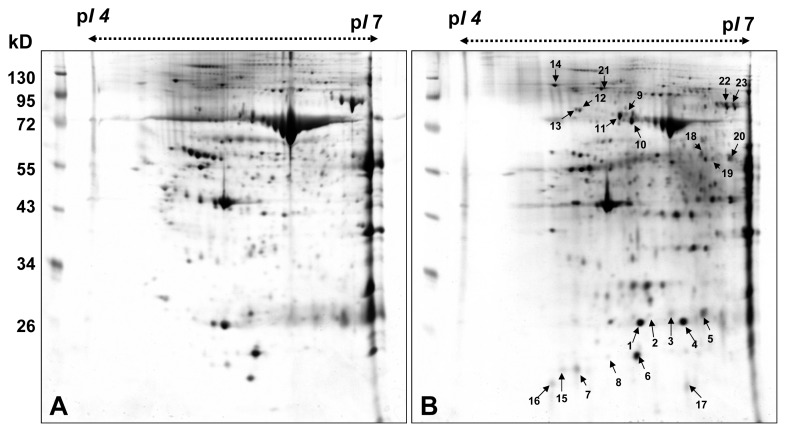
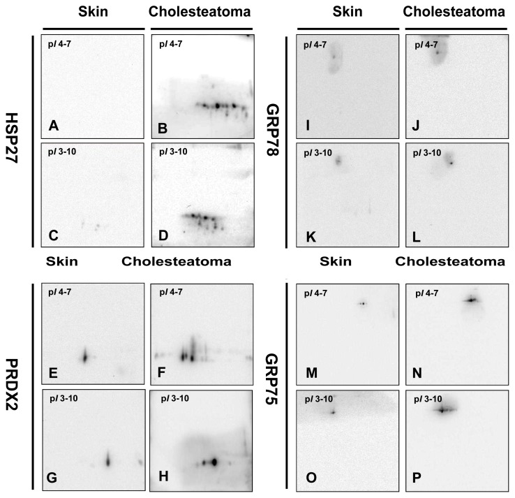
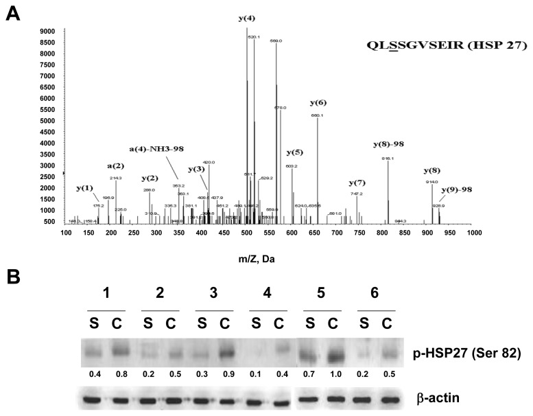
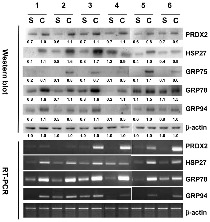
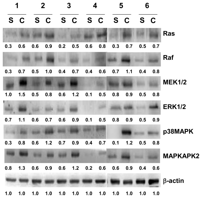
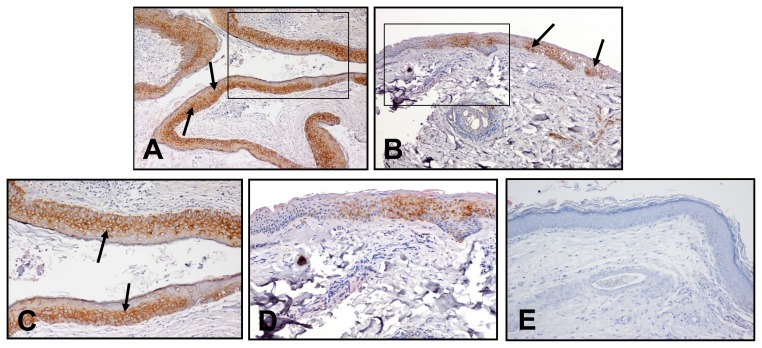
Cholesteatoma is a destructive and expanding growth of keratinizing squamous epithelium in the middle ear or petrous apex. The molecular and cellular processes of the pathogenesis of acquired middle ear cholesteatoma have not been fully understood. In this study, comparative proteomic analysis was conducted to investigate the roles of specific proteins in the pathways regarding keratinocyte proliferation in cholesteatoma. The differential proteins were detected by comparing the two-dimension electrophoresis (2-DE) maps of the epithelial tissues of 12 attic cholesteatomas with those of retroauricular skins. There were 14 upregulated proteins in the epithelial tissues of cholesteatoma in comparison with retroauricular skin. The modulation of five crucial proteins, HSP27, PRDX2, GRP75, GRP78 and GRP94, was further determined by RT-PCR, Western blot and immunohistochemistry. Phosphorylation of HSP27 at Ser-82 was identified by mass spectroscopy. The results of this study suggested that phosphorylated HSP27 is the end expression of two potential signal-transduction pathways, and together with PRDX2, they are very likely involved in the proliferation of keratinocytes in cholesteatoma. Upregulations of GRP75, GRP78 and GRP94 in keratinocytes may be able to counter endoplasmic reticulum stress, to inhibit cell apoptosis, to prevent protein unfolding and to promote cholesteatoma growth.
Figures
Similar articles
-
Heat shock proteins in middle ear cholesteatoma.Otolaryngol Head Neck Surg. 1996 Jan;114(1):77-83. doi: 10.1016/S0194-59989670287-5. Otolaryngol Head Neck Surg. 1996. PMID: 8570255
-
TFIIB-related factor 2 regulates glucose-regulated protein 78 expression in acquired middle ear cholesteatoma.Biochem Biophys Res Commun. 2021 Feb 12;540:95-100. doi: 10.1016/j.bbrc.2020.12.052. Epub 2021 Jan 13. Biochem Biophys Res Commun. 2021. PMID: 33453679
-
Cholesteatoma growth and proliferation: relevance with serpin B3.Laryngoscope. 2012 Dec;122(12):2818-23. doi: 10.1002/lary.23547. Laryngoscope. 2012. PMID: 23239141
-
Keratinocyte growth factor signaling promotes stem/progenitor cell proliferation under p63 expression during middle ear cholesteatoma formation.Curr Opin Otolaryngol Head Neck Surg. 2020 Oct;28(5):291-295. doi: 10.1097/MOO.0000000000000655. Curr Opin Otolaryngol Head Neck Surg. 2020. PMID: 32796271 Review.
-
[Current concepts of the pathogenesis of acquired middle ear cholesteatoma].Laryngorhinootologie. 2011 Jan;90(1):38-48; quiz 49-50. doi: 10.1055/s-0030-1268464. Epub 2011 Jan 10. Laryngorhinootologie. 2011. PMID: 21225533 Review. German.
Cited by
-
Increased Acquired Cholesteatoma Risk in Patients with Osteoporosis: A Retrospective Cohort Study.PLoS One. 2015 Jul 14;10(7):e0132447. doi: 10.1371/journal.pone.0132447. eCollection 2015. PLoS One. 2015. PMID: 26171780 Free PMC article.
-
Large-scale proteomics differentiates cholesteatoma from surrounding tissues and identifies novel proteins related to the pathogenesis.PLoS One. 2014 Aug 5;9(8):e104103. doi: 10.1371/journal.pone.0104103. eCollection 2014. PLoS One. 2014. PMID: 25093596 Free PMC article.
-
GRP94 Inhabits the Immortalized Porcine Hepatic Stellate Cells Apoptosis under Endoplasmic Reticulum Stress through Modulating the Expression of IGF-1 and Ubiquitin.Int J Mol Sci. 2022 Nov 14;23(22):14059. doi: 10.3390/ijms232214059. Int J Mol Sci. 2022. PMID: 36430538 Free PMC article.
-
Sinulariolide induced hepatocellular carcinoma apoptosis through activation of mitochondrial-related apoptotic and PERK/eIF2α/ATF4/CHOP pathway.Molecules. 2013 Aug 22;18(9):10146-61. doi: 10.3390/molecules180910146. Molecules. 2013. PMID: 23973991 Free PMC article.
-
Proteomic characterization of primary cultured myocytes in a fish model at different myogenesis stages.Sci Rep. 2019 Oct 1;9(1):14126. doi: 10.1038/s41598-019-50651-w. Sci Rep. 2019. PMID: 31576009 Free PMC article.
References
-
- Kuczkowski J., Sakowicz-Burkiewicz M., Iżycka-Œwieszewska E., Mikaszewski B., Pawełczyk T. Expression of tumor necrosis factor-α, interleukin-1α, interleukin-6 and interleukin-10 in chronic otitis media with bone osteolysis. ORL J. Otorhinolaryngol. Relat. Spec. 2011;73:93–99. - PubMed
-
- Haruyam T., Furukawa M., Kusunoki T., Onoda J., Ikeda K. Expression of IL-17 and its role in bone destruction in human middle ear cholesteatoma. ORL J. Otorhinolaryngol. Relat. Spec. 2010;72:325–331. - PubMed
-
- Kuczkowski J., Sakowicz-Burkiewicz M., Iżycka-Œwieszewska E. Expression of the receptor activator for nuclear factor-κB ligand and osteoprotegerin in chronic otitis media. Am. J. Otolaryngol. 2010;31:404–409. - PubMed
Publication types
MeSH terms
Substances
LinkOut - more resources
Full Text Sources
Other Literature Sources
Molecular Biology Databases
Research Materials
Miscellaneous
