

Growth hormone and melatonin prevent age-related alteration in apoptosis processes in the dentate gyrus of male rats
- PMID: 23852044
- PMCID: PMC3739870
- DOI: 10.1007/s10522-013-9443-6
Growth hormone and melatonin prevent age-related alteration in apoptosis processes in the dentate gyrus of male rats
Abstract
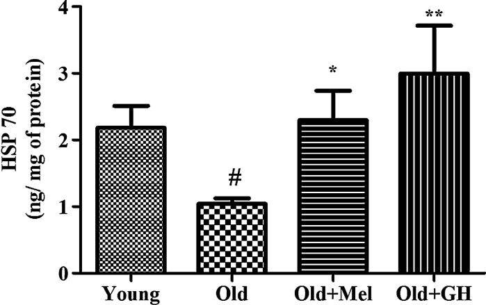
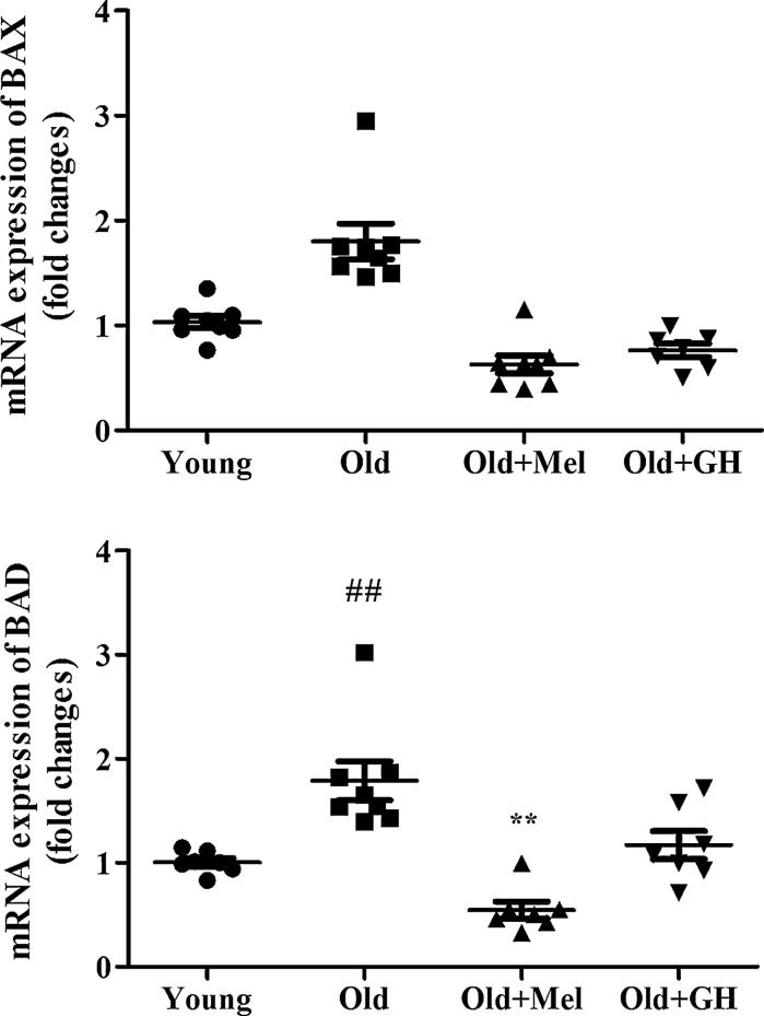
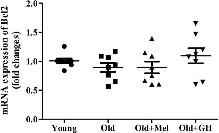
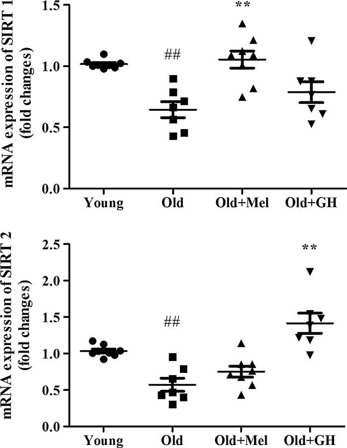
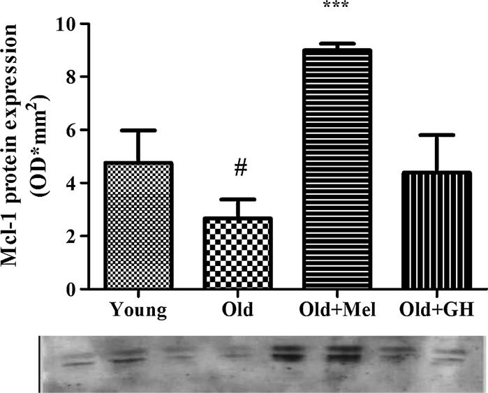
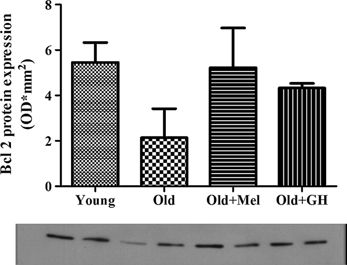
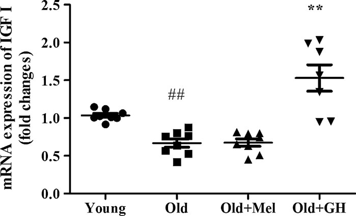
It has been suggested that the age-related decrease in the number of neurons in the hippocampus that leads to alterations in brain function, may be associated with an increase in apoptosis due to the reduced secretion of growth hormone (GH) and/or melatonin in old animals. In order to investigate this possibility, male Wistar rats of 22 months of age were divided into three groups. One group remained untreated and acted as the control group. The second was treated with growth hormone (hGH) for 10 weeks (2 mg/kg/d sc) and the third was subjected to melatonin treatment (1 mg/kg/d) in the drinking water for the same time. A group of 2-months-old male rats was used as young controls. All rats were killed by decapitation at more than 24 month of age and dentate gyri of the hippocampi were collected. Aging in the dentate gyrus was associated with an increase in apoptosis promoting markers (Bax, Bad and AIF) and with the reduction of some anti-apoptotic ones (XIAP, NIAP, Mcl-1). Expressions of sirtuin 1 and 2 (SIRT1 and 2) as well as levels of HSP 70 were decreased in the dentate gyrus of old rats. GH treatment was able to reduce the pro/anti-apoptotic ratio to levels observed in young animals and also to increase SIRT2. Melatonin reduced also expression of pro-apoptotic genes and proteins (Bax, Bad and AIF), and increased levels of myeloid cell leukemia-1 proteins and SIRT1. Both treatments were able to reduce apoptosis and to enhance survival markers in this part of the hippocampus.
Figures


References
-
- Alvira D, Tajes M, Verdaguer E, Acuna-Castroviejo D, Folch J, Camins A, Pallas M. Inhibition of the cdk5/p25 fragment formation may explain the antiapoptotic effects of melatonin in an experimental model of Parkinson’s disease. J Pineal Res. 2006;40:251–258. doi: 10.1111/j.1600-079X.2005.00308.x. - DOI - PubMed
-
- Andrabi SA, Sayeed I, Siemen D, Wolf G, Horn TF. Direct inhibition of the mitochondrial permeability transition pore: a possible mechanism responsible for anti-apoptotic effects of melatonin. FASEB J. 2004;18:869–871. - PubMed
-
- Arbour N, Vanderluit JL, Le Grand JN, Jahani-Asl A, Ruzhynsky VA, Cheung ECC, Kelly MA, MacKenzie AE, Park DS, Opferman JT, Slack RS. Mcl-1 is a key regulator of apoptosis during CNS development and following DNA damage. J Neurosci. 2008;28(24):6068–6078. doi: 10.1523/JNEUROSCI.4940-07.2008. - DOI - PMC - PubMed
Publication types
MeSH terms
Substances
LinkOut - more resources
Full Text Sources
Other Literature Sources
Medical
Research Materials
