. 2013 Aug;16(8):1016-23.
doi: 10.1038/nn.3458. Epub 2013 Jul 14.

Developmental origins of central norepinephrine neuron diversity

Affiliations

Developmental origins of central norepinephrine neuron diversity

Sabrina D Robertson et al. Nat Neurosci. 2013 Aug.

Abstract

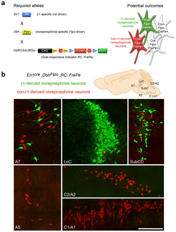
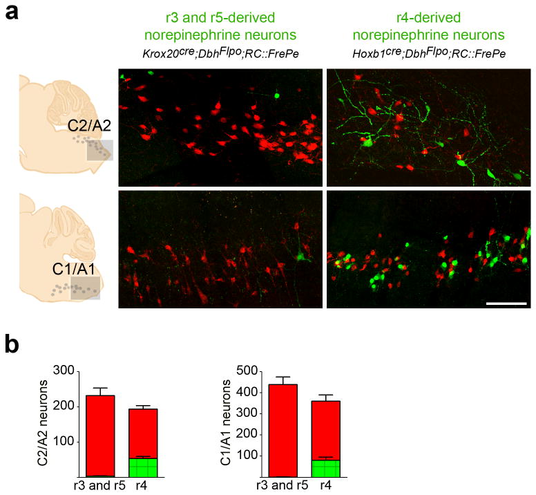
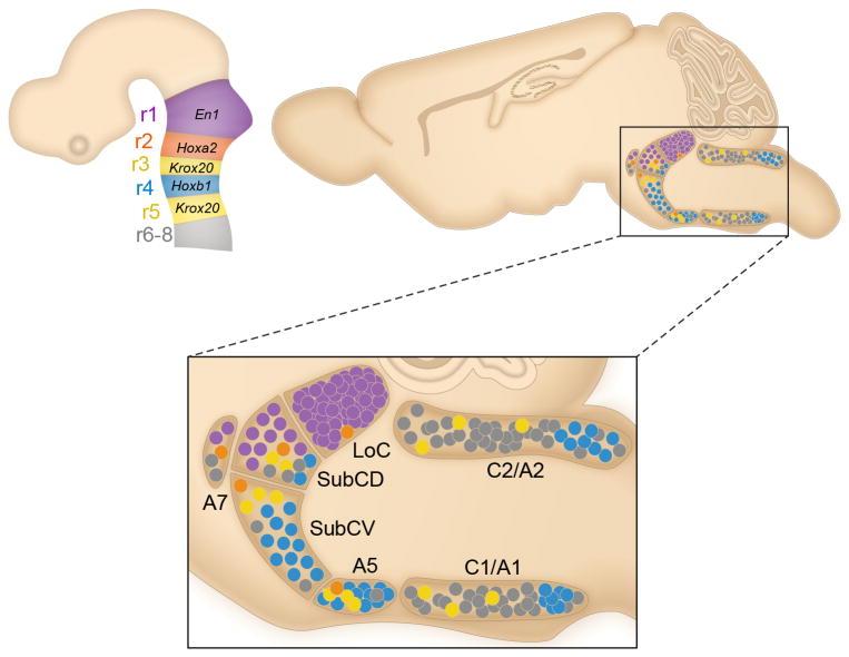
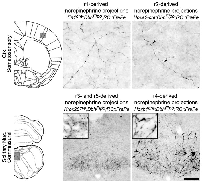
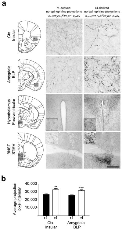
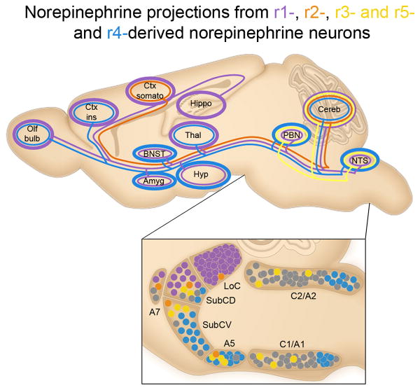
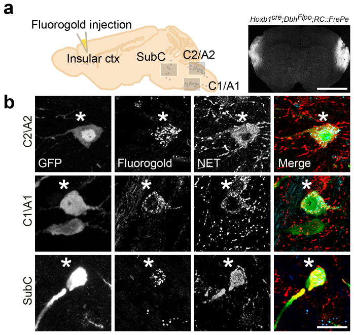
Central norepinephrine-producing neurons comprise a diverse population of cells differing in anatomical location, connectivity, function and response to disease and environmental insult. The mechanisms that generate this diversity are unknown. Here we elucidate the lineal relationship between molecularly distinct progenitor populations in the developing mouse hindbrain and mature norepinephrine neuron subtype identity. We have identified four genetically separable subpopulations of mature norepinephrine neurons differing in their anatomical location, axon morphology and efferent projection pattern. One of the subpopulations showed an unexpected projection to the prefrontal cortex, challenging the long-held belief that the locus coeruleus is the sole source of norepinephrine projections to the cortex. These findings reveal the embryonic origins of central norepinephrine neurons and provide multiple molecular points of entry for future study of individual norepinephrine circuits in complex behavioral and physiological processes including arousal, attention, mood, memory, appetite and homeostasis.


Figures

Fig. 1
Fig. 1. Intersectional genetic fate mapping strategy distinguishes r1(En1cre)-derived from non-r1-derived norepinephrine neurons
(a) Visualization of r1-derived norepinephrine neurons in isolation requires a r1-specific cre driver line (En1cre), a norepinephrine-specific Flpo driver line (DbhFlpo, see Supplementary Fig. 1), and a dual recombinase-responsive indicator line (RC∷FrePe). In mice that inherit all three alleles, norepinephrine neurons from r1 will express both Flpo and cre resulting in eGFP expression (green neuron in schematic), and all non-r1-derived norepinephrine neurons will express only Flpo resulting in mCherry expression (red neuron in schematic). Non-norepinephrine neurons will not express Flpo and are not marked by a fluorophore regardless of Cre expression (white cells in schematic). (b) Sections from En1Cre;DbhFlpo;RC∷FrePe adult mouse brainstem immunostained for eGFP and mCherry reveal the contribution of r1(En1cre)-derived norepinephrine neurons (green) to A7, LoC, and SubCD nuclei, and non-r1-derived norepinephrine neurons (red) to A7, LoC, SubCD, A5, C2/A2 and C1/A1 nuclei. Scale bar indicates 200 μm (A7, LoC, SubCD, and A5 coronal images) and 218 μm (C2/A2 and C1/A1 sagittal images).
Fig. 2
Fig. 2. Complementary fate maps reveal the distribution of r-derived norepinephrine subpopulations in the pontine norepinephrine nuclei
(a) Coronal sections from adult mouse brainstems reveal the contribution of r1(En1cre;DbhFlpo;RC∷FrePe)-, r2(Hoxa2-cre;DbhFlpo;RC∷FrePe)-, r3&5(Krox20cre;DbhFlpo;RC∷FrePe)-, and r4(Hoxb1cre;DbhFlpo;RC∷FrePe)-derived norepinephrine neurons to the SubCV, LoC, and A5 nuclei. eGFP (green) marks the r-derived norepinephrine population and mCherry (red) marks all other norepinephrine neurons in the representative sections corresponding to the boxed areas within the schematics (left). Scale bar indicates 200 μm (LoC) and 166 μm (SubCV and A5). (b) Cell counts of eGFP and mCherry positive neurons reveal the contribution of norepinephrine neurons derived from r1, r2, r3&5 and r4 to the LoC, SubCD, SubCV, A7, and A5 pontine nuclei. Numbers are the sum of bilateral counts from 30 μm sections spaced 120 μm apart from the brainstem of four animals for each fate map (error bars are mean ± s.e.m). Green bars represent counts of eGFP positive r-derived norepinephrine neurons, and red bars represent the counts of mCherry positive norepinephrine neurons (all other non-r-derived norepinephrine neurons). Total cell counts (mCherry plus eGFP positive cells) for each norepinephrine nucleus were not significantly different between fate maps (One-way ANOVA analysis p > 0.05; df=3 and 12; LoC F=0.6073; SubCD F=2.727; SubCV F=2.203; A7 F=0.7999; A5 F=2.694).
Fig. 3
Fig. 3. r3&5(Krox20cre)- and r4(Hoxb1cre)-derived norepinephrine neurons populate the medullary C1/A1 and C2/A2 brainstem nuclei
(a) Sagittal sections from adult mouse brainstems reveal the contribution of norepinephrine neurons derived from r3&5(Krox20cre;DbhFlpo;RC∷FrePe) and r4(Hoxb1cre;DbhFlpo;RC∷FrePe) to the caudal regions of the C1/A1 and C2/A2 nuclei. Our analyses do not distinguish epinephrine neurons in C2 and C1 from norepinephrine neurons in A2 and A1. eGFP (green) marks the r-derived population and mCherry (red) marks all other norepinephrine and epinephrine neurons in the representative sections corresponding to the boxed areas within the schematics (left). Scale bar indicates 100 μm. (b) Cell counts of r-derived norepinephrine and epinephrine neurons in the C1/A1 and C2/A2 medullary nuclei (r3&5 n=5; r4 n=6 mice; error bars are mean ± s.e.m.). Unpaired, two-tailed t-tests demonstrate that total numbers of norepinephrine and epinephrine neurons (sum of eGFP- and mCherry-positive cells) were not significantly different between genotypes (p>0.05; df=9; C2/A2 t=1.571; C1/A1 t=1.631).
Fig. 4
Fig. 4. Distribution of central norepinephrine neurons defined by genetic lineage differs from the traditional anatomical subdivisions
Schematic sagittal views of the embryonic brain (left), and the adult brain (right) compressed along the mediolateral axis. The shaded regions of the embryonic brain correspond to r1-8 of the hindbrain, and are labeled with genes used to define each rhombomere. In the adult brainstem the anatomically defined nuclei are designated as LoC, SubCD, SubCV, A7, A5, C2/A2 and C1/A1. r1(En1cre)-derived norepinephrine neurons (purple circles) contribute to the LoC, SubCD, and A7. r2(Hoxa2-cre)-derived norepinephrine neurons (orange circles) contribute to the LoC, SubCD, SubCV, A7, and A5. r3&5(Krox20cre)-derived norepinephrine neurons (yellow circles) contribute to SubCD, SubCV, A5, C2/A2, and C1/A1. r4(Hoxb1cre)-derived norepinephrine neurons (blue circles) contribute to the SubCD, SubCV, A5, C2/A2, and C1/A1. Gray circles indicate presumptive r6-derived norepinephrine neurons.
Fig. 5
Fig. 5. r2(Hoxa2-cre)- and r3&5(Krox20cre)-derived norepinephrine neurons project to limited targets
Coronal sections from adult mouse brains immunostained for eGFP to detect axonal inputs from r1(En1cre;DbhFlpo;RC∷FrePe)- and r2(Hoxa2-cre;DbhFlpo;RC∷FrePe)- derived norepinephrine neurons to the somatosensory cortex (top panel), and r3&5(Krox20cre;DbhFlpo;RC∷FrePe)- and r4(Hoxb1cre;DbhFlpo;RC∷FrePe)-derived norepinephrine neurons to the commissural solitary nucleus (bottom panel). The representative sections correspond to the boxed areas within the brain schematics (left). Sparse but specific projections from r2(Hoxa2-cre)-derived norepinephrine neurons are seen in the somatosensory cortex (top row, arrowheads in second panel), which is also targeted by the r1(En1cre)-derived subpopulation (top row, first panel). Strong innervation of the solitary nucleus by r3&5(Krox20cre)-derived norepinephrine neurons (bottom row, first panel) and r4(Hoxb1cre)-derived norepinephrine neurons (bottom row, second panel) is observed in the brainstem. Scale bar indicates 25 μm (cortex) and 100 μm (solitary nucleus).
Fig. 6
Fig. 6. r1(En1cre)- and r4(Hoxb1cre)-derived norepinephrine neurons differ in their axon morphology at multiple target sites
(a) Coronal sections from adult mouse brains immunostained for eGFP to detect axonal inputs from r1(En1cre;DbhFlpo;RC∷FrePe)- and r4(Hoxb1cre;DbhFlpo;RC∷FrePe)-derived norepinephrine neurons to the insular cortex, basolateral amygdala posterior part (BLP), paraventricular hypothalamus, and the bed nucleus of the stria terminalis (BNST) medial division ventral part (STMV). In the representative sections, corresponding to the boxed areas within the brain schematics (left), axons from r4(Hoxb1cre)-derived norepinephrine neurons appear thicker and have larger varicosities than those of r1(En1cre)-derived neurons. Scale bar indicates 56 μm (cortex, amygdala), 222 μm (hypothalamus), and 500 μm (BNST). (b) Quantitative comparison of projection fiber type from r1(En1cre)- and r4(Hoxb1cre)-derived norepinephrine neurons, confirms the link between genetic lineage and fiber morphology. The average projection pixel intensity of eGFP-positive axon fibers from r1(En1cre)- and r4(Hoxb1cre)-derived norepinephrine neurons projecting to the insular cortex and BLP amygdala is shown (insular cortex n=15 and BLP amygdala n=10 images from four mice; error bars are mean ± s.e.m.). A two tailed, unpaired t-test was used to determine significance **p=0.009 t=6.801 df =14 (insular Ctx), ***p=0.0006 t=2.808 df=28 (BLP amygdala).
Fig. 7
Fig. 7. Genetically defined norepinephrine subpopulations project to unique sets of targets
Selected axonal projections from r-derived norepinephrine neuron subpopulations located in the brainstem are illustrated as colored lines on a schematic of the adult mouse brain compressed along the mediolateral axis. Approximate positions of the subpopulations from which the projections originate are illustrated as colored circles (bottom panel). Thickness of the colored lines encircling target regions of interest qualitatively represent the density of innervation from the genetically defined subpopulation of the same color (r1 purple; r2 orange; r3&5 yellow; r4 blue). r1(En1cre)-derived neurons (purple) project throughout the neuroaxis, providing the most dense innervation to regions involved in higher order cognition and sensory perception, including the cortex (Ctx), thalamus (Thal), and hippocampus (Hippo). r2(Hoxa2-cre)-derived norepinephrine neurons (orange) project to limited targets, including a sparse projection to the somatosensory cortex (Ctx somato) and the cerebellum (Cereb). Projections from r3&5(Krox20cre)-derived norepinephrine neurons (yellow) are restricted to the hindbrain, including a sparse to moderate input to the parabrachial nucleus (PBN) and Cereb, as well as a substantial input to the NTS. r4(Hoxb1cre)-derived norepinephrine neurons (blue) project to key components of the central autonomic nervous system, including regions of the amygdala (Amyg), hypothalamus (Hyp), bed nucleus of the stria terminalis (BNST), and insular cortex (Ctx ins).
Fig. 8
Fig. 8. Identification of a shared projection to the insular cortex from r4(Hoxb1cre)-derived norepinephrine neurons residing in the C2/A2, C1/A1 and SubC nuclei
(a) Schematic sagittal view of adult mouse brain compressed along the mediolateral axis (left) and representative coronal section from Hoxb1cre;DbhFlpo;RC∷FrePe mouse brain (right) injected with FluoroGold into the insular cortex. Scale bar indicates 2.5 mm. (b) Sagittal sections from adult animals corresponding to the boxed regions in (a). eGFP-positive r4(Hoxb1cre)-derived norepinephrine neurons (green) co-labeled (*) with FluoroGold (blue) and NET (red) in C2/A2 (top panel), C1/A1 (middle panel), and SubC (bottom panel). Scale bar indicates 20 μm.

References

    1. Berridge CW, Waterhouse BD. The locus coeruleus-noradrenergic system: modulation of behavioral state and state-dependent cognitive processes. Brain research Brain research reviews. 2003;42:33–84. - PubMed
    1. Rinaman L. Hindbrain noradrenergic A2 neurons: diverse roles in autonomic, endocrine, cognitive, and behavioral functions. Am J Physiol Regul Integr Comp Physiol. 2011;300:R222–235. - PMC - PubMed
    1. Sara SJ, Bouret S. Orienting and reorienting: the locus coeruleus mediates cognition through arousal. Neuron. 2012;76:130–141. - PubMed
    1. Dahlström A, Fuxe K. Evidence for the existence of monoamine containing neurons in the central nervous system, I. Demonstration of monoamines in the cell bodies of brain stem neurons. Acta Physiol Scand. 1964;62(Suppl. 232):1–55. - PubMed
    1. Grzanna R, Fritschy JM. Efferent projections of different subpopulations of central noradrenaline neurons. Prog Brain Res. 1991;88:89–101. - PubMed

Publication types

MeSH terms