. 2013 Aug;16(8):1008-15.
doi: 10.1038/nn.3460. Epub 2013 Jul 14.

SIRT1 collaborates with ATM and HDAC1 to maintain genomic stability in neurons

Affiliations

SIRT1 collaborates with ATM and HDAC1 to maintain genomic stability in neurons

Matthew M Dobbin et al. Nat Neurosci. 2013 Aug.

Abstract

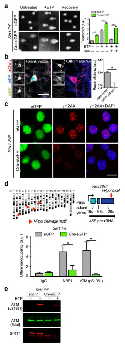
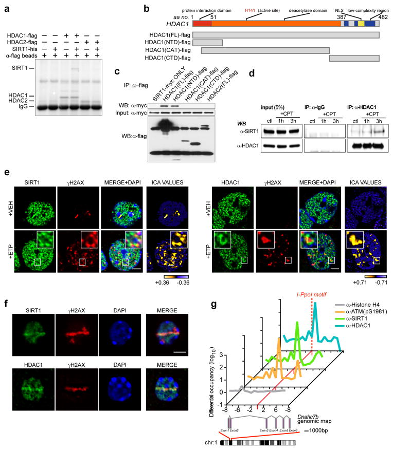
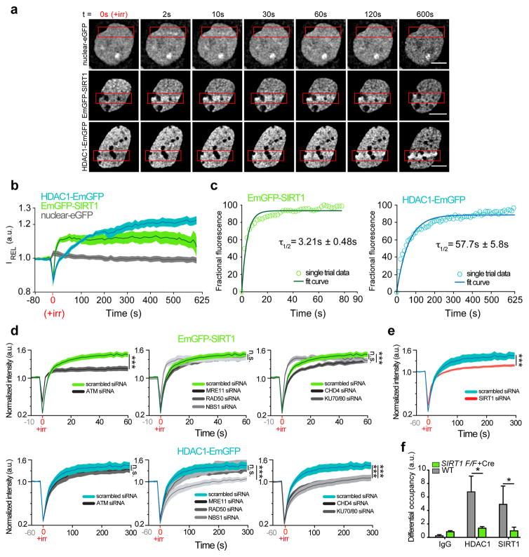
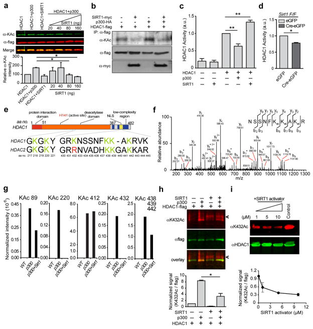
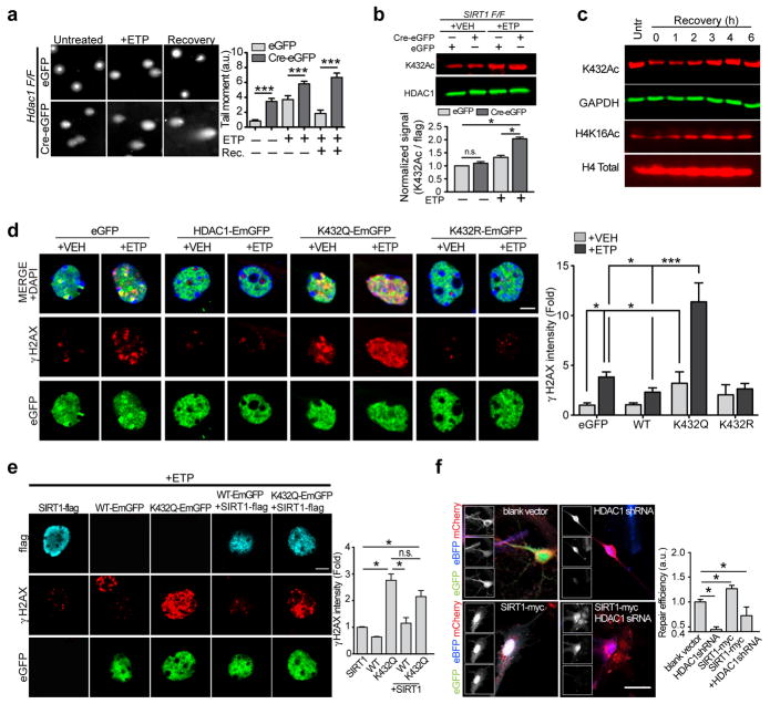
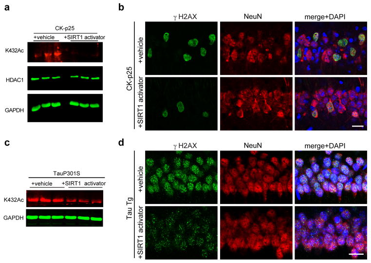
Defects in DNA repair have been linked to cognitive decline with age and neurodegenerative disease, yet the mechanisms that protect neurons from genotoxic stress remain largely obscure. We sought to characterize the roles of the NAD(+)-dependent deacetylase SIRT1 in the neuronal response to DNA double-strand breaks (DSBs). We found that SIRT1 was rapidly recruited to DSBs in postmitotic neurons, where it showed a synergistic relationship with ataxia telangiectasia mutated (ATM). SIRT1 recruitment to breaks was ATM dependent; however, SIRT1 also stimulated ATM autophosphorylation and activity and stabilized ATM at DSB sites. After DSB induction, SIRT1 also bound the neuroprotective class I histone deacetylase HDAC1. We found that SIRT1 deacetylated HDAC1 and stimulated its enzymatic activity, which was necessary for DSB repair through the nonhomologous end-joining pathway. HDAC1 mutations that mimic a constitutively acetylated state rendered neurons more susceptible to DNA damage, whereas pharmacological SIRT1 activators that promoted HDAC1 deacetylation also reduced DNA damage in two mouse models of neurodegeneration. We propose that SIRT1 is an apical transducer of the DSB response and that SIRT1 activation offers an important therapeutic avenue in neurodegeneration.


Figures

Figure 1
Figure 1. SIRT1 is necessary for initial DSB signaling events and DNA repair in neurons
a, Sirt1 F/F neurons were infected with lentiviral vectors carrying either a functional Cre recombinase (Cre-eGFP) or a non-functional Cre (eGFP) were treated with 5μM etoposide for 1h, and were either allowed to recover for 16h in the absence of etoposide or lysed immediately. DNA damage was then assessed using the comet assay. Graph indicates “comet tail moments” (***p< 0.001, n = at least 50 per condition, one-way ANOVA). b, Cultured primary neurons were transfected with a pre-digested NHEJ reporter construct (see also Supplementary Figs. 1b and 1c) together with either scrambled shRNA or SIRT1 shRNA and the number of GFP+ cells were assessed to indicate NHEJ-mediated repair (* p < 0.05, unpaired t-test). c, Sirt1 F/F neurons infected as in a were treated with either vehicle or 2μM etoposide, following which the cells were fixed and stained with antibodies to γH2AX. d, A synthetic, inducible system encoding the rare-cutting homing endonuclease, I-PpoI, was used to generate DSBs at defined regions within the genomes of primary Sirt1 F/F neurons transduced as in a. I-PpoI cleavage sites in the Rna28s1 locus are depicted (top). Results from ChIP experiments measuring the recruitment of ATM (pS1981) and NBS1 to cleavage sites within the Rna28s1 locus (bottom) (*p < 0.05, student’s t-test). e, Sirt1 F/F neurons infected as in a were treated with either vehicle or 5μM etoposide, and levels of phosphorylated ATM were compared by western blotting.
Figure 2
Figure 2. SIRT1 and HDAC1 physically interact and localize to DSB sites in neurons
a, Recombinant SIRT1-his was incubated with either HDAC1-flag or HDAC2-flag, following which HDAC1 and HDAC2 were precipitated with anti-flag conjugated agarose beads, and their ability to retain SIRT1 was assessed. b, Diagram illustrating HDAC1 fragment constructs for interaction mapping. FL – full-length; CTD – carboxy-terminal domain; CAT – catalytic domain; NTD – amino-terminal domain. c, The indicated flag-tagged fragments were expressed together with SIRT1-myc, and the flag-tagged proteins were immunoprecipitated and blotted with antibodies against myc. d, HT22 cells were treated with camptothecin (CPT; 1μM), precipitated with antibodies against HDAC1, and blotted with antibodies against SIRT1. e, Etoposide-treated primary neurons treated were fixed and stained with antibodies against either SIRT1 or HDAC1, and γH2AX. Right-most panel illustrates Intensity Correlation Analysis (ICA). Pixels from input channel co-varying positively with corresponding signal from γH2AX channel are indicated in yellow, while negatively co-varying pixels are indicated in blue. Scale bar = 3μm. f, Primary neurons were subjected to sub-nuclear, laser-generated DNA lesioning and stained with antibodies against either SIRT1 or HDAC1, and γH2AX. Scale bar = 3μm. g, The rare-cutting homing endonuclease, I-PpoI, was used to generate DNA DSBs at defined genetic loci in mouse cortical neurons. Recruitment of the indicated proteins at a unique cleavage site located between exons 2 and 3 in the Dnahc7b gene was then assessed by ChIP. Primers were designed at regular 1kb intervals, spanning 10kb both 3’ and 5’ to the I-PpoI consensus site (red dotted line).
Figure 3
Figure 3. SIRT1 stabilizes HDAC1 at sites of DNA DSBs in neurons
a, Time-lapse images of primary cortical neurons that were transfected with a vector carrying nuclear-eGFP, EmGFP-SIRT1, or HDAC1-EmGFP, and subjected to sub-nuclear, laser-generated DNA lesioning using a confocal microscope equipped with a 405 nm laser. Red boxes indicate damage ROI. Scale bar = 5 μm. b, Quantification of relative fluorescence intensity (IREL) as a function of time at lesioned ROIs for neurons expressing nuclear-eGFP (grey; n=13, **r=0.37 Pearson corr.), HDAC1-EmGFP (blue; n=15, ***r=0.98 Pearson corr.), or EmGFP-SIRT1 (green; n=11, ***r=0.77 Pearson corr.). c, Modeling and regression analysis of time-lapse data. Empty circles (green-SIRT1; blue-HDAC1) indicate data from a single trial plotted against the fitted curve (solid lines). d, Relative fluorescence intensity (IREL) as a function of time at lesioned ROIs for neurons expressing either EmGFP-SIRT1 (Top) or HDAC1-EmGFP (Bottom) together with the indicated siRNAs (* denotes p < 0.05; one-way ANOVA; the scrambled siRNA traces are reproduced in each graph for SIRT1 and HDAC1 respectively for convenience of visualization). e, Quantification of relative fluorescence intensity (IREL) as a function of time at lesioned ROIs for neurons expressing either HDAC1-EmGFP together with either a scrambled siRNA or a SIRT1 siRNA (*** p < 0.001, unpaired t-test). f, DSBs were generated using the I-PpoI-ER system in WT and Sirt1 KO primary neurons, and the recruitment of the indicated proteins to cleavage sites within the Rna28s1 locus was assessed using ChIP as in Fig. 1d (* p < 0.05, unpaired t-test).
Figure 4
Figure 4. SIRT1 deacetylates HDAC1 at residue K432 and stimulates its enzymatic activity
a, Recombinant SIRT1 was titrated over a fixed amount of p300 and HDAC1 and the acetylation of HDAC1 was assessed using western blotting (* p < 0.05, one-way ANOVA). b, HDAC1-flag, p300-HA, and SIRT1-myc were expressed in HT22 cells. HDAC1 was immunoprecipitated, and its acetylation status was probed using an anti-acetyl lysine antibody. c, HDAC1 was pre-incubated with either p300 or with SIRT1 as in (A) and (B), and its enzymatic activity was examined using a fluorescence based HDAC enzymatic activity assay (Supplementary Fig. 3b) (** p < 0.01, one-way ANOVA). d, Sirt1 F/F neurons were infected with Cre-eGFP and eGFP lentiviral vectors, following which HDAC1 was immunoprecipitated, and its activity was measured as in c (*p < 0.05, unpaired t-test). e, Diagram depicting the acetylated lysine residues in HDAC1 and HDAC2. f, Annotated MS/MS spectrum of Lys acetylated peptide “NSSNFKacKacAKacR” that identified lysine acetylation sites on K438, K439 and K441 of HDAC1 upon reaction with p300. b and y ions represents collision-induced peptide fragment ions containing N- or C-terminal respectively. “*” indicates fragment ions with neutral loss of amine. g, Label-free quantification for each lysine acetylation site using protein-abundance normalized peptide precursor ion intensity shows that lysine acetylation abundance on p300-treated HDAC1 decreased upon the addition of SIRT1 to the reaction. h, Recombinant SIRT1 was incubated together with p300 and HDAC1 as in a, and acetylation of HDAC1 was assessed using an antibody specific to acetylated K432. (Arrowhead shows a non-specific cross-reacting band in the lanes containing recombinant SIRT1; * p < 0.05, unpaired t-test). i, HEK293T cells were treated with the indicated concentrations of SIRT1 activator (Compound#10) for 12 h and the HDAC1 acetylation at K432 was assessed.
Figure 5
Figure 5. Deacetylation of HDAC1 is essential for DSB repair in neurons
a, Hdac1 F/F neurons were infected and treated with etoposide and DNA damage was assessed using the comet assay as in Fig. 1a (***p< 0.001, one-way ANOVA). b, Primary neurons cultured from SIRT1F/F embryos were infected as in Fig. 1a. The neurons were then treated with 5μM etoposide for 2 h and the acetylation of HDAC1 at K432 was assessed using western blots (*p < 0.05, one-way ANOVA). c, Cultured primary neurons (DIV7) were treated with 5μM etoposide for 30 min. Cells were then lysed either immediately or following recovery after etoposide washout for the indicated times. The lysates were electrophoresed and the indicated acetylation marks were probed by western blotting. d, Primary neurons were transfected with the indicated vectors and treated with etoposide (2μM) for 1 h, after which the cells were fixed and stained with antibodies against γH2AX. Scale bar = 10μm (* p < 0.05, *** p < 0.001, one-way ANOVA). e, Cultured primary neurons expressing flag-SIRT1 together with either HDAC1-EmGFP or HDAC1K432Q-EmGFP were treated with etoposide (2μM) for 1 h, following which the cells were fixed and stained as in d. Scale bar = 15μm; Quantification shown below (* p < 0.05, one-way ANOVA). f, Cultured primary neurons expressing either SIRT1 together with HDAC1 shRNA were transfected with the pre-digested NHEJ reporter construct and the number of GFP+ cells were assessed as a measure of DNA repair using NHEJ (*p < 0.05, one-way ANOVA).
Figure 6
Figure 6. Pharmacological SIRT1 activation can protect neurons against DNA damage in vivo
a, 6-weeks induced CKIIα-p25 mice were administered either vehicle or 30mg/kg compound#10. Brain lysates were then prepared and the acetylation of HDAC1 at K432 was assessed using quantitative western blotting. b, Representative immunohistochemical images depicting γH2AX and NeuN staining in 6-weeks induced CKIIα-p25 mice that were either administered compound#10 or vehicle as described in Methods. c, Two-month old Tau P301S Tg mice were treated with SIRT1 activator and hippocampal lysates were prepared. The acetylation of HDAC1 at K432 was then assessed using western blotting. d, Two-month old P301S Tg mice were administered either vehicle or 30mg/kg compound#10 through oral gavage (5 mice per group). Injections were performed once daily for two weeks. Following this, the mice were sacrificed, and their brains were sectioned and stained with antibodies against γH2AX and NeuN.

References

    1. Lu T, et al. Gene regulation and DNA damage in the ageing human brain. Nature. 2004;429:883–891. - PubMed
    1. Rass U, Ahel I, West SC. Defective DNA repair and neurodegenerative disease. Cell. 2007;130:991–1004. - PubMed
    1. Adamec E, Vonsattel JP, Nixon RA. DNA strand breaks in Alzheimer's disease. Brain Res. 1999;849:67–77. - PubMed
    1. Ferrante RJ, et al. Evidence of increased oxidative damage in both sporadic and familial amyotrophic lateral sclerosis. J Neurochem. 1997;69:2064–2074. - PubMed
    1. Nouspikel T, Hanawalt PC. When parsimony backfires: neglecting DNA repair may doom neurons in Alzheimer's disease. Bioessays. 2003;25:168–173. - PubMed

Publication types

MeSH terms

LinkOut - more resources