. 2013 Oct;20(10):1330-40.
doi: 10.1038/cdd.2013.83. Epub 2013 Jul 12.

The Hippo pathway kinase Lats2 prevents DNA damage-induced apoptosis through inhibition of the tyrosine kinase c-Abl

Affiliations

The Hippo pathway kinase Lats2 prevents DNA damage-induced apoptosis through inhibition of the tyrosine kinase c-Abl

N Reuven et al. Cell Death Differ. 2013 Oct.

Abstract

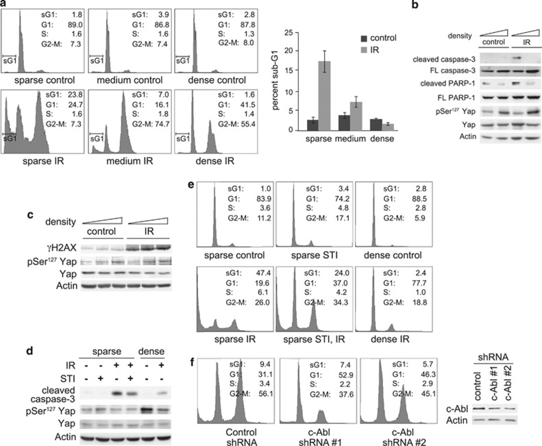
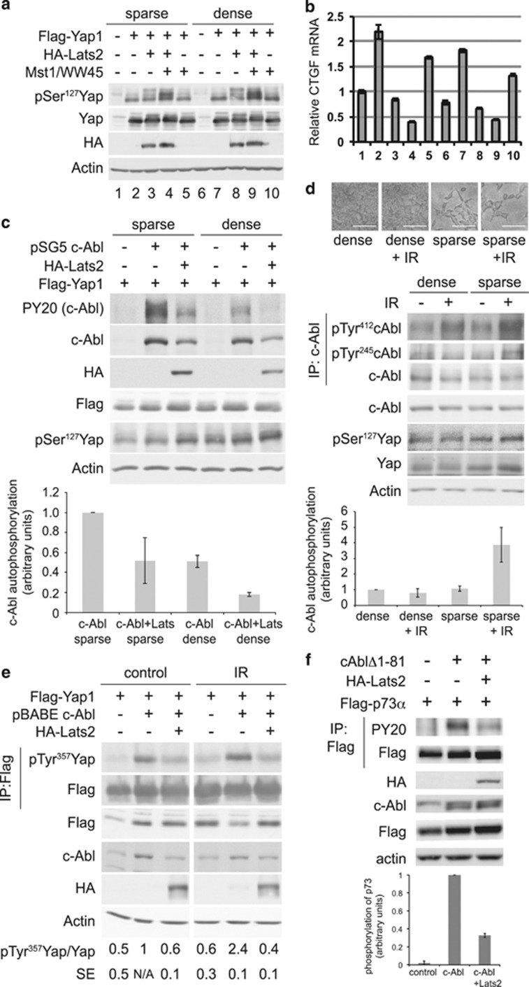
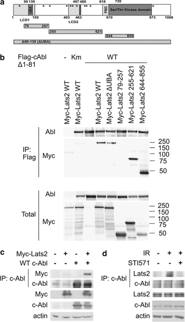
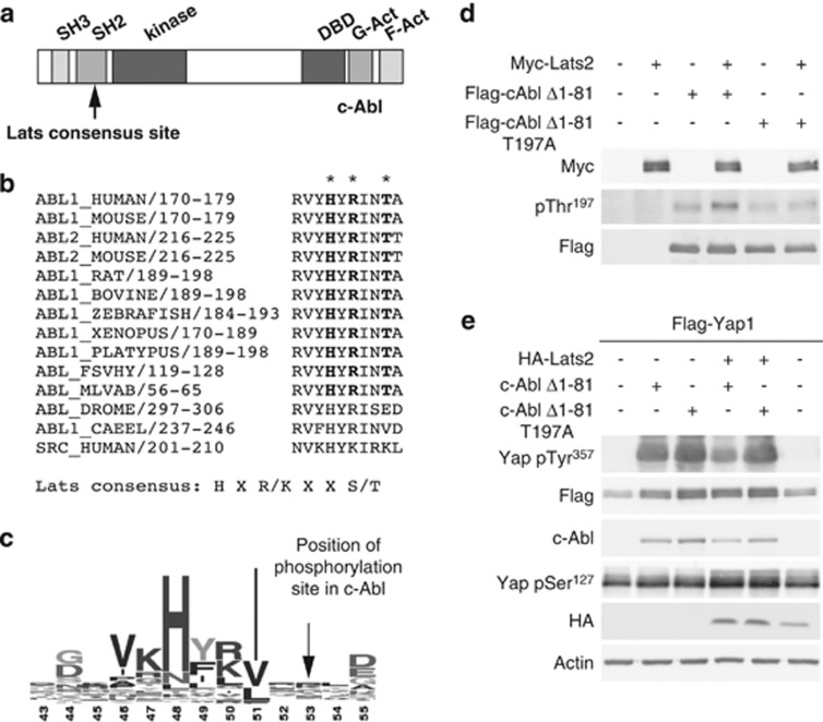
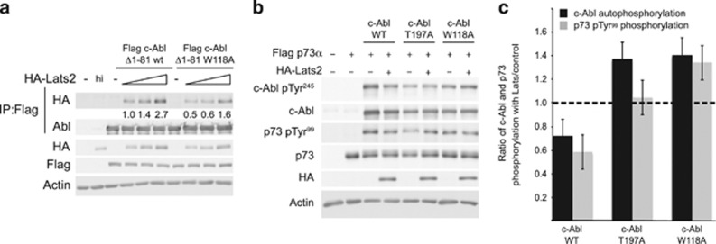
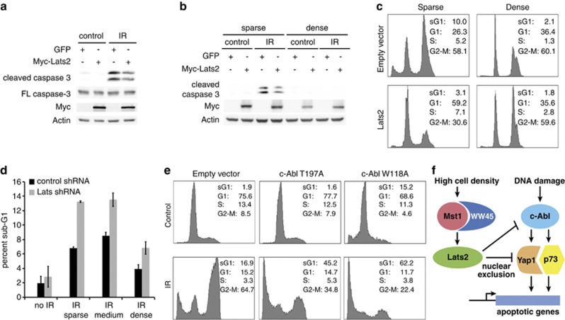
The Hippo pathway is an evolutionarily conserved pathway that controls cell proliferation, organ size, tissue regeneration and stem cell self-renewal. Here we show that it also regulates the DNA damage response. At high cell density, when the Hippo pathway is active, DNA damage-induced apoptosis and the activation of the tyrosine kinase c-Abl were suppressed. At low cell density, overexpression of the Hippo pathway kinase large tumor suppressor 2 (Lats2) inhibited c-Abl activity. This led to reduced phosphorylation of downstream c-Abl substrates, the transcription coactivator Yes-associated protein (Yap) and the tumor suppressor p73. Inhibition of c-Abl by Lats2 was mediated through Lats2 interaction with and phosphorylation of c-Abl. Lats2 knockdown, or expression of c-Abl mutants that escape inhibition by Lats2, enabled DNA damage-induced apoptosis of densely plated cells, while Lats2 overexpression inhibited apoptosis in sparse cells. These findings explain a long-standing enigma of why densely plated cells are radioresistant. Furthermore, they demonstrate that the Hippo pathway regulates cell fate decisions in response to DNA damage.


Figures

Figure 1
Figure 1
DNA damage-induced apoptosis is inhibited at high cell density. (a) MCF10A cells were plated at different cell densities. Cells were irradiated at 20 Gy, and were harvested for fluorescence-activated cell sorting (FACS) analysis 72 h post-irradiation (IR). Left panels, profile plots of representative FACS analysis. Percentages of cells in the different fractions (sub-G1, G1, S and G2-M) are indicated. Right panel, graph showing quantification of three independent experiments. (b) Cells were treated as in (a), and analyzed by immunoblot with the indicated antibodies. (c) Cells were plated and irradiated as in (a), but were harvested 2.5 h post-IR for immunoblot analysis. (d) NIH3T3 cells were plated sparse or dense and were treated with 10 μM STI571 or dimethyl sulfoxide (solvent control) as indicated. Cells were irradiated 2.5 h later at 40 Gy, and were harvested 96 h post-IR, and analyzed by immunoblotting. (e) NIH3T3 cells were treated as in (d), and were analyzed by FACS. Percentages of cells in the different fractions are indicated. (f) MCF10A stably expressing small hairpin RNA (shRNA) to c-Abl or non-targeting control were plated sparse, and were irradiated at 25 Gy. Left: cells were harvested for FACS analysis 96 h post-IR. Percentages of cells in the different fractions are indicated. Right: the expression of c-Abl in the c-Abl-shRNA-expressing cells was analyzed by immunoblotting
Figure 2
Figure 2
Nuclear Yes-associated protein (Yap) does not rescue apoptosis at high cell density. (a) MCF10A cells expressing Flag-Yap1, Flag-Yap1 S127A or vector control were plated sparse and dense, and irradiated at 20 Gy. After 24 h, cells were fixed for immunofluorescence analysis with the anti-Flag antibody. Nuclear staining is denoted by Hoechst 33342. Panels show representative fields of three independent experiments. Arrows indicate some of the cells with nuclear-localized Yap1. (b) The cells used in (a) were plated sparse and dense, irradiated at 20 Gy and harvested for FACS analysis 120 h post-IR. Percentages of cells in the different fractions (sub-G1, G1, S and G2-M) are indicated
Figure 3
Figure 3
High-cell density inhibits c-Abl autophosphorylation. (ab) Activation of Lats by overexpression and high-cell density. HEK293 cells were transfected with the indicated and green fluorescent protein (GFP)-expressing plasmids. Cells were replated 28 h post-transfection to produce sparse and dense plates. Sixteen hours later, cells were harvested and immunoblotted with the indicated antibodies (a). (b) CTGF mRNA levels were analyzed by qPCR and normalized by GFP mRNA. (c) High cell density and Lats2 overexpression inhibit c-Abl autophosphorylation. HEK293 cells were transfected with Flag-Yap1 (all lanes), and indicated plasmids. Transfected cells were replated sparse and dense, harvested 11 h later, and analyzed by immunoblotting. PY20 detects phosphotyrosine. Lower panel: Quantification of c-Abl autophosphorylation, N=2. (d) c-Abl DNA damage-induced activation is inhibited at high cell density. HEK293 cells were plated at high and low cell density. Cells were irradiated at 20 Gy, and were photographed (upper panel, scale bar=0.1 mm) and then harvested 24 h post-IR. Middle panel: c-Abl was immunoprecipitated and samples were analyzed by immunoblotting with the indicated antibodies. Lower panel: Quantification of c-Abl autophosphorylation, N=6. (e) Lats2 inhibits IR-induced c-Abl phosphorylation of Yap1. HEK293 cells were transfected with the plasmids indicated, and 24 h post-transfection, cells were irradiated at 20 Gy. Cells were harvested 4 h post-IR, and Flag-Yap1 was immunoprecipitated. Samples were analyzed by immunoblotting with the indicated antibodies. Quantification of pTyr357 Yap, N=3. (f) Lats2 inhibits c-Abl phosphorylation of p73. Proteins were immunoprecipitated from transfected HEK293 cells with anti-Flag. Upper panel: Samples were analyzed by immunoblotting with the indicated antibodies. Lower panel: Quantification of p73 phosphorylation, N=3
Figure 4
Figure 4
Interaction of c-Abl and Lats2. (a) Schematic representation of human Lats2 and truncation mutants. LCD, Lats Conserved Domain (aa1–160, 403–463; UBA, ubiquitin-binding domain (aa99–139), Ser/Thr kinase domain (aa670–973), PAPA repeat (aa467–480). Consensus SH3 binding motifs (PXXP) are indicated by asterisks. (b) Lats2 interacts with c-Abl through multiple sites. HEK293 cells were transfected with pCDNA Flag Δ1-81Abl or Km (kinase mutant) and pCDNA Myc-Lats2 variants (WT, ΔUBA; 79–257; 255–621; 644–855). Cells were harvested and Flag-Abl was immunoprecipitated by anti-Flag agarose. IP and total extract samples were analyzed by immunoblotting. (c) Lats2 interacts with WT c-Abl. HEK293 cells were transfected with pSLX c-Abl and pCDNA Myc-Lats2. Cells were harvested and c-Abl was immunoprecipitated by anti-c-Abl antibodies and protein A/G agarose. IP and total extract samples were analyzed as in (b). (d) Lats2 interacts with c-Abl following DNA damage. HEK293 cells were treated with 20 μM STI571, where indicated, and irradiated at 40 Gy 1 h later. Cells were harvested 72 h post-IR, and endogenous c-Abl was immunoprecipitated. IP and total extracts were analyzed by SDS-PAGE and immunoblotting
Figure 5
Figure 5
Lats2 inhibits c-Abl activity, and phosphorylates c-Abl at its SH2 domain. (a) Schematic representation of c-Abl. The SH3, SH2, kinase, DNA binding (DBD) and G and F-actin (G-Act, F-Act) binding domains are indicated. The putative Lats phosphorylation site is indicated by an arrow. (b) Lats consensus phosphorylation site is conserved in Abl from higher organisms. Alignment of region of putative Lats phosphorylation sites in Abl from different organisms. The Lats consensus site is shown in bold and its position in the aligned regions is marked by asterisks. The corresponding region of human c-Src is shown for comparison. (c) Lats phosphorylation site is not conserved in all SH2 domains. A consensus sequence logo compiled by Prosite from 444 SH2 domain sequences showing the region containing the Lats phosphorylation site in c-Abl. (d) Lats2 phosphorylates c-Abl at Thr197 in vitro. In vitro kinase assays were performed and analyzed by immunoblot. The anti-phospho c-Abl (Thr197) antibody was used to detect c-Abl phosphorylated by Lats2. (e) c-Abl T197A mutant is resistant to inhibition by Lats2. HEK293 cells were transfected with Flag-Yap1 (all lanes) and the plasmids indicated. Cell extracts were analyzed by immunoblotting
Figure 6
Figure 6
c-Abl SH3 mutants are resistant to inhibition by Lats2. (a) Mutation of c-Abl at W118 impairs binding to Lats2. HEK293 cells were transfected with the constructs indicated, with 0.25, 0.5, and 1 μg of HA-Lats2 plasmids used. Flag-c-Abl constructs were immunoprecipitated and IP and total extracts were analyzed by immunoblotting. Quantification of the amount of HA-Lats2 that co-immunoprecipitated, normalized to the amount of immunoprecipitated c-Abl, is presented. (b,c) c-Abl T197A and W118A mutants are not inhibited by Lats2. HEK293 cells were transfected with plasmids expressing the proteins indicated. (b) Extracts were analyzed by immunoblotting. (c) The ratio of c-Abl autophosphorylation in the presence of Lats2 versus control (black bars), and the ratio of p73 pTyr99 in the presence of Lats2 versus control (gray bars) is presented. The level of c-Abl autophosphorylation was normalized to the amount of total c-Abl, and the level of phosphorylated p73 was normalized to total p73. Ratios were calculated from three independent experiments. The dashed line represents a ratio of 1, where phosphorylation of c-Abl and p73 is the same in the presence or absence of Lats2. Ratios less than 1 indicate inhibition of phosphorylation in the presence of Lats2
Figure 7
Figure 7
Lats2 regulates DNA damage-induced apoptosis. (a-c) Overexpression of Lats2 inhibits DNA damage-induced apoptosis in sparse cells. (a) H1299 cells stably overexpressing myc-Lats2 or GFP control were plated sparse, and were irradiated at 25 Gy. Cells were harvested 2 days post-IR, and were analyzed by immunoblotting. (b) MCF10A cells stably overexpressing myc-Lats2 or GFP control were plated at low and high density, and were irradiated at 20 Gy. Cells were harvested 62 h post-IR, and analyzed as in (a). (c) MCF10A cells stably overexpressing myc-Lats2 or control were plated and irradiated as in (b). Cells were harvested for FACS analysis 120 h post-IR. Percentages of cells in the different fractions (sub-G1, G1, S, and G2-M) are indicated. (d) Knockdown of Lats2 leads to increase in DNA damage-induced apoptosis. H1299 cells stably expressing shRNA to Lats2 or control were plated at different densities, and were irradiated at 25 Gy. Cells were harvested for FACS analysis 24 h post-IR. The percentage of cells in the sub-G1 fraction for control (black bars) and Lats2 knockdown (gray bars) is shown. (e) Expression of c-Abl T197A and W118A mutants rescues DNA damage-induced apoptosis at high cell density. H1299 cells stably overexpressing full-length c-Abl W118A or T197A mutants, or the empty plasmid were plated dense and irradiated at 25 Gy. Cells were analyzed by FACS 72 h post-IR, as in (c). (f) High cell density inhibits DNA damage-induced apoptosis through inhibition of c-Abl by the Hippo pathway; a model. DNA damage induces activation of c-Abl, which phosphorylates Yap and p73, leading to transcription of pro-apoptotic genes. At high cell density, the Hippo pathway is activated, leading to the activation of Mst and WW45. Mst and WW45 in turn activate Lats, which inhibits Yap and c-Abl. Inhibition of c-Abl prevents the phosphorylation of Yap and p73, which inhibits apoptosis

Comment in

  • Hippo signaling: to die or not to die.
    Aqeilan RI. Aqeilan RI. Cell Death Differ. 2013 Oct;20(10):1287-8. doi: 10.1038/cdd.2013.100. Cell Death Differ. 2013. PMID: 24013777 Free PMC article. No abstract available.

References

    1. Vousden KH, Prives C. Blinded by the light: the growing complexity of p53. Cell. 2009;137:413–431. - PubMed
    1. Beckerman R, Prives C. Transcriptional regulation by p53. Cold Spring Harbor Perspect Biol. 2010;2:a000935. - PMC - PubMed
    1. Bar J, Cohen-Noyman E, Geiger B, Oren M. Attenuation of the p53 response to DNA damage by high cell density. Oncogene. 2004;23:2128–2137. - PubMed
    1. Pan D. The hippo signaling pathway in development and cancer. Dev Cell. 2010;19:491–505. - PMC - PubMed
    1. Pan D. Hippo signaling in organ size control. Genes Dev. 2007;21:886–897. - PubMed

Publication types

MeSH terms

LinkOut - more resources