. 2013 Dec;70(24):4825-39.
doi: 10.1007/s00018-013-1416-z. Epub 2013 Jul 13.

Cell-penetrating peptide secures an efficient endosomal escape of an intact cargo upon a brief photo-induction

Affiliations

Cell-penetrating peptide secures an efficient endosomal escape of an intact cargo upon a brief photo-induction

Helin Räägel et al. Cell Mol Life Sci. 2013 Dec.

Abstract

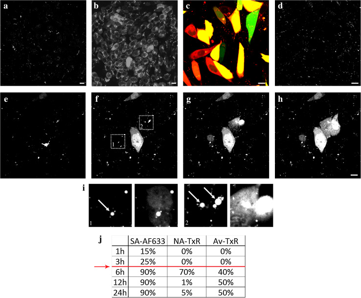
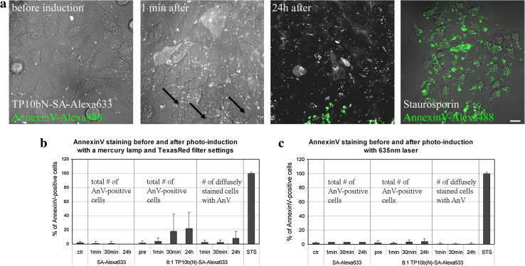
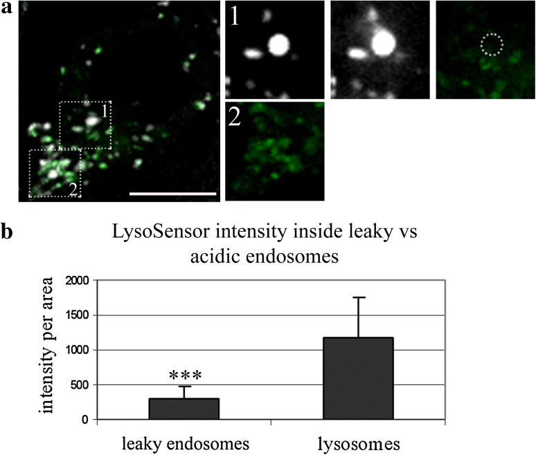
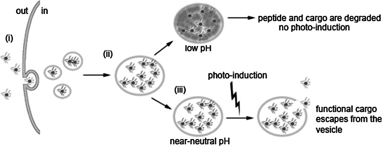
Since their discovery, cell-penetrating peptides (CPPs) have provided a novel, efficient, and non-invasive mode of transport for various (bioactive) cargos into cells. Despite the ever-growing number of successful implications of the CPP-mediated delivery, issues concerning their intracellular trafficking, significant targeting to degradative organelles, and limited endosomal escape are still hindering their widespread use. To overcome these obstacles, we have utilized a potent photo-induction technique with a fluorescently labeled protein cargo attached to an efficient CPP, TP10. In this study we have determined some key requirements behind this induced escape (e.g., dependence on peptide-to-cargo ratio, time and cargo), and have semi-quantitatively assessed the characteristics of the endosomes that become leaky upon this treatment. Furthermore, we provide evidence that the photo-released cargo remains intact and functional. Altogether, we can conclude that the photo-induced endosomes are specific large complexes-condensed non-acidic vesicles, where the released cargo remains in its native intact form. The latter was confirmed with tubulin as the cargo, which upon photo-induction was incorporated into microtubules. Because of this, we propose that combining the CPP-mediated delivery with photo-activation technique could provide a simple method for overcoming major limitations faced today and serve as a basis for enhanced delivery efficiency and a subsequent elevated cellular response of different bioactive cargo molecules.


Figures

Fig. 1
Fig. 1
Photo-induction leads to an effective release of endosome-entrapped CPP-protein complexes. CHO cells were pulsed with 4 μM unlabeled (all, except c) or FAM-labeled (c, green) N-terminally biotinylated TP10 (TP10b(N)) complexed with 500 nM streptavidin (SA)-Alexa Fluor(AF) 633 (white in a, b, di; red in c) for 1 h and chased for additional 6 h (ac, ei) or 24 h (d). d Even after 24 h the complexes are still found inside endosomes, if not induced to escape. Photo-induction was carried out at 545–580 nm for 10 s (b, c) or with a 635-nm laser excitation for 1 min (eh). eh Image sequence of photo-induction with a 635-nm laser. i Close-ups from selected areas 1 and 2 in f illustrate the large intensely fluorescent endosomes (indicated by arrows) that are induced to become leaky upon light excitation. j Percentages of diffusely stained cells pulsed (1 h) and chased (indicated times) with TP10b(N) complexes with SA-AF633, neutravidin(NA)-Texas Red (TxR) or avidin (Av)-TxR after photo-induction at 545–580 nm. Red line indicates the time threshold for effective photo-induction. Bar 10 μm
Fig. 2
Fig. 2
Photo-induction at 545–580 nm, but not with 635-nm laser, causes some cells to become apoptotic. CHO cells were pulsed with 4 μM TP10b(N) complexed with 500 nM SA-Alexa Fluor 633 (white) (ac) or with 500 nM SA-Alexa Fluor 633 alone (b, c) for 1 h and chased for additional 6 h. Exposure of phosphatidylserine to the plasma membrane of treated cells before and after photo-induction at 545–580 nm was detected with annexin V-Alexa Fluor 488 (green in a). Arrows in a indicate cell blebbing. As a positive control 10 μM staurosporine was used. Bar 20 μm. b, c Quantification of the number of annexin V-positive cells before and after photo-induction at 545–580 nm for 10 s (b) or with 635-nm laser excitation for 1 min (c), untreated cells (ctr) were used as negative control
Fig. 3
Fig. 3
Large intensely fluorescent endosomes that become leaky upon photo-induction are not as acidic as lysosomes. a TP10b(N)-SA-Alexa Fluor 633 complexes (white) are found in endosomes marked with pH-sensitive LysoSensor dye (green). A large intensely fluorescent endosome becoming leaky upon photo-induction is indicated in the upper area, and the sequence of endosomal leakage and the LysoSensor signal inside the same endosome are shown in the upper panel of images on the right. The edges of the leaky endosome are marked as a dotted circle in the far right image. The LysoSensor signal in lysosomes of the same cell is highlighted in the lower area; its close-up is on the bottom right image on the right. b Quantification of the LysoSensor signal intensity in leaky and strongly acidic endosomes; p value <0.001 (n = 67). p values <0.001 are marked with triple asterisks. Bar 10 μm
Fig. 4
Fig. 4
Protein cargo in complex with CPP remains intact inside the cells for at least 12 h. CHO cells were pulsed with 1.5, 2.5, or 4 μM TP10b(N) complexed with SA-Alexa Fluor 633 (resulting in 3:1, 5:1, and 8:1 peptide-to-cargo ratios, respectively) for 1 h and chased for the additional time. The soluble fractions of the treated cells were applied to a 10 % SDS-PAGE and the fluorescence signal was visualized. LC marks loading control, which is TP10b(N)-SA complexes in an 8:1 ratio without cells (a). The cargo bands from different lanes were analyzed together as intact (>17 kDa, black) and degraded (≤17 kDa, gray) (b) or separately (c). The ratio of 8:1 gave the highest internalization efficiency as well as displayed the best preservation of the cargo molecule. Additionally, most of the protein cargo remained intact (in a tetrameric form) for at least 12 h. d Fluorescence signals of dual-labeled complexes (FAM-TP10b(N)-SA-Alexa Fluor 633) were also visualized by SDS-PAGE and the intensities of both signals were plotted over time to depict the degradation kinetics of the whole CPP-cargo complex. The peptide deserts the complex rather quickly, nonetheless, a fairly large amount of the peptide is still present in cells after 7 h of incubation
Fig. 5
Fig. 5
Tubulin, as a functional cargo, is efficiently incorporated into tubular structures after photo-induction treatment. CHO cells were pulsed with 4 μM TP10b(N) complexed with 500 nM tubulin-TRITC (white, pseudocolor) and 160 nM (1/3 amount) SA-Alexa Fluor 633 (not shown) for 1 h, chased for an additional 6 h and photo-induced at 545–580 nm. Tubulin-TRITC-positive tubular structures (indicated with arrows) were visualized 3 h after photo-induction. Bar 20 μm
Fig. 6
Fig. 6
Model for photo-induced endosomal escape. The cargo protein in complex with excess peptide (i) enters the cell via endocytosis and over time concentrates to larger intracellular vesicles (ii). Inside vesicles that do not acidify, the peptide is able to remodel the endosomal membrane leading to its destabilization upon photo-induction (iii). This results in the escape of the cargo into cytosol, where it is intact and functional

References

    1. Mäe M, Langel Ü. Cell-penetrating peptides as vectors for peptide, protein and oligonucleotide delivery. Curr Opin Pharmacol. 2006;6(5):509–514. doi: 10.1016/j.coph.2006.04.004. - DOI - PubMed
    1. Järver P, Mäger I, Langel Ü. In vivo biodistribution and efficacy of peptide mediated delivery. Trends Pharmacol Sci. 2010;31(11):528–535. doi: 10.1016/j.tips.2010.07.006. - DOI - PubMed
    1. Morris MC, Deshayes S, Heitz F, Divita G. Cell-penetrating peptides: from molecular mechanisms to therapeutics. Biol Cell. 2008;100(4):201–217. doi: 10.1042/BC20070116. - DOI - PubMed
    1. Margus H, Padari K, Pooga M. Cell-penetrating peptides as versatile vehicles for oligonucleotide delivery. Mol Ther. 2012;20(3):525–533. doi: 10.1038/mt.2011.284. - DOI - PMC - PubMed
    1. Fotin-Mleczek M, Fischer R, Brock R. Endocytosis and cationic cell-penetrating peptides–a merger of concepts and methods. Curr Pharm Des. 2005;11(28):3613–3628. doi: 10.2174/138161205774580778. - DOI - PubMed

Publication types

LinkOut - more resources