

doi: 10.1007/978-1-62703-523-1_15.
Quantification of adipose tissue leukocytosis in obesity
Affiliations
- PMID: 23852606
- PMCID: PMC3972009
- DOI: 10.1007/978-1-62703-523-1_15
Item in Clipboard
Quantification of adipose tissue leukocytosis in obesity
Methods Mol Biol.
2013.
Abstract
The infiltration of immune cell subsets in adipose tissue termed "adipose tissue leukocytosis" is a critical event in the development of chronic inflammation and obesity-associated comorbidities. Given that a significant proportion of cells in adipose tissue of obese patients are of hematopoietic lineage, the distinct adipose depots represent an uncharacterized immunological organ that can impact metabolic functions. Here, we describe approaches to characterize and isolate leukocytes from the complex adipose tissue microenvironment, to aid mechanistic studies to better understand the role of specific pattern recognition receptors (PRRs) such as inflammasomes in adipose-immune cross talk.
Figures
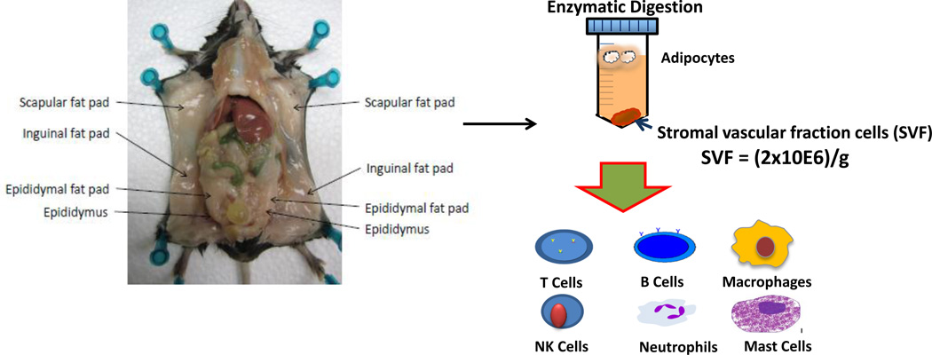
Overview of the separation and analysis of leukocytes from adipose tissue. Mouse adipose tissue is collected and weighed prior to subsequent analysis. Adipose tissue is digested to yield a stromal-vascular fraction. The stromal-vascular fraction contains immune cell subsets including T and B cells, macrophages, NK cells, neutrophils, and mast cells, which are increased by obesity.
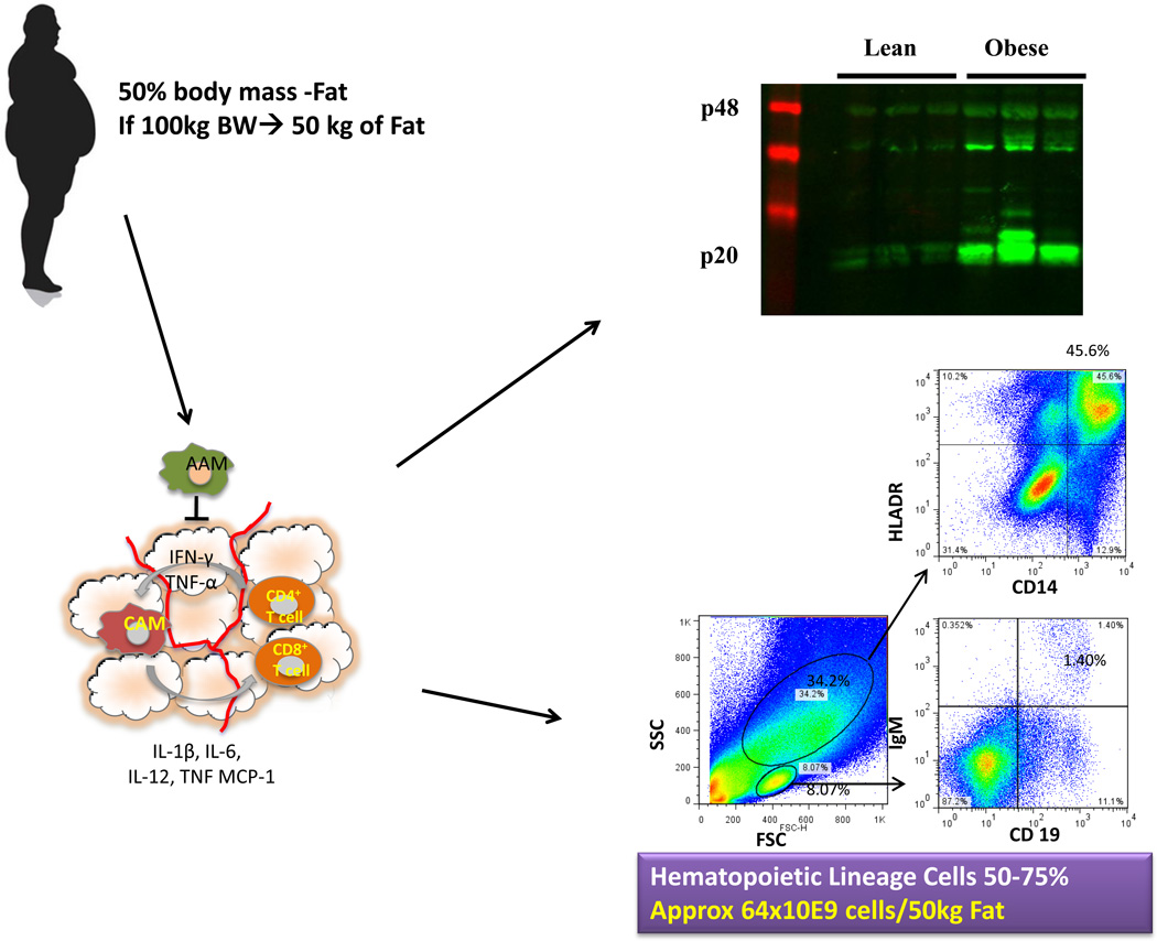
Overview of the analysis of leukocytes from adipose tissue. Human adipose tissue is collected, weighed and digested prior to downstream analysis. FACS plots from human adipose tissue stromal-vascular fraction taken from a female subject (BMI 29.9) showing CD14+HLADR+ macrophages and IGM+CD19+ B cells. Adipose tissue leukocytosis and the accretion of body fat lead to a large increase in the total amount of cells in fat ~64 × 109 cells/50 kg fat. Western blot analysis of caspase-1 activation (p20) in visceral adipose tissue from 9-month WT lean, WT diet-induced obese (DIO) mice.
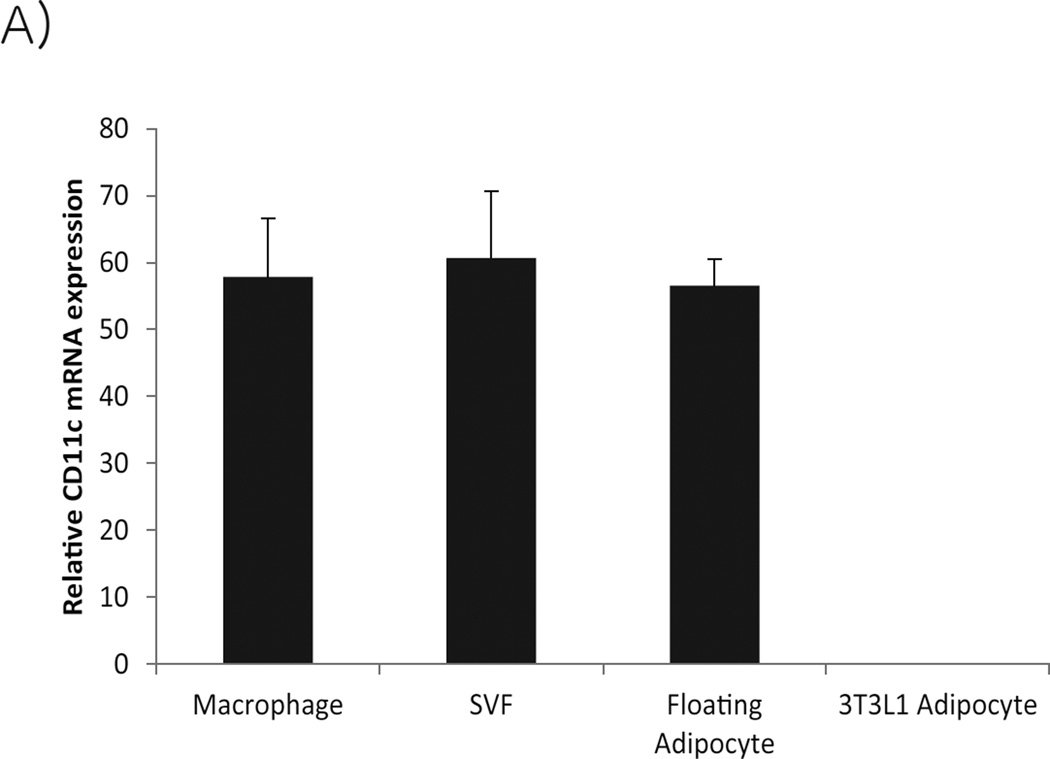
Relative mRNA expression of CD11c in F4/80+ adipose tissue macrophages, stromal vascular and floating adipocyte fractions and 3T3-L1 adipocytes (mean ± SEM).
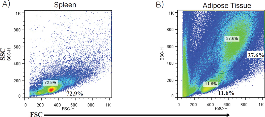
Comparison of forward scatter and side scatter FACS plots from A) splenocytes and B) stromal vascular fraction cells derived from epididymal fat pad of a 4-month-old obese wild type mouse.
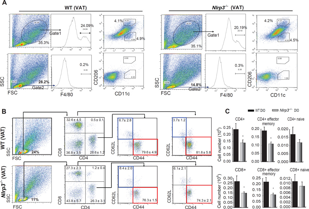
Gate placement for analysis of macrophages and lymphocytes, and the influence of NLRP3 ablation on T cell subpopulations in visceral adipose tissue (VAT) during DIO. (A) Upper row of FACS plots demonstrates that macrophages reside in a population of large SVF cells (Gate 1), while the lower row demonstrates that they do not reside in the small “lymphoid” gate (Gate 2) in VAT of 9-month WT and Nlrp3−/− DIO mice. Starting with the FSC and SSC (left) populations are sequentially gated for macrophage analysis, first by F4/80 expression and then by CD11c (M1 marker) and CD206 (M2 marker). (A) Sequential analysis of lymphocytes starting with Gate 2 (lymphoid gate), then separating lymphocytes by CD4+ and CD8+ T cells, and then evaluating the naïve (CD62L+CD44−, blue boxes) and effector memory (CD62L−CD44+, red boxes) T cells in both the CD4+ and CD8+ populations. (B) Absolute numbers (in million cells) of naïve and effector CD4+ and CD8+ T cells in adipose tissue of 9 month old WT-DIO and Nlrp3−/− DIO mice. (Figure adapted from Vandanmagsar et al. Nat. Med., 10, 179–188: 2011)
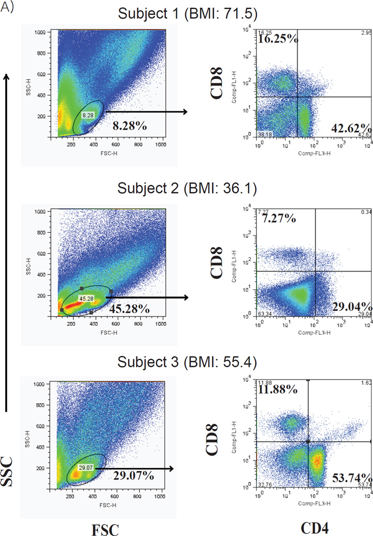
Human subjects display variability in adipose tissue T cell populations. FACS analysis of SVF -T cells from omental adipose tissue obtained from obese patients undergoing bariatric surgery. Lymphocyte populations (left) were gated and then analyzed for CD4+ and CD8+ T cells (right). Subjects were all female with a BMI (from top to bottom) of 71.5, 36.1, 55.4, respectively.
References
-
- Greenberg AS, Obin MS. Obesity and the role of adipose tissue in inflammation and metabolism. The American journal of clinical nutrition. 2006;83(2):461S–465S. - PubMed
-
- Winer DA, Winer S, Shen L, Wadia PP, Yantha J, Paltser G, Tsui H, Wu P, Davidson MG, Alonso MN, Leong HX, Glassford A, Caimol M, Kenkel JA, Tedder TF, McLaughlin T, Miklos DB, Dosch HM, Engleman EG. B cells promote insulin resistance through modulation of T cells and production of pathogenic IgG antibodies. Nature medicine. 2011;17(5):610–617. - PMC - PubMed
-
- Rausch ME, Weisberg S, Vardhana P, Tortoriello DV. Obesity in C57BL/6J mice is characterized by adipose tissue hypoxia and cytotoxic T-cell infiltration. Int J Obes (Lond) 2008;32(3):451–463. - PubMed
-
- Elgazar-Carmon V, Rudich A, Hadad N, Levy R. Neutrophils transiently infiltrate intra-abdominal fat early in the course of high-fat feeding. Journal of lipid research. 2008;49(9):1894–1903. - PubMed
Publication types
MeSH terms
Grants and funding
LinkOut - more resources
Full Text Sources
Other Literature Sources
Medical
