Review
. 2013 Dec 15;521(18):4124-44.
doi: 10.1002/cne.23415.

Cholinergic circuitry of the human nucleus basalis and its fate in Alzheimer's disease

Affiliations
Review

Cholinergic circuitry of the human nucleus basalis and its fate in Alzheimer's disease

M-Marsel Mesulam. J Comp Neurol. .

Abstract

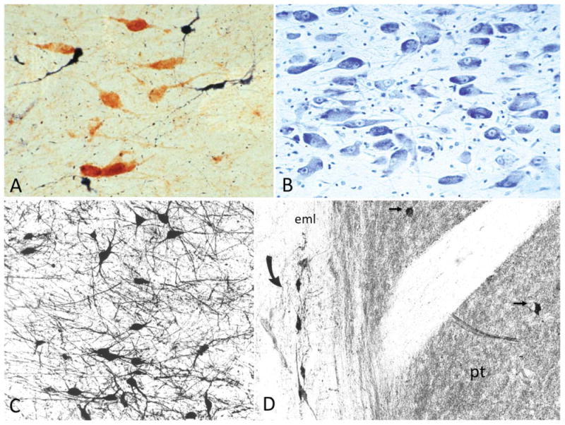
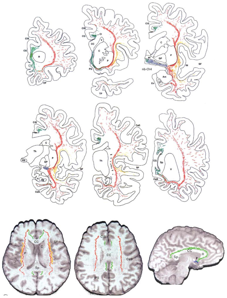
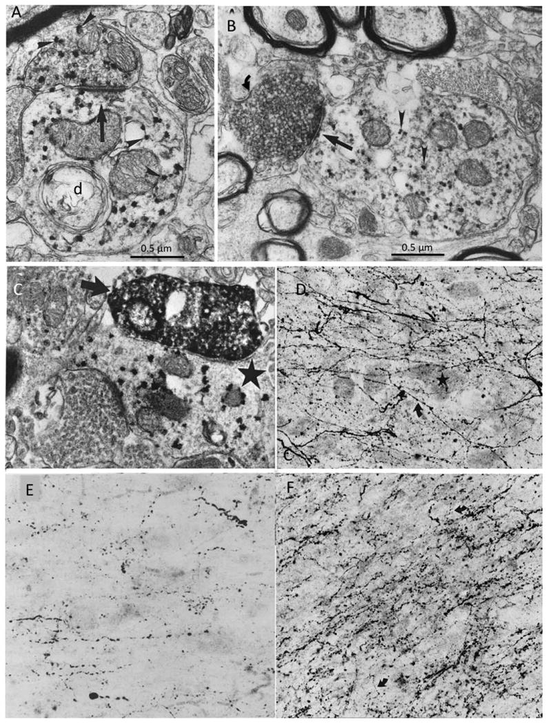
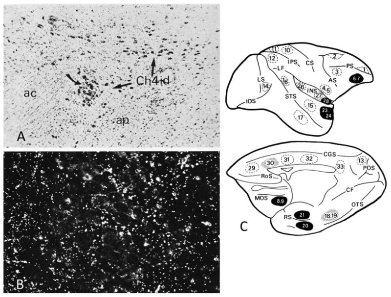
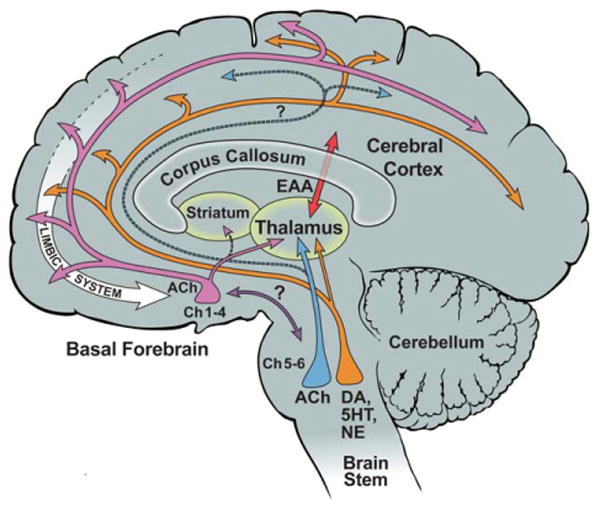
The nucleus basalis is located at the confluence of the limbic and reticular activating systems. It receives dopaminergic input from the ventral tegmental area/substantia nigra, serotonergic input from the raphe nuclei, and noradrenergic input from the nucleus locus coeruleus. Its cholinergic contingent, known as Ch4, provides the principal source of acetylcholine for the cerebral cortex and amygdala. More than half of presynaptic varicosities along its cholinergic axons make traditional synaptic contacts with cortical neurons. Limbic and paralimbic cortices of the brain receive the heaviest cholinergic input from Ch4 and are also the principal sources of reciprocal cortical projections back to the nucleus basalis. This limbic affiliation explains the role of the nucleus basalis in modulating the impact and memorability of incoming sensory information. The anatomical continuity of the nucleus basalis with other basomedial limbic structures may underlie its early and high vulnerability to the tauopathy and neurofibrillary degeneration of Alzheimer's disease. The tauopathy in Ch4 eventually leads to the degeneration of the cholinergic axons that it sends to the cerebral cortex. The early involvement of Ch4 has a magnifying effect on Alzheimer's pathology, because neurofibrillary degeneration in a small number of neurons can perturb neurotransmission in all cortical areas. Although the exact contribution of the Ch4 lesion to the cognitive changes of Alzheimer's disease remains poorly understood, the cholinergic circuitry of the nucleus basalis is emerging as one of the most strategically positioned and behaviorally consequential modulatory systems of the human cerebral cortex. J. Comp. Neurol. 521:4124-4144, 2013. © 2013 Wiley Periodicals, Inc.

Keywords: Alzheimer's disease; cholinergic circuitry; nucleus basalis.


Conflict of interest statement

Conflict of Interest Statement: The author has no conflicts of interest to disclose.

Figures

Figure 1
Figure 1
Three coronal sections from a rhesus monkey with HRP injected into Brodmann areas 4 and 6. A: The cortical areas within the injection site are blackened. Triangles represent nucleus basalis neurons. B: Camera lucida drawing from section 2 in A. Open profiles represent AChE-rich perikarya, blackened ones indicate double labeling of AChE-rich perikarya with retrogradely transported HRP. The original version of this figure was hand drawn in India ink by Gary Van Hoesen. ac, Anterior commissure; amg, amygdala; cgs, cingulate sulcus; gp, globus palidus; h, hypothalamus; ic, internal capsule; oc, optic chiasm; pu, putamen; rs, rhinal sulcus; spd, superior precentral dimple; Syl. f., Sylvian fissure. From Mesulam and Van Hoesen (1976) with permission.
Figure 2
Figure 2
Choline acetyltransferase immunohistochemistry in the macaque monkey showing the Ch1-4 cell groups and anteromedial (am), anterolateral (al), intermediodorsal (id), intermedioventral (iv), and posterior (p) sectors of Ch4. Black dot-like profiles represent ChAT-positive neurons. The coronal sections (A-E) are from progressively more caudal levels of the brain. Medial is to the left, dorsal toward the top. Curved arrows point to interstitial components of Ch4 embedded in the internal capsule, anterior commissure, and borders of the globus pallidus. Note that the caudate, nucleus accumbens, and putamen also contain ChAT-positive cells. These project internally within the striatum and are not part of Ch1-4. ×24. ac, Anterior commissure; amg, amygdala; ap, ansa peduncularis; cd, caudate; ci, islands of Calleja; gp, gpl, gpm, gpv, globus pallidus and its lateral, medial, and ventral segments; ic, internal capsule; iml, internal medullary lamina of the globus pallidus; lg, lateral geniculate nucleus; ot, optic tract; pt, putamen. From Mesulam et al. (1984) with permission.
Figure 3
Figure 3
Bielschowsky myelin staining of a coronal section of the human brain. The region under the anterior commissure (ac) is also known as the substantia innominata (si) and contains the anterior sector of Ch4. GP, globus pallidus; pt, putamen.
Figure 4
Figure 4
Acetylcholinesterase histochemistry was used in a 91-year-old control brain to delineate Ch4 from other components of the fore-brain. A-D represent increasingly more caudal coronal sections and contain the anteromedial (am), anterolateral (al), anterointermediate (ai), intermediodorsal (id), intermedioventral (iv), and posterior (p) sectors of Ch4. Medial is to the left, dorsal toward the top. Section A is at approximately the same level as Figure 3. ×5. ac, Anterior commissure; Am, amygdala; an, ansa lenticularis; ap, ansa peduncularis; bl, basolateral nucleus of the amygdala; GPe, GPi, GPv, external, internal, and medial sectors of the globus pallidus; he, head of the caudate; Hp, hippocampal formation; ic, internal capsule; iml, internal medullary lamina of the globus pallidus; Nhl, nucleus of the horizontal limb; nst, nucleus of the stria terminalis; ot, optic tract; pt, putamen; son, supraoptic nucleus; st, striatal islands; tc, tail of caudate; vt, temporal horn of lateral ventricle. From Mesulam and Geula (1988) with permission.
Figure 5
Figure 5
Cytological detail of the human nucleus basalis and Ch4. A: Cholinergic (ChAT-positive, brown) and noncholinergic (NADPH-positive, blue) neurons are intermingled in the nucleus basalis. The Ch4 designation applies only to the cholinergic contingent, whereas the term nucleus basalis refers to all neuronal populations in the nucleus. Control human brain. ×275. B: Cresyl violet stain of the nucleus basalis at the level of Figure 4A. Note the polymorphism, heterochromia, and prominent nucleoli of the magnocellular neurons. From a 60-year-old control brain. ×150. C: ChAT-positive cholinergic neurons in Ch4a. From an 82-year-old control brain. ×100. D: ChAT-positive interstitial cholinergic neurons (curved arrow) embedded within the white matter or the external medullary lamina of the globus pallidus. The abutting putamen (pt) is characterized by a ChAT-rich matrix and contains ChAT-positive neurons (straight arrows). The interstitial Ch4 neurons are larger and more elongated than the radially more symmetrical putaminal cholinergic neurons. Medial is to the left, dorsal to the top. From a 76-year-old control brain. ×120. eml, External medullary lamina of the globus pallidus.
Figure 6
Figure 6
Trajectory of Ch4 projections. Top two rows show electronic plotter maps of cholinergic fibers in the white matter of the human brain as identified in whole-hemisphere sections processed for AChE histochemistry and ChAT and NGFr immunocytochemistry. Sections represent increasingly more caudal coronal levels. The Ch4 neurons are represented in blue, the medial cholinergic pathway in green, the capsular branch of the lateral pathway in red and the perisylvian branch of the lateral pathway in orange. The bottom row shows a reconstruction of the trajectories in a 3D volume of a control magnetic resonance image (MRI) of the whole brain. The two figures at left show axial views and the third at right a parasaggital view, ac, Anterior commissure; Am, amygdala; C, caudate; CaS, calcarine sulcus; CC, corpus callosum; CeS, central sulcus; Ci, cingulum; CiS, cingulate sulcus; CoS, collateral sulcus; dg, dentate gyrus; En, entorhinal cortex; GP, globus pallidus; H; hippocampus; IN, insula; mb, mammillary body; OF, orbitofrontal cortex; P, putamen; PG, parolfactory gyrus; R, rostrum of corpus callosum; SF, Sylvian fissure; Sp, splenium of corpus callosum; Th, thalamus; TP, temporal pole; V, ventricle. From Selden et al. (1998) with permission.
Figure 7
Figure 7
Transmitter-specific input into the monkey and human Ch4. A: ChAT immunoreactivity is visualized with the punctate VIP red reaction product (arrowheads) in the macaque monkey nucleus basalis. The ChAT axon on top is forming an asymmetric synapse (straight arrow) onto a postsynaptic ChAT dendrite (d) in the monkey Ch4. B: Double labeling of GAD (with diffuse DAB reaction product) and ChAT (with punctate VIP red reaction product) shows a GAD-positive GABAergic bouton (curved arrow) making a symmetric synapse (straight arrow) onto a nucleus basalis dendrite containing ChAT (arrowheads) in the macaque monkey brain. C: Double labeling of TH (with the diffuse DAB reaction product) and ChAT (with the punctate VIP red reaction product) showing a TH-immunoreactive bouton (arrow) forming a fine synapse onto a ChAT-immunoreactive nucleus basalis dendrite (star) in the macaque monkey. ×34,000. D: TH immunohistochemistry in the Ch4i sector of an autopsy specimen from a 27-year-old man showing multiple fine, varicose, TH-positive dopaminergic axons (curved arrow) coursing through unlabeled Ch4 perikarya (star). ×250. E: Serotonin immunohistochemistry showing serotonergic axonal varicosities in the Ch4a sector of the human brain. ×250. F: DBH immunohistochemistry in the Ch4i sector of an 82-year-old brain shows a dense plexus of thick and thin, varicose noradrenergic axons (curved arrows) coursing through the nucleus basalis perikarya. ×250. From Smiley and Mesulam (1999) and Smiley et al. (1999) with permission. Scale bars = 0.5 μm.
Figure 8
Figure 8
Projections into the Ch4 of macaque monkeys following tritiated amino acid injections into entorhinal cortex. A: Brightfield view at ×70 of Ch4id (arrows) and their relationship to the anterior commissure (ac) and ansa peduncularis (ap). B: Darkfield view at ×500 of anterogradely transported tracer (white dots) within the cell island designated by the curved arrow in A. C: Tritiated amino acid injection sites. Those that resulted in definite projections to the nucleus basalis are shown in black, and those that resulted in questionable projections are stippled. Dashed circles represent sites where injections did not cause anterograde transport to the nucleus basalis. Numbers refer to individual cases, not to architectonic designations. AS, arcuate sulcus; CF, calcarine fissure; CGS, cingulate sulcus; CS, central sulcus; IOS, inferior occipital sulcus; IPS, intraparietal sulcus; LF, lateral fissure; LS, lunate sulcus; MOS, medial orbitofrontal sulcus; OTS, occipitotemporal sulcus; POS, parietooccipital sulcus; PS, sulcus principalis; RoS, Rostral sulcus; RS, rhinal sulcus; STS, superior temporal sulcus. From Mesulam and Mufson (1984) with permission.
Figure 9
Figure 9
This photomicrograph of AChE histochemistry based on a modified Karnovsky-Roots method shows cholinergic axons in layer 3 of inferotemporal cortex in the brain of a 22-year-old control specimen. The multiple varicosities represent putative sites of ACh release and are closely associated with AChE-rich cholinoceptive pyramidal neurons. The arrows point to two examples where the varicosities are apposed to the apical dendrite of cholinoceptive neurons. ×415. From Mesulam and Geula (1988) with permission.
Figure 10
Figure 10
Darkfield photomicrograph of ChAT-positive axons in the human cerebral cortex illustrating regional variations in the density of afferents from Ch4. V1 designates primary visual cortex; post. 22 designates the posterior part of Brodman area 22 where initial synaptic relays in the hierarchy of auditory association pathways are located; ant. 22 designates a synaptically more downstream component of this hierarchy. ×45. From Mesulam et al. (1992a) with permission.
Figure 11
Figure 11
Brightfield photomicrograph of ChAT-positive axons in the granular (A) and dysgranular (B) sectors of the human orbitofrontal cortex. The density is higher in the dysgranular sector, which is synaptically closer to core limbic structures. Arrowheads mark laminar boundaries. ×266. From Mesulam et al. (1992a) with permission.
Figure 12
Figure 12
Surgically removed temporal lobectomy specimen from a 20-year-old subject. The electronmicrograph shows a ChAT-positive varicosity labeled with the VIP reaction product (open arrows) forming a fine symmetric synapse (single straight arrow) with an unlabeled (noncholinergic) dendrite. ×32,000. From Smiley et al. (1997) with permission.
Figure 13
Figure 13
Age-related differences in the density of AChE-rich neurons in the banks of the superior temporal sulcus in four human brains that came to postmortem examination at 2, 10, 22, and 91 years of age (A-D, respectively). The histochemical reaction was obtained with a modified Koelle-Friedenwald method, which provides better visualization of cell bodies than of axons. Straight arrows point to AChE-rich neurons. ×100. From Mesulam and Geula (1991) with permission.
Figure 14
Figure 14
Early neurofibrillary degeneration of Ch4. A,B: Raw data obtained through the electronic plotter mapping of thioflavin-S stained whole-hemisphere sections in two autopsy cases aged 71 (A) and 72 (B). Neither had dementia or neurological disease. Each red cross marks a single NFT. These plots show that the nucleus basalis is at least as vulnerable to NFT formation as the entorhinal cortex and the amygdala. In A, there were no NFTs outside of the medial temporal lobe in that section. In B, the one additional NFT was encountered in the dorsal insula. C: Double immunostaining for ChAT (brown) and early neurofibrillary degeneration labeled with AT-8 (blue), an antibody that recognizes abnormally phosphorylated tau at early stages of neurofibrillary degeneration. The neuron on top is alive but undergoing neurofibrillary degeneration, whereas the one on the bottom is spared. The tauopathy has extended into the processes of the affected neuron (arrows). D: AChE histochemistry with a modified Karnovsky-Roots method in peristriate cortex of a 99-year-old woman who had no known dementia. Varicose cholinergic axons display dystrophic swollen profiles, which could represent early stages of cortical cholinergic degeneration and perhaps also aberrant attempts at regeneration. ×650. Amg, amygdala; En, entorhinal cortex; GP, globus pallidus; nb, nucleus basalis.
Figure 15
Figure 15
Cognitive correlates of tangles in the nucleus basalis. A and B are from a patient who died at the age of 95 years. A shows her test scores 3 years (black bars) and 4 months (gray bars) before death. On this graph, a level of 1 or 2 indicates performance that is impaired for age, levels 3 and 4 performance that is normal for age, and level 5 or 6 performance that is superior for age. In this patient, none of the scores was abnormal at either of the testing sessions. She was considered cognitively unimpaired. B, based on electronic plotting of stained tissue sections, shows normal neurons in her nucleus basalis (green circles) as well as those that contain NFT (red stars). C and D are from a patient who died at the age of 91 years. He was tested 2 years (black bars) and then 1 year (gray bars) before death. C shows that there has been a decline in performance and that he had multiple cognitive impairments (scores at level 2) at the last testing. Clinical notes indicate that the patient had progressed from a stage of mild cognitive impairment (MCI) at the first testing to early dementia of the AD type in the second. D shows his nucleus basalis at postmortem. Normal neurons are shown in green and neurons with NFT in red. Abbreviations for tests in frames A and C (from left to right): MMSE, Mini-Mental State Examination; Trail Making A and B, tests of executive function; CFL, test of verbal fluency; BNT, Boston naming test; CERAD Acquisition and Delay, tests of memory for words; Log Mem II, recall of a short story; BVRT, Benton visual retention test; CERAD constr., visuomotor test. AC, anterior commissure; Amg, amygdala; GP, globus pallidus; HY, hypothalamus; OT, optic tract. From Mesulam et al. (2004) with permission.
Figure 16
Figure 16
Neurofibrillary tangles and cholinergic innervation in AD. A: Nucleus basalis of a woman who died at the age of 84 years with clinical dementia and postmortem evidence of AD pathology. Histofluorescence in this thioflavin-S-stained section indicates that nearly all nucleus basalis neurons have been invaded by NFTs (arrow). Most are intracellular; others are ghost tangles. B: AChE histochemistry with a modified Karnovsky-Roots method shows nearly complete loss of cholinergic fibers in the middle temporal gyrus of the same patient. The arrow points to the one remaining axon in the field of view. C: Same region of the brain stained in the same fashion as in B but from a control subject who died at the age of 89 years with no evidence of dementia. The arrows point to two examples of cholinergic axons. ×100.
Figure 17
Figure 17
Cholinergic circuitry of the human nucleus basalis. ACh, acetylcholine; DA, dopamine; EAA, excitatory amino acids; NE, norepinephrine; 5HT, serotonin. Question marks indicate that the connection has not been confirmed in the human brain.

References

    1. Arendt T, Bigl V, Tennstedt A, Arendt A. Neuronal loss in different parts of the nucleus basalis is related to neuritic plaque formation in cortical target areas in Alzheimer's disease. Neuroscience. 1985;14:1–14. - PubMed
    1. Aston-Jones G, Shaver R, Dinan TG. Nucleus basalis neurons exhibit axonal branching with decreased impulse conduction velocity in rat cerebrocortex. Brain Res. 1985;325:271–285. - PubMed
    1. Berger-Sweeney J, Heckers S, Mesulam MM, Wiley RG, Lappi DA, Sharma M. Differential effects upon spatial navigation of immunotoxin-induced cholinergic lesions of the medial septal area and nucleus basalis magnocellularis. J Neurosci. 1994;14:4507–4519. - PMC - PubMed
    1. Braak H, Braak E. Evolution of the neuropathology of Alzheimer's disease. Acta Neurol Scand. 1996;165(Suppl):3–12. - PubMed
    1. Croxson PL, Kyriazis DA, Baxter MG. Cholinergic modulation of a specific memory function of prefrontal cortex. Nat Neurosci. 2011;14:1510–1512. - PMC - PubMed

Publication types