. 2013;9(7):e1003440.
doi: 10.1371/journal.ppat.1003440. Epub 2013 Jul 4.

The interactomes of influenza virus NS1 and NS2 proteins identify new host factors and provide insights for ADAR1 playing a supportive role in virus replication

Affiliations

The interactomes of influenza virus NS1 and NS2 proteins identify new host factors and provide insights for ADAR1 playing a supportive role in virus replication

Benoît de Chassey et al. PLoS Pathog. 2013.

Abstract

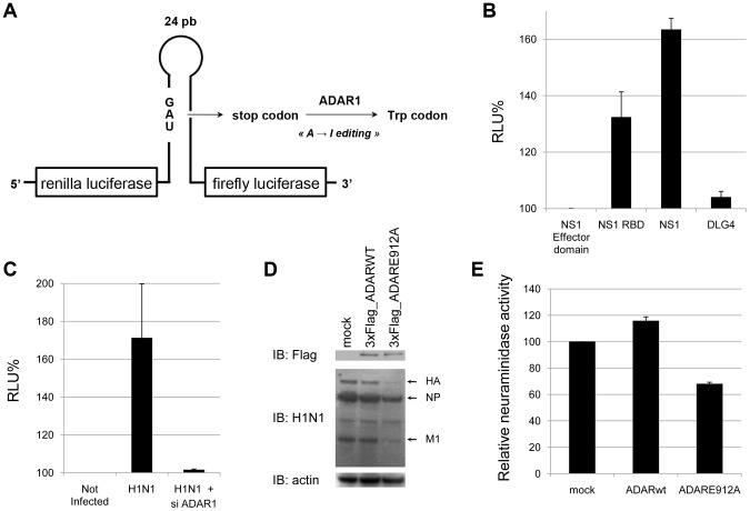
Influenza A NS1 and NS2 proteins are encoded by the RNA segment 8 of the viral genome. NS1 is a multifunctional protein and a virulence factor while NS2 is involved in nuclear export of viral ribonucleoprotein complexes. A yeast two-hybrid screening strategy was used to identify host factors supporting NS1 and NS2 functions. More than 560 interactions between 79 cellular proteins and NS1 and NS2 proteins from 9 different influenza virus strains have been identified. These interacting proteins are potentially involved in each step of the infectious process and their contribution to viral replication was tested by RNA interference. Validation of the relevance of these host cell proteins for the viral replication cycle revealed that 7 of the 79 NS1 and/or NS2-interacting proteins positively or negatively controlled virus replication. One of the main factors targeted by NS1 of all virus strains was double-stranded RNA binding domain protein family. In particular, adenosine deaminase acting on RNA 1 (ADAR1) appeared as a pro-viral host factor whose expression is necessary for optimal viral protein synthesis and replication. Surprisingly, ADAR1 also appeared as a pro-viral host factor for dengue virus replication and directly interacted with the viral NS3 protein. ADAR1 editing activity was enhanced by both viruses through dengue virus NS3 and influenza virus NS1 proteins, suggesting a similar virus-host co-evolution.


Conflict of interest statement

The authors have declared that no competing interests exist.

Figures

Figure 1
Figure 1. Interactions between NS1 and NS2 proteins and human host factors.
(A) Yeast two-hybrid array. The 33 NS1-specific interactors are indicated in blue, 28 NS2-specific interactors in grey and shared interactors in yellow. The 11 NS1 and the single NS2 interactors described earlier are highlighted with bold letters. DRBD-containing proteins (DRBPs) are indicated with a star. (B) Frequency of interactions between individual host cell factors and NS1 and/or NS2 proteins of the 9 different influenza virus strains. (C) Degree distribution of human proteins and human proteins targeted by NS1 and/or NS2 proteins in the human interactome. P(k) is the probability of a node to connect k other nodes in the network. Solid lines represent linear regression fits. Vertical dashed lines indicate the mean degree of each distribution. (D) Betweenness distribution of human proteins and human proteins targeted by NS1 and/or NS2 proteins in the human interactome. P(b) is the probability for a node to have a betweenness value of b in the network. Solid lines represent linear regression fits. Vertical dashed lines indicate the mean betweenness value for each distribution.
Figure 2
Figure 2. Impact of silencing of NS1 and NS2 interactors on influenza A virus replication.
A549 cells were transfected with indicated siRNAs, infected at a MOI of 0.5 with A/H1N1/Puerto Rico/8/34 virus strain (A) or A/H1N1/New Caledonia/2006 virus strain (B) and the neuraminidase activity (NA) was measured in the supernatant 48 h post infection. Values are normalized to neuraminidase activity measured in supernatants of control siRNA-transfected cells and represent the mean +/− standard deviation (triplicates). ATP6V1G1 is a host dependency factor, CSNK2B is an anti-viral host factor. Both served as controls.
Figure 3
Figure 3. Overview of NS1 and NS2 interactors on the influenza A virus life cycle.
The framework of this picture is from Brass et al. . NS1 and NS2 interactors identified in the present study and described in the literature were placed according to information from Gene Ontology, Human Protein Atlas and HPRD. When multiple annotations were available, the most relevant for the virus was chosen. Circle node: NS1 interactor; Square node: NS2 interactor; Diamond-shaped node: NS1 and NS2 interactor. A red node is a pro-viral host factor and a green node is an anti-viral host factor according to the siRNA data from this study and genome-wide screens. The node size is proportional to the number of virus strains interacting with the host factors (interactors from the literature are often tested against a single virus strain hence appearing with a small node size).
Figure 4
Figure 4. ADAR1 is a pro-viral host factor for influenza A virus replication.
(A) ADAR1 expression in A549 cells upon interferon treatment or H1N1 infection. A549 cells were incubated with indicated concentration of interferon-(IFN)-α2b or infected with influenza A virus and analysed 24 h later by western blot for expression of ADAR1 and actin that served as loading control. (B) Silencing of ADAR1 impairs viral protein expression. A549 cells were transfected with negative control siRNA (Ctrl) or two distinct siRNA targeting ADAR1 for 48 h and infected with A/H1N1/New Caledonia/2006 virus strain. ADAR1 and viral protein expression were assessed in cell lysates by western blot at indicated time points. (C) Silencing of ADAR1 reduces virus titers. Determination of NA in supernatants 8 h, 12 h, 24 h and 48 h post infection. Values are normalized to cells transfected with control siRNA.
Figure 5
Figure 5. Mapping of ADAR1-NS1 interaction sites.
(A) Subcellular localization of ADAR1 (green), HA and NS1 (red) 8 h post infection of A549 cells. (B) Schematic representation of full-length and truncated ADAR1 proteins. Zα DNA-binding domain (grey boxes), dsRNA binding domain (DRBD, purple boxes), adenosine deaminase domain (yellow boxes). (C) ADAR1 interaction mapping. Yeast diploids co-expressing full length A/H1N1/Puerto Rico/8/34 virus NS1 protein fused to the Gal4 DNA-binding domain and full-length ADAR1 or ADAR1 truncated mutants fused to Gal4 activating domain were spotted onto a plate containing medium without histidine. STAU1 is a positive control for interaction with NS1 . Controls with empty vectors (Gal4-AD and Gal4DB) show no auto-activation induced by the different constructs. (D) GST pull-downs were performed with GST alone or GST fused to NS1 after co-expression of a 3×Flag-tagged DRBD1 peptide in HEK293T cells and incubation of cell lysates with glutathione-Sepharose beads. Cell lysates and pull-downs were analyzed by western blot using antibodies against GST or 3×Flag. (E) Schematic representation of the NS1 protein with its two domains: the N-terminal RNA-binding domain (RBD, 1-73) and the effector domain (74-230). (F.) NS1 interaction mapping. GST alone or GST fused to NS1, NS1 RBD or NS1 effector were co-expressed with 3×Flag-tagged ADAR1 in HEK293T cells and their interaction was assessed after co-affinity purification with glutathione sepharose beads and immunoblotting.
Figure 6
Figure 6. Enhancement of ADAR1 editing activity by influenza virus NS1.
(A) The RNA editing reporter system is composed of the hepatitis D virus minimal sequence edited by ADAR1 and positioned in-between the Renilla luciferase and the Firefly luciferase coding sequence, respectively. The unedited reporter has a stop codon that is converted into Trp codon upon A to I editing by ADAR. Hence, editing is correlated with Firefly luciferase expression while Renilla luciferase expression is used as an internal control. (B) HEK293T were co-transfected with the editing reporter, ADAR1 and NS1 (full-length, RNA-binding domain or effector domain) or the control protein DLG4. Two days post transfection, luciferase activities were determined by luminescence measurement. Data are expressed as percentage of the luciferase activity detected in cells expressing the NS1 effector domain (relative light unit, RLU). (C) Editing activity in HEK293T expressing or not ADAR1, transfected with the editing reporter and infected with influenza virus H1N1. (D, E) A549 cells were transfected with plasmids encoding for wild type or catalytically inactive ADAR1 (ADAR1 E912A, Text S1). Forty eight hours later, cells were infected with the A/H1N1/New Caledonia/2006 virus strain at a MOI of 0.5. After an additional 48 h incubation period, expression of ADAR1 and viral proteins was assessed in cell lysates by using western blot (D) and neuraminidase activities were measured in supernatants (E). Values are normalized to mock-transfected cells.
Figure 7
Figure 7. Dengue virus NS3 protein also interacts with ADAR1.
(A) Pairwise Yeast diploids co-expressing dengue virus type 2 NS3 helicase fused to Gal4 DNA-binding domain and ADAR1 fused to Gal4 transactivation domain were plated onto a selective medium lacking histidine to determine interaction-dependent transactivation of the HIS3 reporter gene. Negative controls are vectors without insert (pPC97 and pPC86 for bait and prey respectively). (B) HEK293T cells were co-transfected with 3×Flag-tagged ADAR1 and GST-tagged full-length NS3 of dengue virus (GST-DV-NS3) or its helicase domain (GST-DV-NS3 helicase) or GST-tagged full-length NS1 of influenza virus (GST-FLUAV-NS1) as a positive control. Proteins bound to glutathione sepharose beads were analyzed by western blot using antibodies against GST or 3×Flag. (C) ADAR1 expression in Huh-7 cells upon interferon treatment or dengue virus infection. Huh-7 cells were incubated with 0, 100 or 1000 IU/ml of interferon- (IFN)-α2b or infected with dengue virus and analyzed 24 h later for expression of ADAR1, NS3 and GAPDH. (D) Impact of ADAR1 silencing on dengue virus replication in Huh-7. Data are expressed as the percentage of virus titer obtained with control siRNA-transfected cells. A siRNA targeting dengue virus NS1-coding region (siDV-NS1) was used as positive control for the silencing. (E) ADAR editing activity in Huh-7 cells infected with dengue virus. (F) Dengue virus NS3 contribution to ADAR1 editing activity. The NS1 effector domain of influenza virus, full-length NS3 of dengue virus or its helicase domain were co-expressed with ADAR1 and the editing reporter in HEK293T cells. Luminescence reflecting Firefly and Renilla luciferase activities was measured 48 h post-transfection. The influenza virus NS1 effector domain does not interact with ADAR1 and was used as negative control. Data are normalized to the values obtained with the NS1 effector domain. RLU, relative light unit.

References

    1. Chen W, Calvo PA, Malide D, Gibbs J, Schubert U, et al. (2001) A novel influenza A virus mitochondrial protein that induces cell death. Nat Med 7: 1306–1312. - PubMed
    1. Wise HM, Foeglein A, Sun J, Dalton RM, Patel S, et al. (2009) A complicated message: Identification of a novel PB1-related protein translated from influenza A virus segment 2 mRNA. J Virol 83: 8021–8031. - PMC - PubMed
    1. Jagger BW, Wise HM, Kash JC, Walters KA, Wills NM, et al. (2012) An overlapping protein-coding region in influenza A virus segment 3 modulates the host response. Science 337: 199–204. - PMC - PubMed
    1. Gack MU, Albrecht RA, Urano T, Inn KS, Huang IC, et al. (2009) Influenza A virus NS1 targets the ubiquitin ligase TRIM25 to evade recognition by the host viral RNA sensor RIG-I. Cell Host Microbe 5: 439–449. - PMC - PubMed
    1. Tan SL, Katze MG (1998) Biochemical and genetic evidence for complex formation between the influenza A virus NS1 protein and the interferon-induced PKR protein kinase. J Interferon Cytokine Res 18: 757–766. - PubMed

Publication types

MeSH terms