

Comparing the neural correlates of affective and cognitive theory of mind using fMRI: Involvement of the basal ganglia in affective theory of mind
- PMID: 23853676
- PMCID: PMC3709103
- DOI: 10.2478/v10053-008-0129-6
Comparing the neural correlates of affective and cognitive theory of mind using fMRI: Involvement of the basal ganglia in affective theory of mind
Abstract
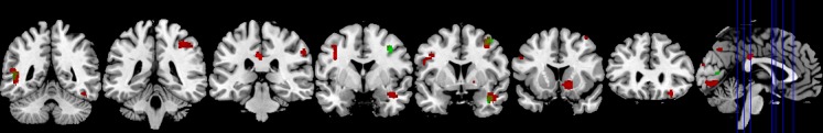
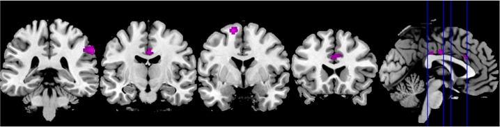
Theory of Mind (ToM) is the ability to infer other people's mental states like intentions or desires. ToM can be differentiated into affective (i.e., recognizing the feelings of another person) and cognitive (i.e., inferring the mental state of the counterpart) subcomponents. Recently, subcortical structures such as the basal ganglia (BG) have also been ascribed to the multifaceted concept ToM and most BG disorders have been reported to elicit ToM deficits. In order to assess both the correlates of affective and cognitive ToM as well as involvement of the basal ganglia, 30 healthy participants underwent event-related fMRI scanning, neuropsychological testing, and filled in questionnaires concerning different aspects of ToM and empathy. Directly contrasting affective (aff) as well as cognitive (cog) ToM to the control (phy) condition, activation was found in classical ToM regions, namely parts of the temporal lobe including the superior temporal sulcus, the supplementary motor area, and parietal structures in the right hemisphere. The contrast aff > phy yielded additional activation in the orbitofrontal cortex on the right and the cingulate cortex, the precentral and inferior frontal gyrus and the cerebellum on the left. The right BG were recruited in this contrast as well. The direct contrast aff > cog showed activation in the temporoparietal junction and the cingulate cortex on the right as well as in the left supplementary motor area. The reverse contrast cog > aff however did not yield any significant clusters. In summary, affective and cognitive ToM partly share neural correlates but can also be differentiated anatomically. Furthermore, the BG are involved in affective ToM and thus their contribution is discussed as possibly providing a motor component of simulation processes, particularly in affective ToM.
Keywords: ToM; affective and cognitive theory of mind; basal ganglia; fMRI; mentalizing; simulation; social cognition.
Figures


References
-
- Adolphs R. Social cognition and the human brain. Trends in Cognitive Sciences. 1999;3:469–479. - PubMed
-
- Adolphs R. Neural systems for recognizing emotion. Current Opinion in Neurobiology. 2002;12:169–177. - PubMed
-
- Adolphs R. Cognitive neuroscience of human social behaviour. Nature Reviews Neuroscience. 2003;4:165–178. - PubMed
-
- Alegre M., Rodríguez-Oroz M. C., Valencia M., Pérez-Alcázar M., Guridi J., Iriarte J., et al. Changes in subthalamic activity during movement observation in Parkinson’s disease: Is the mirror system mirrored in the basal ganglia? Neurophysiology. 2010;121:414–425. - PubMed
LinkOut - more resources
Full Text Sources