Ventricular dyssynchrony and function improve following catheter ablation of nonseptal accessory pathways in children
- PMID: 23853767
- PMCID: PMC3703375
- DOI: 10.1155/2013/158621
Ventricular dyssynchrony and function improve following catheter ablation of nonseptal accessory pathways in children
Abstract
Introduction: Paradoxical or hypokinetic interventricular septal motion has been described in patients with septal or paraseptal accessory pathways. Data regarding nonseptal pathways is limited.
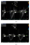
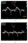
Methods and results: We quantified left ventricular dyssynchrony and function in 16 consecutive children, 14.2 ± 3.7 years, weighing 53 ± 17 kg, prior to and following catheter ablation of bidirectional septal (N = 6) and nonseptal (N = 10) accessory pathways. Following ablation, the left ventricular ejection fraction increased by 4.9 ± 2.1% (P = 0.038) from a baseline value of 57.0% ± 7.8%. By tissue Doppler imaging, the interval between QRS onset and peak systolic velocity (Ts) decreased from a median of 33.0 ms to 18.0 ms (P = 0.013). The left ventricular ejection fraction increased to a greater extent following catheter ablation of nonseptal (5.9% ± 2.6%, P = 0.023) versus septal (2.5% ± 4.1%, P = 0.461) pathways. The four patients with an ejection fraction <50%, two of whom had left lateral pathways, improved to >50% after ablation. Similarly, the improvement in dyssynchrony was more marked in patients with nonseptal versus septal pathways (difference between septal and lateral wall motion delay before and after ablation 20.6 ± 7.1 ms (P = 0.015) versus 1.4 ± 11.4 ms (P = 0.655)).
Conclusion: Left ventricular systolic function and dyssynchrony improve after ablation of antegrade-conducting accessory pathways in children, with more pronounced changes noted for nonseptal pathways.
Figures


References
-
- Hishida H, Sotobata I, Koike Y. Echocardiographic patterns of ventricular contraction in the Wolff-Parkinson-White syndrome. Circulation. 1976;54(4):567–570. - PubMed
-
- DeMaria AN, Vera Z, Neumann A, Mason DT. Alterations in ventricular contraction pattern in the Wolff-Parkinson-White syndrome. Detection by echocardiography. Circulation. 1976;53(2):249–257. - PubMed
-
- Ticzon AR, Damato AN, Caracta AR, Russo G, Foster JR, Lau SH. Interventricular septal motion during preexcitation and normal conduction in Wolff-Parkinson-White syndrome. Echocardiographic and electrophysiologic correlation. The American Journal of Cardiology. 1976;37(6):840–847. - PubMed
-
- Francis GS, Theroux P, O’Rourke RA, Hagan AD, Johnson AD. An echocardiographic study of interventricular septal motion in the Wolff-Parkinson-White syndrome. Circulation. 1976;54(2):174–178. - PubMed
MeSH terms
LinkOut - more resources
Full Text Sources
Other Literature Sources
Medical

