

The microsporidian Enterocytozoon hepatopenaei is not the cause of white feces syndrome in whiteleg shrimp Penaeus (Litopenaeus) vannamei
- PMID: 23856195
- PMCID: PMC3717009
- DOI: 10.1186/1746-6148-9-139
The microsporidian Enterocytozoon hepatopenaei is not the cause of white feces syndrome in whiteleg shrimp Penaeus (Litopenaeus) vannamei
Abstract
Background: The microsporidian Enterocytozoon hepatopenaei was first described from Thailand in 2009 in farmed, indigenous giant tiger shrimp Penaeus (Penaeus) monodon. The natural reservoir for the parasite is still unknown. More recently, a microsporidian closely resembling it in morphology and tissue preference was found in Thai-farmed, exotic, whiteleg shrimp Penaeus (Litopenaeus) vannamei exhibiting white feces syndrome (WFS). Our objective was to compare the newly found pathogen with E. hepatopenaei and to determine its causal relationship with WFS.
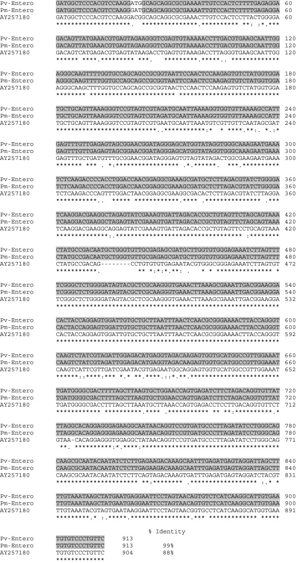
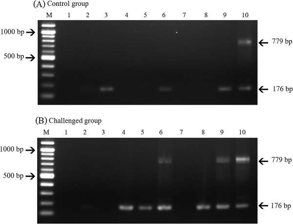
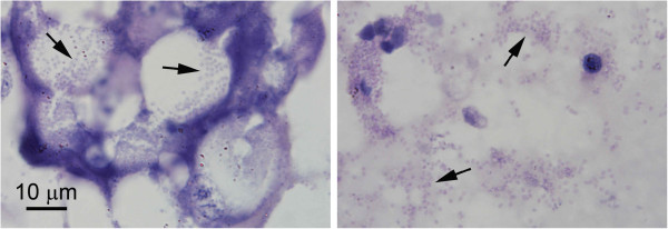
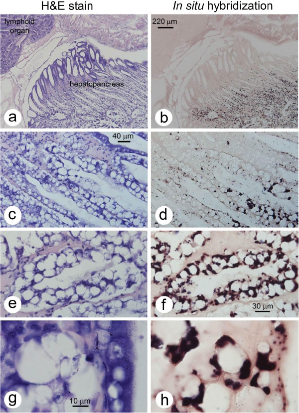
Results: Generic primers used to amplify a fragment of the small subunit ribosomal RNA (ssu rRNA) gene for cloning and sequencing revealed that the new parasite from WFS ponds had 99% sequence identity to that of E. hepatopenaei, suggesting it was conspecific. Normal histological analysis using tissue sections stained with hematoxylin and eosin (H&E) revealed that relatively few tubule epithelial cells exhibited spores, suggesting that the infections were light. However, the H&E results were deceptive since nested PCR and in situ hybridization analysis based on the cloned ssu rRNA gene fragment revealed very heavy infections in tubule epithelial cells in the central region of the hepatopancreas in the absence of spores. Despite these results, high prevalence of E. hepatopenaei in shrimp from ponds not exhibiting WFS and a pond that had recovered from WFS indicated no direct causal association between these infections and WFS. This was supported by laboratory oral challenge trials that revealed direct horizontal transmission to uninfected shrimp but no signs of WFS.
Conclusions: The microsporidian newly found in P. vannamei is conspecific with previously described E. hepatopenaei and it is not causally associated with WFS. However, the deceptive severity of infections (much greater than previously reported in P. monodon) would undoubtedly have a negative effect on whiteleg shrimp growth and production efficiency and this could be exacerbated by the possibility of horizontal transmission revealed by laboratory challenge tests. Thus, it is recommended that the PCR and in situ hybridization methods developed herein be used to identify the natural reservoir species so they can be eliminated from the shrimp rearing system.
Figures
Similar articles
-
Evidences supporting Enterocytozoon hepatopenaei association with white feces syndrome in farmed Penaeus vannamei in Venezuela and Indonesia.Dis Aquat Organ. 2020 Sep 17;141:71-78. doi: 10.3354/dao03522. Dis Aquat Organ. 2020. PMID: 32940252
-
Loop-mediated isothermal amplification combined with colorimetric nanogold for detection of the microsporidian Enterocytozoon hepatopenaei in penaeid shrimp.J Appl Microbiol. 2013 May;114(5):1254-63. doi: 10.1111/jam.12160. Epub 2013 Mar 6. J Appl Microbiol. 2013. Retraction in: J Appl Microbiol. 2023 Mar 1;134(3):lxad027. doi: 10.1093/jambio/lxad027. PMID: 23387348 Retracted.
-
Dense populations of the microsporidian Enterocytozoon hepatopenaei (EHP) in feces of Penaeus vannamei exhibiting white feces syndrome and pathways of their transmission to healthy shrimp.J Invertebr Pathol. 2016 Oct;140:1-7. doi: 10.1016/j.jip.2016.08.004. Epub 2016 Aug 13. J Invertebr Pathol. 2016. PMID: 27530403
-
White feces syndrome in shrimp: Comprehensive understanding of immune system responses.Fish Shellfish Immunol. 2024 Aug;151:109704. doi: 10.1016/j.fsi.2024.109704. Epub 2024 Jun 14. Fish Shellfish Immunol. 2024. PMID: 38880362 Review.
-
Historic emergence, impact and current status of shrimp pathogens in Asia.J Invertebr Pathol. 2012 Jun;110(2):166-73. doi: 10.1016/j.jip.2012.03.004. Epub 2012 Mar 10. J Invertebr Pathol. 2012. PMID: 22429834 Review.
Cited by
-
CRISPR-Cas fluorescent cleavage assay coupled with recombinase polymerase amplification for sensitive and specific detection of Enterocytozoon hepatopenaei.Biotechnol Rep (Amst). 2020 Jun 6;27:e00485. doi: 10.1016/j.btre.2020.e00485. eCollection 2020 Sep. Biotechnol Rep (Amst). 2020. PMID: 32577410 Free PMC article.
-
A Nested PCR Assay to Avoid False Positive Detection of the Microsporidian Enterocytozoon hepatopenaei (EHP) in Environmental Samples in Shrimp Farms.PLoS One. 2016 Nov 10;11(11):e0166320. doi: 10.1371/journal.pone.0166320. eCollection 2016. PLoS One. 2016. PMID: 27832178 Free PMC article.
-
Dynamic Interplay of Metabolic and Transcriptional Responses in Shrimp during Early and Late Infection Stages of Enterocytozoon hepatopenaei (EHP).Int J Mol Sci. 2023 Nov 25;24(23):16738. doi: 10.3390/ijms242316738. Int J Mol Sci. 2023. PMID: 38069062 Free PMC article.
-
Microsporidia-Emergent Pathogens in the Global Food Chain (Trends in Parasitology 32, 336-348; April 2, 2016).Trends Parasitol. 2016 Aug;32(8):657. doi: 10.1016/j.pt.2016.06.002. Epub 2016 Jun 27. Trends Parasitol. 2016. PMID: 27365191 No abstract available.
-
Quantification of Enterocytozoon hepatopenaei (EHP) in Penaeid Shrimps from Southeast Asia and Latin America Using TaqMan Probe-Based Quantitative PCR.Pathogens. 2019 Nov 12;8(4):233. doi: 10.3390/pathogens8040233. Pathogens. 2019. PMID: 31726681 Free PMC article.
References
-
- Lightner DV. A handbook of pathology and diagnostic procedures for diseases of penaeid shrimp. Baton Rouge, LA: World Aquaculture Society; 1996.
-
- Flegel TW, Boonyaratpalin S, Fegan DF, Guerin M, Sriurairatana S. In: Diseases in Asian Aquaculture I. Shariff M, Subasinghe RP, Arthur JR, editor. Manila: Fish Health Section, Asian Fisheries Society; 1992. High mortality of black tiger prawns from cotton shrimp disease in Thailand; pp. 181–197.
-
- Pasharawipas T, Flegel TW, Chaiyaroj S, Mongkolsuk S, Sirisinha S. Comparison of amplified RNA gene sequences from microsporidian parasites (Agmasoma or Thelohania) in Penaeus merguiensis and P. monodon. Asian Fisheries Science. 1994;7:169–178.
-
- Pasharawipas T, Flegel TW. A specific DNA probe to identify the intermediate host of a common microsporidian parasite of Penaeus merguiensis and P. monodon. Asian Fish Sci. 1994;7:157–167.
-
- Kelly JF. Tissue specificities of Thelohania duorara, Agmasoma penaei, and Pleistophora sp., microsporidian parasites of pink shrimp, Penaeus duorarum. J Invertebr Pathol. 1979;33:331–339. doi: 10.1016/0022-2011(79)90035-1. - DOI
Publication types
MeSH terms
Substances
LinkOut - more resources
Full Text Sources
Other Literature Sources
Molecular Biology Databases
