. 2013 Aug 16;113(5):517-26.
doi: 10.1161/CIRCRESAHA.113.301678. Epub 2013 Jul 15.

Phospholamban knockout breaks arrhythmogenic Ca²⁺ waves and suppresses catecholaminergic polymorphic ventricular tachycardia in mice

Affiliations

Phospholamban knockout breaks arrhythmogenic Ca²⁺ waves and suppresses catecholaminergic polymorphic ventricular tachycardia in mice

Yunlong Bai et al. Circ Res. .

Abstract

Rationale: Phospholamban (PLN) is an inhibitor of cardiac sarco(endo)plasmic reticulum Ca²⁺ ATPase. PLN knockout (PLN-KO) enhances sarcoplasmic reticulum Ca²⁺ load and Ca²⁺ leak. Conversely, PLN-KO accelerates Ca²⁺ sequestration and aborts arrhythmogenic spontaneous Ca²⁺ waves (SCWs). An important question is whether these seemingly paradoxical effects of PLN-KO exacerbate or protect against Ca²⁺-triggered arrhythmias.

Objective: We investigate the impact of PLN-KO on SCWs, triggered activities, and stress-induced ventricular tachyarrhythmias (VTs) in a mouse model of cardiac ryanodine-receptor (RyR2)-linked catecholaminergic polymorphic VT.

Methods and results: We generated a PLN-deficient, RyR2-mutant mouse model (PLN-/-/RyR2-R4496C+/-) by crossbreeding PLN-KO mice with catecholaminergic polymorphic VT-associated RyR2-R4496C mutant mice. Ca²⁺ imaging and patch-clamp recording revealed cell-wide propagating SCWs and triggered activities in RyR2-R4496C+/- ventricular myocytes during sarcoplasmic reticulum Ca²⁺ overload. PLN-KO fragmented these cell-wide SCWs into mini-waves and Ca²⁺ sparks and suppressed the triggered activities evoked by sarcoplasmic reticulum Ca²⁺ overload. Importantly, these effects of PLN-KO were reverted by partially inhibiting sarco(endo)plasmic reticulum Ca²⁺ ATPase with 2,5-di-tert-butylhydroquinone. However, Bay K, caffeine, or Li⁺ failed to convert mini-waves to cell-wide SCWs in PLN-/-/RyR2-R4496C+/- ventricular myocytes. Furthermore, ECG analysis showed that PLN-KO mice are not susceptible to stress-induced VTs. On the contrary, PLN-KO protected RyR2-R4496C mutant mice from stress-induced VTs.

Conclusions: Our results demonstrate that despite severe sarcoplasmic reticulum Ca²⁺ leak, PLN-KO suppresses triggered activities and stress-induced VTs in a mouse model of catecholaminergic polymorphic VT. These data suggest that breaking up cell-wide propagating SCWs by enhancing Ca²⁺ sequestration represents an effective approach for suppressing Ca²⁺-triggered arrhythmias.

Keywords: Ca2+ leak; Ca2+ waves; Ca2+-triggered arrhythmias; phospholamban; ryanodine receptor calcium release channel; sarcoplasmic reticulum.


Figures

Figure 1
Figure 1. PLN-KO breaks cell-wide propagating spontaneous Ca2+ waves in isolated ventricular myocytes
Ventricular myocytes were isolated from RyR2-R4496C+/− mutant mice, the PLN deficient, RyR2-R4496C+/− mice (PLN−/−/RyR2-R4496C+/−) or the PLN−/− mice, and loaded with the fluorescent Ca2+ indicator dye fluo-4, AM. The fluo-4 loaded cells were perfused with KRH buffer containing 6 mM extracellular Ca2+ to induce SR Ca2+ overload. Store overload induced spontaneous SR Ca2+ release events were detected by line-scan confocal Ca2+ imaging. Representative line-scan images of spontaneous Ca2+ release in isolated RyR2-R4496C+/− (n=39) (A), PLN−/−/RyR2-R4496C+/− (n=43) (B), and PLN−/− (n=9) (C) ventricular myocytes are shown.
Figure 2
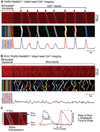
Figure 2. PLN-KO fragments cell-wide propagating spontaneous Ca2+ waves in intact hearts
Intact hearts isolated from RyR2-R4496C+/− or PLN−/−/RyR2-R4496C+/− mice were loaded with Rhod-2-AM and Langendorff-perfused with 6 mM extracellular Ca2+ and paced at 6 Hz to induce SR Ca2+ overload. Spontaneous SR Ca2+ release in epicardial ventricular myocytes in intact hearts was monitored by line-scan confocal Ca2+ imaging. Representative line-scan images (top) and the corresponding digitized images (middle) of cell-wide propagating spontaneous Ca2+ waves in intact RyR2-R4496C+/− hearts (A) and of mini-waves and Ca2+ sparks in intact PLN−/−/RyR2-R4496C+/− hearts (B) are shown. Panels A and B (bottom) show the spatial average of fluorescence signal along the scan-line. (C) Definition of wave fluorescence, full duration at half maximum (FDHM), time to peak, amplitude, and rate of rise.
Figure 3
Figure 3. Distribution of spontaneous Ca2+ release events in intact RyR2-R4496C mutant hearts with or without PLN
Line-scan confocal images were digitized and spontaneous Ca2+ release events were detected and classified using a custom-made program as described in the supplementary methods. (A, B, C) Distribution of spontaneous Ca2+ release events in RyR2-R4496C+/− mutant (A), PLN−/−/RyR2-R4496C+/− (B), and PLN−/− (C) hearts according to their total fluorescence. Three types of spontaneous Ca2+ release events (Ca2+ sparks, mini-waves, and waves) were classified based on the size of the total fluorescence (see Supplementary Methods). The red line represents a Gaussian fit of the distribution of Ca2+ waves in RyR2-R4496C+/− hearts. (D) The overall contribution (%) of sparks, mini-waves, and waves in RyR2-R4496C+/−, PLN−/−/RyR2-R4496C+/−, and PLN−/− hearts. (E, F, G) The occurrence (events/scan) of Ca2+ waves (E), mini-waves (F), and Ca2+ sparks (G) in RyR2-R4496C+/−, PLN−/−/RyR2-R4496C+/−, and PLN−/− hearts. Data shown are mean ± SEM from 19 (PLN−/−), 39 (RyR2-R4496C+/−), and 43 (PLN−/−/RyR2-R4496C+/−) line-scan images (*P < 0.001).
Figure 4
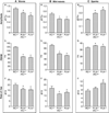
Figure 4. Effect of PLN-KO on spontaneous Ca2+ release in intact RyR2-R4496C mutant hearts
Spontaneous Ca2+ release events in intact RyR2-R4496C+/−, PLN−/−/RyR2-R4496C+/−, and PLN−/− hearts were divided into Ca2+ waves (A), mini-waves (B), and Ca2+ sparks (C) as described in the legend of Fig. 3, and their amplitude (top), full duration at half maximum (FDHM) (middle), and rate of rise (bottom) were compared. Data shown are mean ± SEM from 19–43 line-scan images (#P < 0.01, *P < 0.001, vs RC+/−).
Figure 5
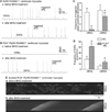
Figure 5. Effect of PLN-KO on delayed afterdepolarizations and triggered activities
Ventricular myocytes were isolated from RyR2-R4496C+/− and PLN−/−/RyR2-R4496C+/− hearts and perfused with 6 mM extracellular Ca2+ to induce spontaneous Ca2+ release. Membrane potentials in RyR2-R4496C+/− (A) or PLN−/−/RyR2-R4496C+/− (B) myocytes before (a) and after (b) the treatment with tBHQ, a SERCA2a inhibitor, were recorded using the perforated patch current clamp technique. (C, D) The frequency of spontaneously triggered APs (C) and delayed afterdepolarizations (DADs) (D) in RyR2-R4496C+/− and PLN−/−/RyR2-R4496C+/− ventricular myocytes before (a) and after (b) tBHQ treatment. Data shown are mean ± SEM from 8–12 cells (#P < 0.05, vs RC mice; *P < 0.05, vs -tBHQ). (E) Representative line-scan images of spontaneous Ca2+ release in isolated PLN−/−/RyR2-R4496C+/− ventricular myocytes before (a) and after (b) the treatment with 5 µM tBHQ (n = 21–22 cells) are shown.
Figure 6
Figure 6. Role of RyR2, LTCC, NCX, and SR Ca2+ load in the generation of mini-waves in PLN−/−/RyR2-R4496C+/− ventricular myocytes
(A) Whole heart homogenates were prepared from RyR2-R4496C+/− and PLN−/−/RyR2-R4496C+/− mutant mice and used for immunoblotting analysis using antibodies against RyR2, LTCC, SERCA2a, NCX or β-actin. Data shown are mean ± SEM (n=3, *P < 0.05, vs RyR2-R4496C+/−). (B, C, D) Ventricular myocytes were isolated from PLN−/−/RyR2-R4496C+/− hearts and loaded with the fluorescent Ca2+ indicator dye fluo-4-AM. Representative line-scan images of spontaneous Ca2+ release induced by elevated extracellular Ca2+ (6 mM) in PLN−/−/RyR2-R4496C+/− cells before (a) and after (b) the treatment with Bay K (B), caffeine (C), and LiCl (replacing NaCl in KRH) (D) (n = 14–18 cells) are shown. Note that none of these treatments converted mini-waves to cell-wide SCWs. (E) SR Ca2+ contents in fluo-4-AM loaded RyR2-R4496C+/− (a), PLN−/−/RyR2-R4496C+/− (b), and PLN−/− (c) ventricular myocytes were estimated by measuring the amplitude of caffeine (20mM) induced Ca2+ transients (d). Data shown are mean ± SEM (n=12–18) (*P < 0.05, vs RyR2-R4496C+/−).
Figure 7
Figure 7. RyR2-R4496C+/− mice are susceptible to CPVT
Representative ECG recordings of WT littermates (A) and RyR2-R4496C+/− (B) mice before (a) and after (b) the injection of epinephrine (1.6 mg/kg) and caffeine (120 mg/kg). VT duration (%) in WT littermates and RyR2-R4496C+/− mutant mice within each 3-min (C) or 30-min (D) period of ECG recordings. Data shown are mean ± SEM from 9–11 mice (*P < 0.05, vs WT).
Figure 8
Figure 8. PLN-KO protects against stress-induced VTs in mice
Representative ECG recordings of PLN−/− (A), PLN−/−/RyR2-R4496C+/− (B), and PLN−/−/RyR2-R4496C+/+ (C) mice before (a) and after (b) the injection of epinephrine (1.6 mg/kg) and caffeine (120 mg/kg). VT duration (%) in PLN−/−, PLN−/−/RyR2-R4496C+/−, and PLN−/−/RyR2-R4496C+/+ mice within each 3-min (D) or 30-min (E) period of ECG recordings. Data shown are mean ± SEM from 9–10 mice (*P < 0.05, vs PLN−/−).

References

    1. Fabiato A. Calcium-induced release of calcium from the cardiac sarcoplasmic reticulum. Am J Physiol. 1983;245:C1–C14. - PubMed
    1. Bers DM. Cardiac excitation-contraction coupling. Nature. 2002;415:198–205. - PubMed
    1. Fill M, Copello JA. Ryanodine receptor calcium release channels. Physiol Rev. 2002;82:893–922. - PubMed
    1. Kass RS, Tsien RW. Fluctuations in membrane current driven by intracellular calcium in cardiac purkinje fibers. Biophys J. 1982;38:259–269. - PMC - PubMed
    1. Stern M, Kort A, Bhatnagar G, Lakatta E. Scattered-light intensity fluctuations in diastolic rat cardiac muscle caused by spontaneous Ca2+-dependent cellular mechanical oscillations. J Gen Physiol. 1983;82:119–153. - PMC - PubMed

Publication types

MeSH terms

Substances