Review
. 2013 Oct;24(5):940-947.
doi: 10.1016/j.copbio.2013.05.007. Epub 2013 Jul 12.

Engineering cell-cell signaling

Affiliations
Review

Engineering cell-cell signaling

Katarina Blagovic et al. Curr Opin Biotechnol. 2013 Oct.

Abstract

Juxtacrine cell-cell signaling mediated by the direct interaction of adjoining mammalian cells is arguably the mode of cell communication that is most recalcitrant to engineering. Overcoming this challenge is crucial for progress in biomedical applications, such as tissue engineering, regenerative medicine, immune system engineering and therapeutic design. Here, we describe the significant advances that have been made in developing synthetic platforms (materials and devices) and synthetic cells (cell surface engineering and synthetic gene circuits) to modulate juxtacrine cell-cell signaling. In addition, significant progress has been made in elucidating design rules and strategies to modulate juxtacrine signaling on the basis of quantitative, engineering analysis of the mechanical and regulatory role of juxtacrine signals in the context of other cues and physical constraints in the microenvironment. These advances in engineering juxtacrine signaling lay a strong foundation for an integrative approach to utilize synthetic cells, advanced 'chassis' and predictive modeling to engineer the form and function of living tissues.


Figures

Figure 1
Figure 1
Engineering cell-cell signaling. Juxtacrine signals, such as cadherins, ephrins and Notch-Delta, are cues intrinsic to the cells in contrast to paracrine soluble signals and ECM proteins that provide extrinsic stimuli. The focus of this review (highlighted in red) is on engineering approaches to manipulate juxtacrine cues and associated intracellular regulatory signals and on the emerging design strategies to tune juxtacrine signals in the context of other microenvironmental cues that cumulatively affect cell functions with implications for biomedical applications.
Figure 2
Figure 2
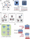
Devices and materials for modulating juxtacrine cell-cell signaling. (a) Juxtacrine cues are affixed to a scaffold, such as a PEG-based polymer network. (b) Chromium barriers restrict the movement of ephrin A1 on the supported membrane (bottom surface), thereby restricting the movement of EphA2-ephrin A1 complexes. (c) Controlling the direction of flow and using cell traps, isolated heterotypic cell pairs are induced. (d) Bowtie-shaped alginate-walled wells either accommodate one cell (half bowtie) or two cells (complete bowtie), thereby simulating cells with or without juxtracrine cell-cell interactions. (e) By controlling the gap size, juxtacrine signaling and the length scale for paracrine signaling are tunable in a dynamic manner.
Figure 3
Figure 3
The influence of juxtacrine cell-cell interactions on proliferation in epithelial cell systems depends on the microenvironmental context, including cell density (top three panels), soluble growth factors and a compliant ECM. The state diagram portrays the design space for shifting the cell system between a contact-inhibited state in which proliferation (green) occurs at the periphery of clusters and a contact-independent state in which proliferation occurs throughout the cluster. Matrix stiffening (leftward red arrows) perturbs cell-cell contacts, shifting the cell system (blue dots) closer to the transition line (solid black line) and enabling a switch from contact-inhibited to contact-independent proliferation at lower growth factor concentration.
Figure 4
Figure 4
Analysis of cell-cell forces. (a) Traction forces (red arrows) generated by the cell on the ECM are inferred from the measured displacements of fluorescent beads or microposts. The cell-cell force (green arrow) is calculated from a Newtonian force balance that requires the sum of the forces acting on a single cell to be zero. (b) Traction forces (red) cause displacement of fluorescent beads (blue arrows). In-plane (2D) characterization of bead displacement likely underestimates traction forces in the regions between cells because of counterbalancing forces (indicated by blue-boxed ‘X’). Bead displacements in the out-of-plane direction are reinforcing and can be captured using 3D confocal microscopy. (c) The extent of bending of microposts is proportional to the in-plane force applied at the top of the post. Because the posts bend independently from each other, they are more likely to capture the effect of in-plane traction forces in regions near the cell-cell interface.

References

    1. Mohr JC, de Pablo JJ, Palecek SP. 3-D microwell culture of human embryonic stem cells. Biomaterials. 2006;27:6032–6042. - PubMed
    1. Shin Y, Han S, Jeon JS, Yamamoto K, Zervantonakis IK, Sudo R, Kamm RD, Chung S. Microfluidic assay for simultaneous culture of multiple cell types on surfaces or within hydrogels. Nature Protocols. 2012;7:1247–1259. - PMC - PubMed
    1. Tumarkin E, Tzadu L, Csaszar E, Seo M, Zhang H, Lee A, Peerani R, Purpura K, Zandstra PW, Kumacheva E. High-throughput combinatorial cell co-culture using microfluidics. Integrative Biology. 2011;3:653–662. - PubMed
    1. Nagaoka M, Ise H, Akaike T. Immobilized E-cadherin model can enhance cell attachment and differentiation of primary hepatocytes but not proliferation. Biotechnology Letters. 2002;24:1857–1862.
    1. Liu CY, Apuzzo ML, Tirrell DA. Engineering of the extracellular matrix: working toward neural stem cell programming and neurorestoration--concept and progress report. Neurosurgery. 2003;52:1154–1165. discussion 1165-1157. - PubMed

Publication types

LinkOut - more resources