Review
. 2013 Oct;10(10):607-20.
doi: 10.1038/nrgastro.2013.120. Epub 2013 Jul 16.

Mucins in pancreatic cancer and its microenvironment

Affiliations
Review

Mucins in pancreatic cancer and its microenvironment

Sukhwinder Kaur et al. Nat Rev Gastroenterol Hepatol. 2013 Oct.

Abstract

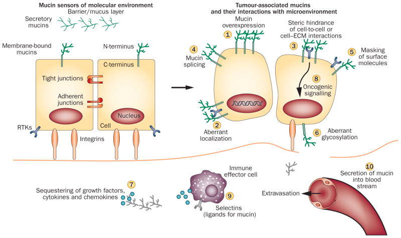
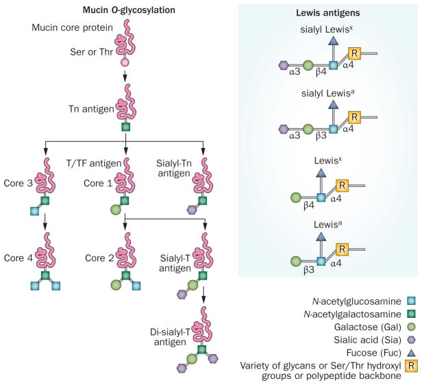
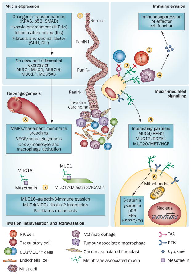
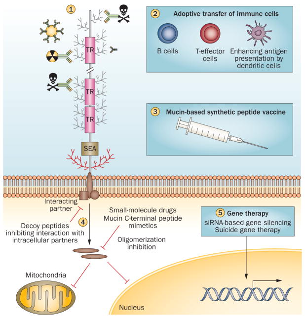
Pancreatic cancer remains a lethal malignancy with poor prognosis owing to therapeutic resistance, frequent recurrence and the absence of treatment strategies that specifically target the tumour and its supporting stroma. Deregulated cell-surface proteins drive neoplastic transformations and are envisioned to mediate crosstalk between the tumour and its microenvironment. Emerging studies have elaborated on the role of mucins in diverse biological functions, including enhanced tumorigenicity, invasiveness, metastasis and drug resistance through their characteristic O-linked and N-linked oligosaccharides (glycans), extended structures and unique domains. Multiple mucin domains differentially interact and regulate different components of the tumour microenvironment. This Review discusses: the expression pattern of various mucins in the pancreas under healthy, inflammatory, and cancerous conditions; the context-dependent attributes of mucins that differ under healthy and pathological conditions; the contribution of the tumour microenvironment in pancreatic cancer development and/or progression; diagnostic and/or prognostic efficacy of mucins; and mucin-based therapeutic strategies. Overall, this information should help to delineate the intricacies of pancreatic cancer by exploring the family of mucins, which, through various mechanisms in both tumour cells and the microenvironment, worsen disease outcome.


Conflict of interest statement

Competing interests

The authors declare no competing interests.

Figures

Figure 1
Figure 1
Domain structure of transmembrane and secretory mucins. The characteristic domains of transmembrane mucins (represented by MUC1 as well as domains present exclusively in MUC4) and secretory mucins (represented by MUC5AC). See Box 1 for further details on the individual domains. Abbreviations: AMOP, adhesion-associated domain in MUC4 and other proteins; EGF, epidermal growth factor; NIDO, nidogen-like domain, SEA, sea urchin sperm protein– enterokinase–agrin; TM, transmembrane; TR, tandem repeat, vWD, von Willebrand factor D domain; vWF-like, von Willebrand factor like domain.
Figure 2
Figure 2
Mucins in transition from normal to malignant cells. Mucins with their extended structure, hygroscopic nature and gelation abilities act as both sensor and defensive barrier to foreign insults under normal conditions. Cancer cells manipulate mucins at multiple levels to promote tumorigenicity. Events initiated by mucins (circled numbers) mediate the interactions between tumour cells and the surrounding stroma to create conditions favourable for tumour progression.
Figure 3
Figure 3
Antigenic and differently expressed O-glycans of pancreatic cancer. Tn and sialyl-Tn antigen are the simplest O-glycans formed by the addition of N-acetylgalactosamine to the –OH group of serine/threonine residues. These antigens are expressed in >80% of human carcinomas and are involved in mediating invasion and metastasis of tumour cells., Thomsen-Friedenreich (TF)/T antigen/core 1 are formed by the addition of galactose to Tn antigen through β1-3 linkage; these glycans are expressed in >90% of human malignancies. MUC1-associated TF and galectin-3 interactions promote metastasis by facilitating adhesion to endothelial cells. Core 3 glycans are synthesized by the addition of N-acetylglucosamine to Tn antigen; further addition of β-1,6-N-acetylglucosamine to the core 3 forms core 4 glycans. These glycans are tumour suppressive and are predominantly expressed in healthy cells., Lewis antigen: Fucosylated carbohydrate antigen sialyl Lewisa (or CA19-9, prognostic marker for PC) and sialyl Lewisx (or NCC-ST-439, prognostic marker for breast cancer) are overexpressed during neoplastic developments, whereas disialyl Lewisa and sialyl-6-sulpho-Lewisx are expressed in healthy cells. By acting as ligands for E-selectin, sialyl Lewisx mediates the adhesion of tumour cells to vascular endothelial cells and facilitates their metastasis.
Figure 4
Figure 4
Tumour cells crosstalk with stromal cells via mucins. Neoplastic developments elicit a proinflammatory and hypoxic environment leading to a desmoplastic and fibrotic reaction and enhanced mucin expression (1).,,– Aberrantly upregulated mucins sterically hide the tumour-associated antigens from invading macrophages, neutrophils and cytotoxic T cells (2), and indirectly protect tumour cells from the cytotoxic effects of activated immune effector cells (3). TAG72 and CA125 modulate M2 macrophages to an immunosuppressive phenotype (4). Aberrantly localized mucins interact and impart stability to various RTKs and aggravate oncogenic signalling (5). MUC1 cytoplasmic tail, through interactions with a variety of proteins, mediates oncogenic signalling (6); its interaction with galectins promotes tumour cell survival during metastasis by avoiding killing by NK cells and helps in extravasation through interaction with ICAM1 (7). Similarly, MUC4 and MUC16 interactions facilitate metastasis (7). Overexpression of mucins and the hypoxic environment in pancreatic tumour cells leads to the production of various factors, that in turn, remodel the extracellular matrix and induce neoangiogenesis (8). Abbreviations: MMP, matrix metalloproteinase; PanIN, pancreatic intraepithelial neoplasias; RTK, receptor tyrosine–protein kinase.
Figure 5
Figure 5
Mucins as therapeutic targets. Mucin-based immunotherapies being used in clinical and pre-clinical studies include antibody-targeted therapies (antibodies conjugated to radionuclides, immunotoxins and antibody-labelled gold nanoparticles) (1); cell-based therapies comprising adoptive transfer of CTLs, antigen pulsed dendritic cells, or a combination of both dendritic cells and CTLs] (2); recombinant peptide vaccines that enhance the mucin-antigen presentation by dendritic cells stimulating the immune response and tumour cell killing (3); and small-size inhibitory peptides that block mucin cytoplasmic tail interaction with other signalling molecules or mucin mimetic inhibiting cytoplasmic tail oligomerization, thus preventing its translocation to the mitochondria or nucleus (4). Mucin silencing by RNA interference and mucin promoter driven suicide gene therapy are other approaches to develop mucin-based targeted therapy (suicide gene therapy) (5). Abbreviations: CTL, cytotoxic T lymphocyte; TAA, tumour-associated antigen.

References

    1. Cancer Facts and Statistics. American Cancer Society; 2013. online http://www.cancer.org/research/cancerfactsstatistics/
    1. Kufe DW. Mucins in cancer: function, prognosis and therapy. Nat Rev Cancer. 2009;9:874–885. - PMC - PubMed
    1. Hollingsworth MA, Swanson BJ. Mucins in cancer: protection and control of the cell surface. Nat Rev Cancer. 2004;4:45–60. - PubMed
    1. Pflugfelder SC, et al. Detection of sialomucin complex (MUC4) in human ocular surface epithelium and tear fluid. Invest Ophthalmol Vis Sci. 2000;41:1316–1326. - PubMed
    1. Moniaux N, Escande F, Porchet N, Aubert JP, Batra SK. Structural organization and classification of the human mucin genes. Front Biosci. 2001;6:D1192–D1206. - PubMed

Publication types

MeSH terms