. 2013 Oct;43(10):2613-25.
doi: 10.1002/eji.201243018. Epub 2013 Aug 12.

Interleukin-6 limits influenza-induced inflammation and protects against fatal lung pathology

Affiliations

Interleukin-6 limits influenza-induced inflammation and protects against fatal lung pathology

Sarah N Lauder et al. Eur J Immunol. 2013 Oct.

Abstract

Balancing the generation of immune responses capable of controlling virus replication with those causing immunopathology is critical for the survival of the host and resolution of influenza-induced inflammation. Based on the capacity of interleukin-6 (IL-6) to govern both optimal T-cell responses and inflammatory resolution, we hypothesised that IL-6 plays an important role in maintaining this balance. Comparison of innate and adaptive immune responses in influenza-infected wild-type control and IL-6-deficient mice revealed striking differences in virus clearance, lung immunopathology and generation of heterosubtypic immunity. Mice lacking IL-6 displayed a profound defect in their ability to mount an anti-viral T-cell response. Failure to adequately control virus was further associated with an enhanced infiltration of inflammatory monocytes into the lung and an elevated production of the pro-inflammatory cytokines, IFN-α and TNF-α. These events were associated with severe lung damage, characterised by profound vascular leakage and death. Our data highlight an essential role for IL-6 in orchestrating anti-viral immunity through an ability to limit inflammation, promote protective adaptive immune responses and prevent fatal immunopathology.

Keywords: Adaptive immunity · Heterosubtypic immunity · IL-6 · Innate immunity · Pulmonary damage.


Figures

Figure 1
Figure 1. IL-6−/− mice experience enhanced mortality and morbidity with delayed viral clearance during acute influenza infection
(A, B) WT and IL-6−/− mice were infected i.n. with 50 pfu of PR8 and monitored daily for weight loss. The (A) percentage of initial weight and (B) Kaplan-Meier survival curves are shown and significance determined using the log-rank test (**p<0.01). (C) Core body temperature was determined on a daily basis as an indication of morbidity. (D) Lungs were harvested from the mice at the time-points indicated and the pulmonary viral load quantified. **p<0.01, Mann Whitney test. Results are expressed as the mean ± SEM, n = 6 mice/group from one experiment representative of three performed.
Figure 2
Figure 2. Acute influenza infection induces an enhanced proinflammatory cytokine response in IL-6−/− mice
BALF was aspirated at day 2, 4 or 7 post infection from WT and IL-6−/− mice infected with 50 pfu of PR8. (A) TNF-α, (B) IFN-α, (C) IL-1β and (D) IFN-γ levels present in the BALF were determined by ELISA. Results are expressed as the mean ± SEM, n = 6 mice/group from one experiment representative of two performed. The significances of differences between WT and IL-6−/− mice was assessed using Mann-Whitney test, * p < 0.05, ** p < 0.01.
Figure 3
Figure 3. IL-6−/− mice exhibit exacerbated influenza induced airway inflammation
BALF was aspirated from WT and IL-6−/− mice at days 2, 4 and 7 and the airway infiltrating cells were isolated. (A) The total numbers of cells and the numbers of (B) NK cells, (C) neutrophils, and (D) inflammatory monocytic cells, at the selected time points were determined by flow cytometry. The mean score ± SEM for each group is given, n = 6 mice/group, from one experiment representative of three performed. The significances of differences between WT and IL-6−/− mice were determined using Mann-Whitney test, *** p ≤ 0.001
Figure 4
Figure 4. Pulmonary inflammatory monocytic cells exhibit an altered phenotype in IL-6−/− mice
(A) BALF was aspirated from WT and IL-6−/− mice at day 7 and the airway infiltrating cells isolated. BAL cells were stimulated for five hours in vitro with LPS, the numbers of inflammatory monocytic cells secreting TNF-α were determined by flow cytometry. (B) The presence of the chemokine CCL2 was determined in BALF at day 7 by ELISA. Results are expressed as the mean ± SEM, n = 6 mice/group from one experiment representative of two performed. The significances of differences between WT and IL-6−/− mice was assessed using Mann-Whitney test, * p < 0.05, *** p < 0.001.
Figure 5
Figure 5. IL-6−/− mice exhibit increased pulmonary immunopathology during acute influenza infection
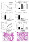
Lungs were harvested from mice at the days indicated post infection and following perfusion were formalin fixed, embedded in paraffin wax, sectioned and stained with haematoxylin and eosin. Representative sections from influenza-infected, (A, D) WT and (B, E) IL-6−/− mice were compared with those from (C, F) uninfected WT mice at day 7 (top) and day 9 (bottom). Arrowheads highlight areas of lymphocytic perivascular aggregate, asterisks highlight areas of leukocyte infiltration into the airway spaces, and H indicates areas of haemorrhage (original magnification ×20, scale bar 100 μm). (G) A representative section illustrating the extent of haemorrhage observed in IL-6−/− mice at day 9 post infection is shown (original magnification x 20). (H) The mean total histopathology score ± SEM for WT and IL-6−/− mice is given, n = 6 mice/group from one experiment representative of three performed. (I) The level of serum albumin, a protein indicative of damage to the pulmonary epithelium, was determined in BALF at day 7 by ELISA. Results are expressed as the mean ± SEM, n = 6 mice/group from one experiment representative of two performed. The significances of differences between WT and IL-6−/− mice was assessed using Mann-Whitney test. * p < 0.05.
Figure 6
Figure 6. Adaptive immune responses are diminished in IL-6−/− mice
(A, B) BALF was aspirated from WT and IL-6−/− mice at day 7 post infection and the airway infiltrating cells isolated. (A) The numbers of CD4+ (left axis), IFN-γ-producing CD4+ cells (right axis) and (B) CD8+ (left axis) and IFN-γ–producing CD8+ cells (right axis) were determined by flow cytometry. (C) Total numbers of CD8+ NP34-tetramer+ cells were determined from the BALF. (D) The total number of CD8+ NP34-tetramer+ cells was determined in the mediastinal lymph node that drains the pulmonary cavity. Results are expressed as the mean ± SEM , n = 6 mice/group from one experiment representative of three performed. The significances of differences between WT and IL-6−/− mice was assessed using Mann-Whitney test, * p < 0.05.
Figure 7
Figure 7. T-cell proliferation and survival characteristics are altered in the absence of IL-6
BALF was aspirated from WT and IL6−/− mice at day 7 post infection and the airway infiltrating cells isolated. Representative flow cytometry plots depict the expression of (A) Ki67 on CD8+ T Cells, and (B) Bcl2 on CD8+ T cells. (C) The numbers of CD8+, (D) NP-34+ tetramer+ CD8+ T cells and (E) CD4+ T cells expressing the proliferation marker Ki67 were determined by flow cytometry. Total numbers of (F) CD8+ T cells, (G) NP-34+ tetramer+ CD8+ T cells and (H) CD4+ T cells expressing the anti-apoptosis gene Bcl2 were also determined by flow cytometry. The significance of differences between WT and IL-6−/− mice was assessed using the Mann-Whitney test, * p < 0.05, ** p < 0.01.
Figure 8
Figure 8. Protective immune responses were significantly reduced in IL-6−/− mice
(A) At 7 – 9 weeks post infection all mice were rechallenged with 200 pfu of X31. Mice were weighed prior to secondary infection to provide a baseline 100% starting weight and for 4 days thereafter. Each data point represents the mean ± SEM for each group, n = 3 – 6 mice/group. (B) At day 4 post secondary infection mice were sacrificed and the lungs harvested following perfusion with PBS. Lungs were homogenized and the viral load determined. Plots represent the mean of each group ± SEM, n = 6 mice/group, 4 mice/group for PR8 naïve mice. (C, D) BALF was aspirated from WT and IL-6−/− mice at 4 days post secondary infection and the airway infiltrating cells isolated. The numbers of (C) CD8+ Influenza-specific tetramer+ cells, and (D) CD4+ cells were determined by flow cytometry. Plots represent the mean ± SEM, n = 6 mice/group, 4 mice/group for PR8 naïve mice. (E) To determine the proliferative capacity of antigen-specific CD4+ T cells, spleens were isolated from WT and IL-6−/− mice between 7 – 9 weeks post infection. CD4+ T cells were isolated by magnetic separation and stimulated by the addition of splenocytes loaded with PR8. The antigen specific proliferation of WT and IL-6−/− CD4+ T cells was determined by thymidine incorporation at 72 hours. Each data point represents the mean ± SEM, n = 4 mice/group. The significances of differences between WT and IL-6−/− mice was assessed using one-way ANOVA with Tukey-Kramers post-hoc multicomparison test, * p < 0.05, ** p < 0.01, *** p < 0.001. (F-H) Lungs were harvested from mice at day 4 post-secondary infection and following perfusion were formalin fixed, embedded in paraffin wax, sectioned and stained with haematoxylin and eosin. Representative sections from influenza infected, (G) WT and (H) IL-6−/− mice are shown. (F) Sections were scored for the extent and severity of haemorrhage present within the lungs of WT and IL-6−/− infected with PR8 and rechallenged with X31 4 days previously and PR8 naïve WT and IL-6−/− mice infected with X31 4 days earlier. Plots represent the mean haemorrhage score ± SEM, n = 4 - 6 mice/group. All data shown are from one experiment representative of two performed. Statistical significance between WT and IL-6−/− mice was determined using Mann-Whitney test, * p < 0.05.

References

    1. Murray CJ, Lopez AD, Chin B, Feehan D, Hill KH. Estimation of potential global pandemic influenza mortality on the basis of vital registry data from the 1918-20 pandemic: a quantitative analysis. Lancet. 2006;368:2211–2218. - PubMed
    1. de Jong MD, Simmons CP, Thanh TT, Hien VM, Smith GJ, Chau TN, Hoang DM, et al. Fatal outcome of human influenza A (H5N1) is associated with high viral load and hypercytokinemia. Nat Med. 2006;12:1203–1207. - PMC - PubMed
    1. Peiris JS, Yu WC, Leung CW, Cheung CY, Ng WF, Nicholls JM, Ng TK, et al. Re-emergence of fatal human influenza A subtype H5N1 disease. Lancet. 2004;363:617–619. - PMC - PubMed
    1. Yuen KY, Chan PK, Peiris M, Tsang DN, Que TL, Shortridge KF, Cheung PT, et al. Clinical features and rapid viral diagnosis of human disease associated with avian influenza A H5N1 virus. Lancet. 1998;351:467–471. - PubMed
    1. Kaiser L, Fritz RS, Straus SE, Gubareva L, Hayden FG. Symptom pathogenesis during acute influenza: interleukin-6 and other cytokine responses. J Med Virol. 2001;64:262–268. - PubMed

Publication types

MeSH terms