

Giant peripheral ossifying fibroma: a case report and clinicopathologic review of 10 cases from the literature
- PMID: 23857548
- PMCID: PMC3824796
- DOI: 10.1007/s12105-013-0452-1
Giant peripheral ossifying fibroma: a case report and clinicopathologic review of 10 cases from the literature
Abstract
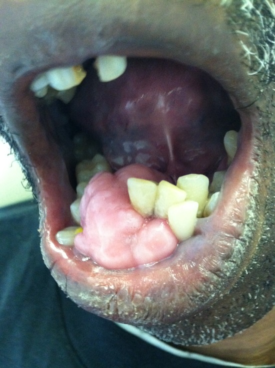
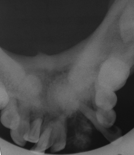
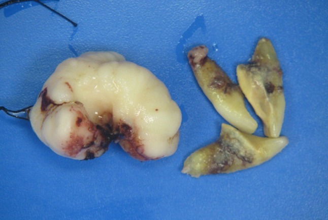
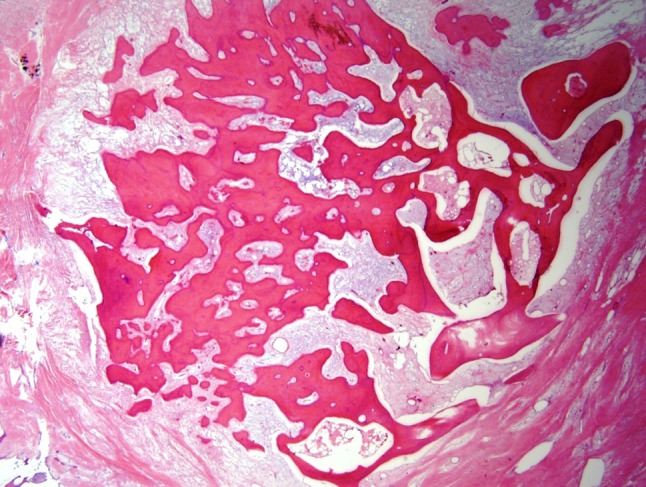
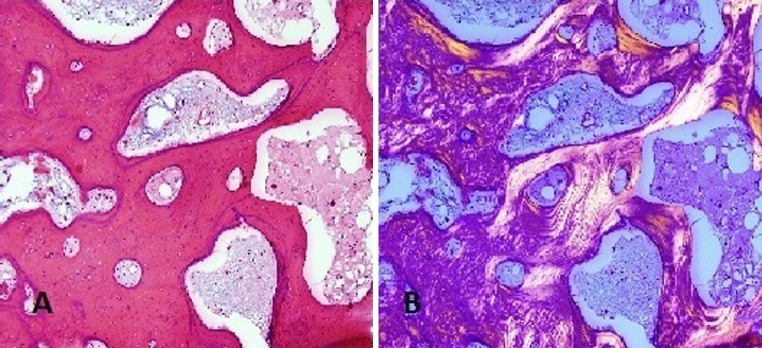
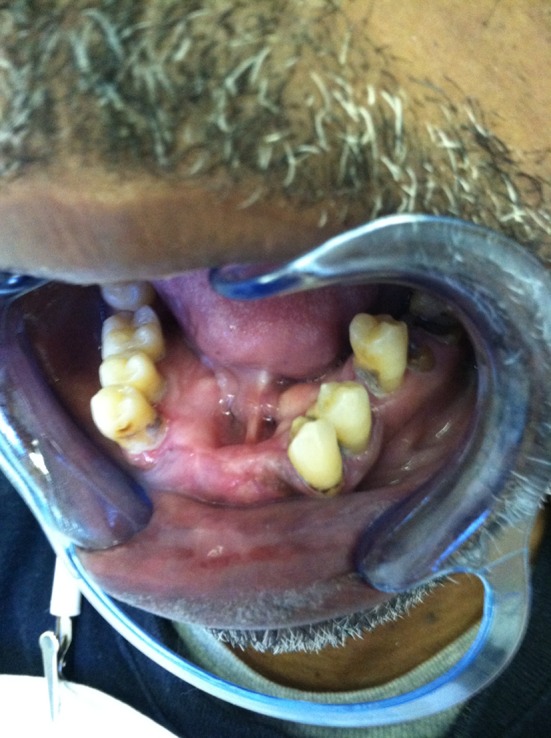
Peripheral ossifying fibroma (POF) is most often a self-limiting, sessile or pedunculated, gingival nodule that is believed to be a reactive rather than neoplastic pathologic process. The lesion is typically <2cm, however it has been recognized that some examples may grow quite large and may displace teeth. The mass-like clinical presentation and radiographic appearance of soft tissue calcification may lead to misclassification; however the histologic appearance is diagnostic. Giant POFs (GPOF) have been referred to in the literature by several other names (large, atypical, huge, gigantiform). The distinguishing characteristics of GPOFs and the factors that contribute to their growth have primarily been explored through case reports. We present a new case of POF that was giant and review 10 previously reported giant lesions, with focus on the clinical presentation, radiographic features, and outcome to explore the possibility that this represents a distinct clinical subset of lesion, with a unique set of features that warrant recognition for accurate diagnosis.
Figures
References
-
- Neville BW, Damm DD, Allen CM, Bouquot JE. Oral and maxillofacial pathology. 3rd ed. St. Louis, MO: Saunders Elsevier; 2009. p. 521–2.
-
- Kim J, Kim ES. Huge peripheral ossifying fibroma of the lower posterior edentulous ridge. J Korean Assoc Maxillofac Plast Reconstr Surg. 2009;31(5):435–439.
Publication types
MeSH terms
LinkOut - more resources
Full Text Sources
Other Literature Sources
