. 2013 Aug 23;288(34):24753-63.
doi: 10.1074/jbc.M113.491985. Epub 2013 Jul 15.

Hepatitis C virus NS5A inhibits mixed lineage kinase 3 to block apoptosis

Affiliations

Hepatitis C virus NS5A inhibits mixed lineage kinase 3 to block apoptosis

Yutaka Amako et al. J Biol Chem. .

Abstract

Hepatitis C virus (HCV) infection results in the activation of numerous stress responses including oxidative stress, with the potential to induce an apoptotic state. Previously we have shown that HCV attenuates the stress-induced, p38MAPK-mediated up-regulation of the K(+) channel Kv2.1, to maintain the survival of infected cells in the face of cellular stress. We demonstrated that this effect was mediated by HCV non-structural 5A (NS5A) protein, which impaired p38MAPK activity through a polyproline motif-dependent interaction, resulting in reduction of phosphorylation activation of Kv2.1. In this study, we investigated the host cell proteins targeted by NS5A to mediate Kv2.1 inhibition. We screened a phage-display library expressing the entire complement of human SH3 domains for novel NS5A-host cell interactions. This analysis identified mixed lineage kinase 3 (MLK3) as a putative NS5A interacting partner. MLK3 is a serine/threonine protein kinase that is a member of the MAPK kinase kinase (MAP3K) family and activates p38MAPK. An NS5A-MLK3 interaction was confirmed by co-immunoprecipitation and Western blot analysis. We further demonstrate a novel role of MLK3 in the modulation of Kv2.1 activity, whereby MLK3 overexpression leads to the up-regulation of channel activity. Accordingly, coexpression of NS5A suppressed this stimulation. Additionally we demonstrate that overexpression of MLK3 induced apoptosis, which was also counteracted by NS5A. We conclude that NS5A targets MLK3 with multiple downstream consequences for both apoptosis and K(+) homeostasis.

Keywords: Apoptosis; Hepatitis c Virus; Ion Channels; Kv2.1; Mixed Lineage Kinase 3; NS5A; Oxidative Stress; SH3 Domains.


Figures

FIGURE 1.
FIGURE 1.
A, molecular organization of MLK3 and HCV NS5A. Schematic illustration of the domain structure of the two proteins. Amino acid residue numbers indicated. The sequence of the P2 motif and the mutations introduced to generate the PA2 mutant are illustrated. The black box on NS5A is the N-terminal membrane-associating amphipathic helix. B, structure of bicistronic vectors used for transfection in this study. The construction of these vectors is detailed in the “Experimental Procedures.”
FIGURE 2.
FIGURE 2.
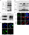
NS5A binds to MLK3 in a polyproline motif (P2)-dependent manner. A, lysates from Huh7 cells harboring a JFH-1 derived subgenomic replicon (SGR-JFH1) were immunoprecipitated with an anti-MLK3 monoclonal antibody and analyzed by Western blot for the presence of MLK3 and NS5A. The top panel shows the lysates and the bottom panel the immunoprecipitates, IgG LC: light chain of the immunoprecipitating antibody. B, lysates from Huh7 cells harboring an SGR with a One-STrEP-tagged NS5A were purified by affinity column capture with Strep-Tactin beads, prior to Western blotting for the presence of NS5A and MLK3. C, SGR-JFH1 cells were analyzed by indirect immunofluorescence with antibodies to NS5A (red) and MLK3 (green). The merged image shows extensive colocalization of the two proteins. Scale bar, 20 μm. D, ectopic expression of NS5A and MLK3, co-immunoprecipitation/Western blotting to demonstrate protein-protein interaction between NS5A and MLK3. Huh7 cells were co-transfected with a combination of plasmid vectors expressing NS5A (wild type or PA2 mutant) and FLAG-tagged MLK3 (wild type or K144E kinase-dead mutant, MLK3 KD) as indicated. Lysates were immunoprecipitated with an anti-FLAG antibody and Western blotted for the presence of NS5A or MLK3. IgG LC: light chain of the immunoprecipitating antibody. E, Huh7 cells were transfected with plasmid vectors expressing NS5A (top row), FLAG-tagged MLK3 (middle row), or both (bottom row). Indirect immunofluorescence microscopy reveals colocalization of NS5A and MLK3 in the cotransfected cells.
FIGURE 3.
FIGURE 3.
Exogenous MLK3 expression induces apoptosis. A, Huh7 cells were seeded on coverslips and transfected with the indicated expression vectors. 24 h after transfection, cells were fixed and stained with DAPI. Fluorescence microscopy was performed to visualize transfected cells (GFP fluorescence) and the nucleus (DAPI). B, multiple fields were counted (n = 4). GFP-positive cells, more than 60 cells for each sample point, were counted as transfected cells and the percentage of GFP-positive cells showing fragmented nuclei are presented. C, Huh7 cells were transfected with the indicated plasmid vectors. Lysates were subjected to Western blotting to detect caspase-cleaved PARP (as a marker of apoptosis), as well as NS5A-GFP and FLAG-MLK3.
FIGURE 4.
FIGURE 4.
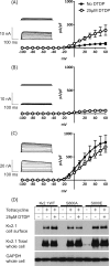
Kv2.1 ion channel activity is regulated by serine 800 phosphorylation. Whole-cell patch clamp recordings of Kv2.1 K+ currents. HEK293 cell lines expressing wild type Kv2.1 (A), S800A (non-phosphorylatable mutant: B) or S800E (phosphomimetic mutant: C) were subjected to whole cell patch clamp in the presence (○) or absence (●) of the oxidant DTDP (n = 3). Insets show representative traces of K+ currents in patch-clamp recordings obtained by step depolarizations applied from a holding potential of −70 mV to between −100 mV and +60 mV, in 10-mV increments. Top; trace without DTDP stimulation, below: with DTDP stimulation (D) HEK293 Kv2.1 cells were incubated with tetracycline to induce Kv2.1 expression, and/or DTDP to induce oxidative stress. Proteins expressed on the plasma membrane of HEK293 Kv2.1 cells were biotin-labeled and harvested samples were probed by Western blot with anti-Kv2.1 antibody (top panel). Whole cell lysates were also probed with the total Kv2.1 (middle). Comparable amount of Kv2.1 proteins were expressed following addition of tetracycline. GAPDH detection was performed as a control (bottom).
FIGURE 5.
FIGURE 5.
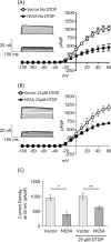
NS5A suppresses Kv2. 1 channel activity. HEK293 cells expressing Kv2.1 were transfected with bicistronic vectors encoding NS5A and GFP, or GFP alone (shown in Fig. 1B). At 24 h after transfection, Kv2.1 expression was induced by tetracycline treatment for 12 h, prior to electrophysiological analysis either without (A), or following (B) of DTDP stimulation (n = 6). Insets show representative traces of outward K+ currents in patch-clamp recordings. Top; control trace in GFP-expressing cells, below: trace from cells expressing NS5A and GFP. C, comparison of current density measurements from A and B at +40 mV. *, p < 0.002, **, p < 0.02, unpaired t test.
FIGURE 6.
FIGURE 6.
Endogenous MLK3 contributes to phosphorylation activation of Kv2. 1 HEK293 cells expressing Kv2.1 were infected with lentiviral vectors expressing shRNA targeting either MLK3 or GFP. After 6 days, cells were analyzed by quantitative-RT-PCR (A) or Western blotting (B). A, absolute quantitation of both MLK3 and GAPDH mRNA was performed and shown as a ratio. B, Western blotting with indicated antibodies. Cells were infected as follows: lanes 1: control (empty) lentiviral vector infection, lanes 2: lentiviral vector expressing shGFP, lanes 3: lentiviral vector expressing shMLK3. GAPDH was detected as a control. Western blots were quantitated by densitometry to show a reduction in Kv2.1 S800 phosphorylation following MLK3 knock-down. C, electrophysiological measurements of Kv2.1-mediated outward K+ currents from shRNA-expressing cells. D, comparison of current density measurements at +40 mV from shRNA-expressing cells. *, p < 0.001, unpaired t test.
FIGURE 7.
FIGURE 7.
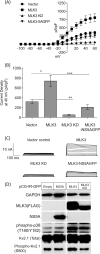
MLK3 acts as an upstream regulator of Kv2. 1 channel via Ser800 phosphorylation. A, HEK293 cells expressing Kv2.1 were transfected with bicistronic vectors expressing MLK3 (wild type or K144E) together with GFP or NS5A-GFP as shown in Fig. 1B. 16 h post-transfection, Kv2.1 expression was induced by tetracycline treatment, and 8 h later GFP-positive cells were identified by fluorescent microscopy and subjected to electrophysiological recordings. Overexpression of MLK3 wild type augments Kv2.1 channel activity (■), whereas, the K144E kinase-inactive mutant suppressed channel activity (▴). Co-expression of NS5A-GFP abrogated the MLK3 mediated enhancement of Kv2.1 activity (▾). B, comparison of current density measurements at +40 mV. *, p < 0.02, **, p < 0.001, and ***, p < 0.005, unpaired t test. C, representative traces of outward K+ currents in patch-clamp recordings. D, lysates from HEK293 cells expressing Kv2.1 wild type were analyzed by Western blotting with the indicated antibodies. Cells were transfected as follows: lane 1: empty vector, lane 2: NS5A-IRES-GFP, lane 3: MLK3 wild type-IRES-GFP, and lane 4: MLK3 K144E-IRES-GFP.
FIGURE 8.
FIGURE 8.
A model of NS5A interference with MLK3/p38/Kv2.1 signaling. HCV infection and RNA replication triggers the elevated production of reactive oxygen species (ROS), which activates MLK3, leading to activation of p38MAPK. In turn p38MAPK phosphorylates serine 800 of Kv2.1, which is a key phosphorylation event involved in the insertion of Kv2.1 into the plasma membrane. The upsurge in the efflux of K+ lowers the intracellular concentration of K+, causing an alteration of ionic homeostasis. At the same time MLK3 stimulates the induction of apoptosis. In this study, we show that NS5A inhibits MLK3 activation, thereby blocking both the phosphorylation and activation of Kv2.1, and MLK3-mediated apoptosis.

Similar articles

Cited by

References

    1. Pawlotsky J. M. (2004) Pathophysiology of hepatitis C virus infection and related liver disease. Trends Microbiol. 12, 96–102 - PubMed
    1. Tellinghuisen T. L., Evans M. J., von Hahn T., You S., Rice C. M. (2007) Studying hepatitis C virus: making the best of a bad virus. J. Virol. 81, 8853–8867 - PMC - PubMed
    1. Evans M. J., Rice C. M., Goff S. P. (2004) Phosphorylation of hepatitis C virus nonstructural protein 5A modulates its protein interactions and viral RNA replication. Proc. Natl. Acad. Sci. U.S.A. 101, 13038–13043 - PMC - PubMed
    1. Appel N., Zayas M., Miller S., Krijnse-Locker J., Schaller T., Friebe P., Kallis S., Engel U., Bartenschlager R. (2008) Essential role of domain III of nonstructural protein 5A for hepatitis C virus infectious particle assembly PLoS Path. 4, e1000035 - PMC - PubMed
    1. Masaki T., Suzuki R., Murakami K., Aizaki H., Ishii K., Murayama A., Date T., Matsuura Y., Miyamura T., Wakita T., Suzuki T. (2008) Interaction of hepatitis C virus nonstructural protein 5A with core protein is critical for the production of infectious virus particles. J. Virol. 82, 7964–7976 - PMC - PubMed

Publication types

MeSH terms