Review
. 2013 Oct;7(9-10):618-31.
doi: 10.1002/prca.201300024. Epub 2013 Sep 9.

Tn and sialyl-Tn antigens, aberrant O-glycomics as human disease markers

Affiliations
Review

Tn and sialyl-Tn antigens, aberrant O-glycomics as human disease markers

Tongzhong Ju et al. Proteomics Clin Appl. 2013 Oct.

Abstract

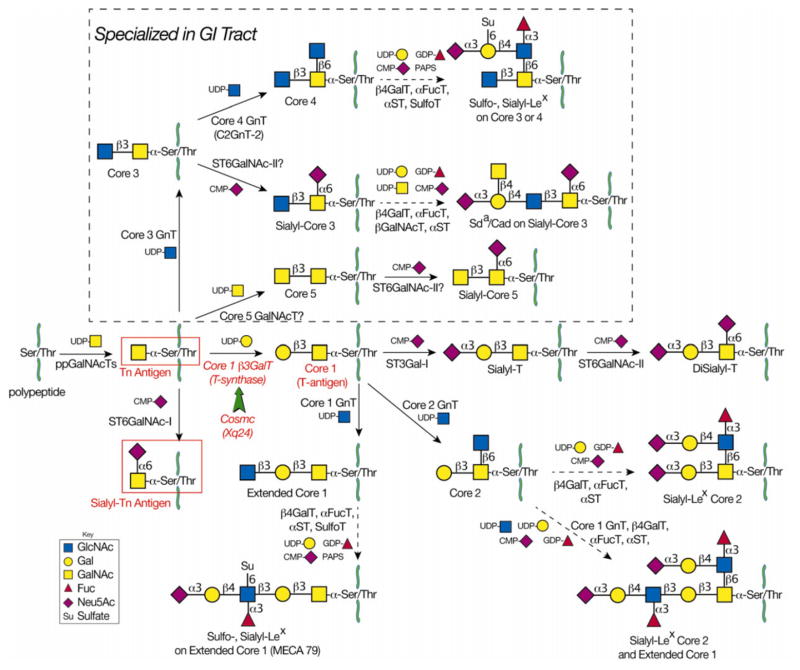
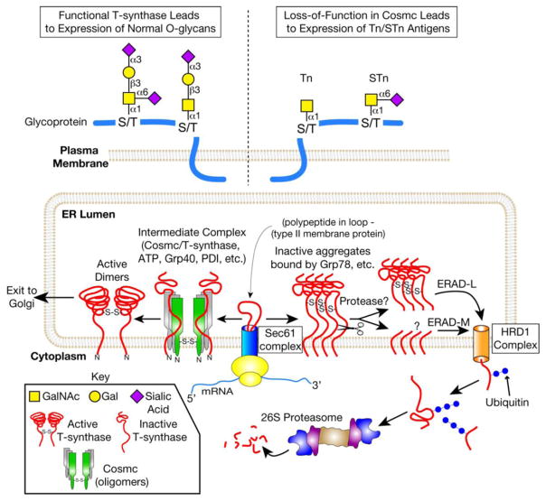
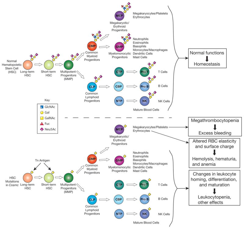
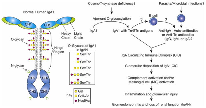
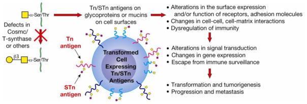
In many different human disorders, the cellular glycome is altered. An interesting but poorly understood alteration occurs in the mucin-type O-glycome, in which there is aberrant expression of the truncated O-glycans Tn (GalNAcα1-Ser/Thr) and its sialylated version sialyl-Tn (STn) (Neu5Acα2,6GalNAcα1-Ser/Thr). Both Tn and STn are tumor-associated carbohydrate antigens and tumor biomarkers, since they are not expressed normally and appear early in tumorigenesis. Moreover, their expression is strongly associated with poor prognosis and tumor metastasis. The Tn and STn antigens are also expressed in other human diseases and disorders, such as Tn syndrome and IgA nephropathy. The major pathological mechanism for expression of the Tn and STn antigens is compromised T-synthase activity, resulting from alteration of the X-linked gene that encodes for Cosmc, a molecular chaperone specifically required for the correct folding of T-synthase to form active enzyme. This review will summarize our current understanding of the Tn and STn antigens in terms of their biochemistry and role in pathology.

Keywords: Cancer; Disease; Glycosylation; IgA nephropathy; Tn antigen.


Conflict of interest statement

The authors have declared no conflict of interest.

Figures

Figure 1
Figure 1
The mucin-type O-glycosylation pathways. Mucin-type O-glycosylation begins in the Golgi when polypeptide GalNAc-transferases transfer GalNAc from UDP-Gal to Ser/Thr on a polypeptide chain to form Tn antigen (GalNAc-α-Ser/Thr). In nondiseased tissue, Tn antigen is further modified to form complex O-glycans. The T-synthase (Core 1 β3GalT) transfers Gal from UDP-Gal to GalNAc to form core 1 (T-antigen) in all cell types. Cosmc is the unique molecular chaperone for the T-synthase. Loss of Cosmc or T-synthase activity results in pathological expression of Tn antigen and sialyl-Tn (STn) antigen. The latter results from the actions of ST6GalNAc-I, which transfers Neu5Ac from CMP-Neu5Ac to the Tn antigen to form STn. Due to poor efficiency of ST6GalNAc-I, it is not likely that high expression of ST6GalNAc-I could outcompete functional T-synthase to result in pathologic STn expression. In GI epithelia, core 3 GnT transfers GlcNAc from UDP-GlcNAc to Tn antigen to form core 3. It is not known whether the T-synthase and core 3 GnT compete for the same substrates in GI epithelial cells. The T antigen is further modified to form extended core 1 structures. Similarly, core 3 is further modified to form extended core 3 structures, including sulfo-Lex, sialyl-Lex, or Sda on core 3 or core 4 O-glycans.
Figure 2
Figure 2
The major molecular mechanism for the expression of Tn and STn antigens in cells lacking a functional Cosmc. Cosmc is the unique molecular chaperone for the T-synthase. Nascent T-synthase is translocated to the ER possibly via the Sec61 complex. Cosmc interacts cotranslationally with nascent, nonnative T-synthase to form active, dimeric T-synthase. The T-synthase is subsequently transported to the Golgi. In the Golgi, the T-synthase transfers Gal from UDP-Gal to Tn antigen to form the T antigen on polypeptide chains. Defective Cosmc due to genetic or epigenetic alterations in Cosmc (e.g. ORF mutations, promoter methylation, or loss of heterozygosity) results in aggregation and proteosomal degradation of the T-synthase. Mis-folded T-synthase interacts with Grp78, is cleaved in its lumenal domain by an unknown protease, and is retrotranslocated to the cytosol. In the cytosol, soluble T-synthase is polyubiquitinated and degraded by the 26S proteasome. Loss of T-synthase activity results in expression of Tn and sialylTn antigens, which are not present in normal, non-transformed tissue.
Figure 3
Figure 3
Tn and STn antigens on blood cells in Tn syndrome. Tn syndrome is a rare autoimmune disorder in which populations of blood cells from all lineages express Tn antigen. Genetic/epigenetic alterations in Cosmc in an early blood progenitor result in Tn expression and associated pathology, including hemolytic anemia, thrombocytopenia, and bleeding disorders. Pathology is thought to be due to formation of anti-Tn IgM antibodies and/or dysfunction of O-glycosylated proteins found on blood cells. A targeted deletion of Cosmc in murine endothelial/hematopoietic cells was recently observed to result in macrothrombocytopenia, prolonged tail-bleeding times, and dysregulation of multiple platelet integrins.
Figure 4
Figure 4
Tn and STn antigens on IgA1 in IgAN. IgA nephropathy (IgAN) is the most common glomerulonephritis and results in renal failure in 20–40% of patients over 25 years of age. Deposition of IgA1 in the glomerular mesangium is thought to drive the disease. Reduced galactose and increased Tn/STn have been observed on IgA1 isolated from renal biopsies of patients. Normal O-glycans in the IgA1 hinge region contain mono- or disialylated core 1 structures. Although the role of Cosmc/T-synthase in IgAN is controversial, a reduction in T-synthase transcript and activity has been observed in B cells isolated from IgAN patients. It is thought that Tn/STn expression could lead to IgA1 aggregation by anti-Tn/STn antibodies or nonimmunologic methods, e.g. by increasing the propensity of IgA1 to self-aggregate. Circulating IgA1 immune complexes deposit in the glomerular mesangium, activating mesangial cells and the complement cascade, which leads to glomerulonephritis and loss of renal function.
Figure 5
Figure 5
Tn and STn antigens on tumor cells. Tn and STn antigens are tumor-associated carbohydrate antigens. They are not expressed on normal, nontransformed tissues. Defects in Cosmc or T-synthase can result in Tn/STn expression, although, to date, only defects in Cosmc have been observed in human tumors or cancer cell lines. Tn/STn expression is found on the majority of carcinomas and its expression correlates with progression of disease. However, the role for Tn/STn expression in tumorigenesis is unknown. Loss of O-glycosylation could lead to surface receptor dysregulation, changes in cell–cell and cell–matrix contacts, and/or immunoregulation. Subsequent changes in gene expression, signal transduction, and/or physicochemical interactions could facilitate tumor initiation, progression, and/or metastasis.

References

    1. Ju T, Otto VI, Cummings RD. The Tn antigen-structural simplicity and biological complexity. Angew Chem. 2011;50:1770–1791. - PMC - PubMed
    1. Steentoft C, Vakhrushev SY, Vester-Christensen MB, Schjoldager KT, et al. Mining the O-glycoproteome using zinc-finger nuclease-glycoengineered simple cell lines. Nat Methods. 2011;8:977–982. - PubMed
    1. Ellies LG, Tsuboi S, Petryniak B, Lowe JB, et al. Core 2 oligosaccharide biosynthesis distinguishes between se-lectin ligands essential for leukocyte homing and inflammation. Immunity. 1998;9:881–890. - PubMed
    1. Homeister JW, Thall AD, Petryniak B, Maly P, et al. The alpha(1,3)fucosyltransferases FucT-IV and FucT-VII exert collaborative control over selectin-dependent leukocyte recruitment and lymphocyte homing. Immunity. 2001;15:115–126. - PubMed
    1. Yeh JC, Hiraoka N, Petryniak B, Nakayama J, et al. Novel sulfated lymphocyte homing receptors and their control by a Core1 extension beta 1,3-N-acetylglucosaminyltransferase. Cell. 2001;105:957–969. - PubMed

Publication types