. 2013 Jul 15;126(Pt 14):3095-104.
doi: 10.1242/jcs.122788.

Ku70 and non-homologous end joining protect testicular cells from DNA damage

Affiliations

Ku70 and non-homologous end joining protect testicular cells from DNA damage

Emad A Ahmed et al. J Cell Sci. .

Abstract

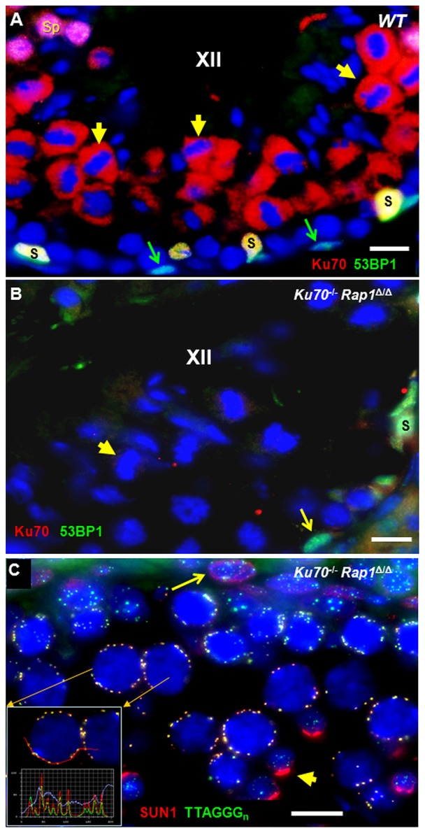
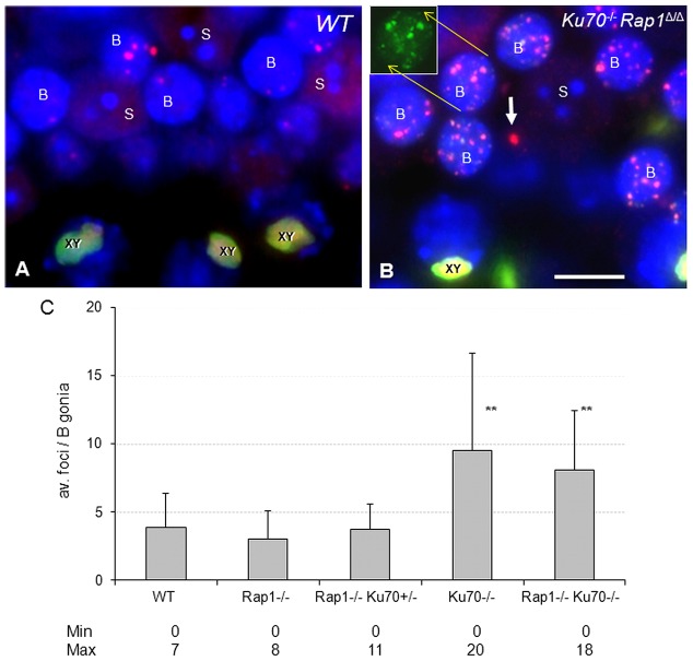
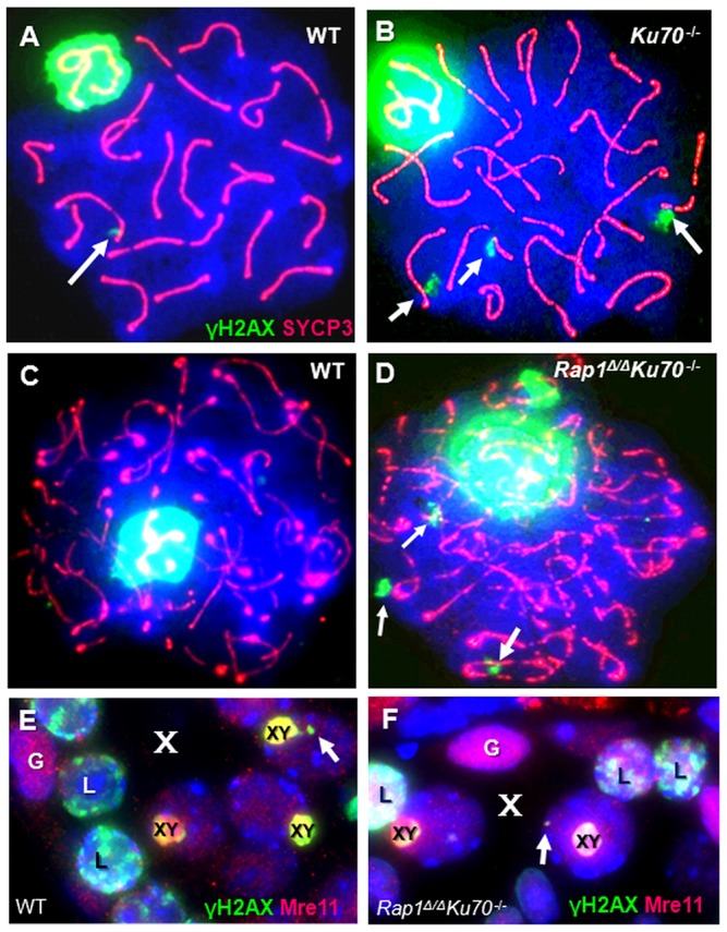
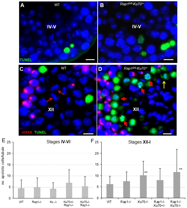
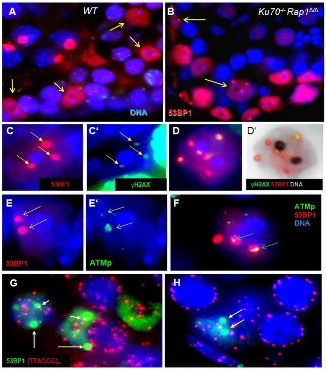
Spermatogenesis is a complex process that generates haploid germ cells or spores and implements meiosis, a succession of two special cell divisions that are required for homologous chromosome segregation. During prophase to the first meiotic division, homologous recombination (HR) repairs Spo11-dependent DNA double-strand breaks (DSBs) in the presence of telomere movements to allow for chromosome pairing and segregation at the meiosis I division. In contrast to HR, non-homologous end joining (NHEJ), the major DSB repair mechanism during the G1 cell cycle phase, is downregulated during early meiotic prophase. At somatic mammalian telomeres, the NHEJ factor Ku70/80 inhibits HR, as does the Rap1 component of the shelterin complex. Here, we investigated the role of Ku70 and Rap1 in meiotic telomere redistribution and genome protection in spermatogenesis by studying single and double knockout mice. Ku70(-/-) mice display reduced testis size and compromised spermatogenesis, whereas meiotic telomere dynamics and chromosomal bouquet formation occurred normally in Ku70(-/-) and Ku70(-/-)Rap1(Δ/Δ) knockout spermatocytes. Elevated mid-preleptotene frequencies were associated with significantly increased DNA damage in Ku-deficient B spermatogonia, and in differentiated Sertoli cells. Significantly elevated levels of γH2AX foci in Ku70(-/-) diplotene spermatocytes suggest compromised progression of DNA repair at a subset of DSBs. This might explain the elevated meiotic metaphase apoptosis that is present in Ku70-deficient stage XII testis tubules, indicating spindle assembly checkpoint activation. In summary, our data indicate that Ku70 is important for repairing DSBs in somatic cells and in late spermatocytes of the testis, thereby assuring the fidelity of spermatogenesis.

Keywords: Bouquet formation; DNA damage; Ku70; Meiosis; Mid-preleptotene; NHEJ; Rap1; Recombination; Sertoli cell; Shelterin; Spermatogenesis; Telomere attachment.


Figures

Fig. 1.
Fig. 1.
Spermatogenesis in Ku70 and Ku70 Rap1 knockout mice. (A) The testes of Ku70−/− and Ku70−/−Rap1Δ/Δ mice are of reduced size, as shown for wild-type (left) and Ku70−/− (right) testes. Scale bar: 5 mm. (B) Ku70−/− and Ku70−/−Rap1Δ/Δ (dko) mice display a significant (P<0.001) reduction in testis size compared with wild type (WT) and heterozygotes (het). Whiskers represent s.d. (≥3 mice). (C–F) Spermatogenesis appears grossly normal in tubule cross-sections of (C) wild type and (D) a Ku70+/−Rap1+/Δ heterozygote. Sperm production and release is reduced in (E) Ku70/− and (F) Ku70−/−Rap1Δ/Δ stage VI–VII tubules (asterisks). DAPI staining is shown in inverted gray scale. Scale bar: in F, 50 µm for C–F. (G–I) Epididymides of (G) wild type, (H) Ku70−/− and (I) Ku70−/−Rap1Δ/Δ mice (gray scale images of H&E staining). Ku70 knockout mice produce less sperm, as indicated by reduced diameter of epididymal tubules and their emptiness. Scale bar: in I, 100 µm for G–I.
Fig. 2.
Fig. 2.
Ku70 (red) and 53BP1 (green) immunofluorescence (IF) staining pattern of wild-type (WT) and Rap1Δ/ΔKu70−/− testes tubules. (A) WT stage XII tubule showing Ku70 protein IF (red) in the cytoplasm of metaphase I and II cells (examples highlighted by short yellow arrows) near the tubule center. Sertoli cells (S) at the tubule periphery appear yellow owing to colocalization of red Ku70 and green 53BP1 fluorescence. A-type spermatogonia show strong greenish 53BP1 expression (green arrows). Spermatids (Sp) appear pink owing to strong fluorescence for Ku70 and 53BP1 at the upper left corner of the image. (B) Ku70−/− stage XII tubule showing absence of red Ku70 protein signals (example indicated by short yellow arrow), whereas an A-type spermatogonium (long arrow) and a Sertoli cell (S) show green 53BP1 fluorescence. (C) IF of SUN1 (red) and (TTAGGG)n telomere FISH (green) revealing the colocalization of SUN1 with meiotic telomeres at the nuclear periphery of pachytene spermatocytes in Rap1Δ/Δ Ku70−/− testis tissue section. The inset shows fluorescence profiles across several telomeres that highlight colocalizing SUN1 (red line) and telomere (green line) fluorescence peaks. This staining is identical to that in wild type (not shown) (cf. Scherthan et al., 2011). A punctate NUP-like somatic distribution pattern of SUN1 (red) is seen at the meiotic NE of a spermatogonium at the tubule periphery (arrow), whereas haploid spermatids (short yellow arrow) display a strong red acrosomal signal. Scale bars: 10 µm.
Fig. 3.
Fig. 3.
DNA damage in B spermatogonia of Ku70-deficient testes revealed by 53BP1 (red) and γH2AX staining (green). (A) Testicular section of a wild-type mouse showing a few 53BP1 foci (red) in B spermatogonia (indicated by letter B). Wild-type Sertoli cells (S) show no 53BP1 foci. XY denotes the 53BP1- and γH2AX-positive sex body (yellow) of pachytene spermatocytes. (B) Numerous 53BP1 DNA damage foci in B spermatogonia of a Ku70−/−Rap1Δ/Δ testis section. These colocalize with γH2AX foci (green) as shown by the green channel image of one nucleus in the inset. The white arrow denotes a large 53BP1 DNA damage focus in the DAPI-faint chromatin of a double knockout Sertoli cell nucleus. (C) γH2AX and 53BP1 foci numbers are significantly increased (**P<0.001) in Ku70-deficient B spermatogonia of single and double knockout testes relative to the Rap1Δ/Δ, heterozygous and wild-type testes. Error bars indicate s.d.
Fig. 4.
Fig. 4.
Immunofluorescence of γ-H2AX (green) and SYCP3 (red) in surface-spread late spermatocytes of control, single and double knockout mice reveal normal synapsis. (A,B) Wild-type (A) and Ku70−/− (B) late pachytene spermatocytes. Arrows indicate a few large γH2AX foci. (C,D) Wild-type (C) and Ku70−/−Rap1Δ/Δ (D) diplotene spermatocytes showing a few large γH2AX foci (arrows) on SYCP3 axes (red threads). (E,F) Mre11 (red) and γH2AX (green) staining in details of control (E) and Ku70−/−Rap1Δ/Δ (F) stage IX–X tubules. Large γH2AX foci are seen as dots (arrows) in the chromatin of diplotene spermatocyte nuclei (marked by their XY body; XY) in stage X stage tubules (X). The patchy green-labeled nuclei represent leptotene spermatocytes (L). Spermatogonia (G) display strong Mre11 expression.
Fig. 5.
Fig. 5.
XY chromosome pairing as studied by X (green) and Y (red) chromosome painting in tissue sections. (A) Stage VI tubule of a Ku70−/−Rap1Δ/Δ testis showing pairing (touching) of XY signals in pachytene spermatocytes (arrow). (B) Stage XII tubule of a double knockout testis showing XY bivalents with close signal apposition in metaphase I plates. Blue, DNA stain DAPI. Signal distribution in wild type and heterozygotes was essentially the same (not shown). Scale bars: 10 µm.
Fig. 6.
Fig. 6.
Apoptosis in wild-type and Ku70−/−Rap1Δ/Δ testes sections. (A) Wild-type stage IV tubule showing two TUNEL-positive apoptotic spermatocytes (green). (B) Ku70−/−Rap1Δ/Δ stage IV tubule with three brightly labeled apoptotic cells. (C) Wild-type stage XII tubule showing two apoptotic meiosis I cells (green) and well developed metaphase I plates (blue; arrows). The red fluorescence stems from patchy γH2AX distribution in zygotene spermatocytes. Red arrows indicate examples of metaphase plates. (D) Double knockout stage XII tubule showing high number of TUNEL-positive apoptotic meiosis I cells. The red arrows denote apoptotic metaphase cells without chromosome congression. The long yellow arrow denotes an apoptotic metaphase with two non-aligned chromosomes. (E,F) Quantification of TUNEL-positive apoptotic cells in stages XII-I and stages IV-VI in control, single and double knockout mice. Although apoptosis was similar in stage IV-VI tubules (E), there was a significant increase of metaphase apoptosis in stage XII tubules (F) of Ku70-deficient testes relative to wild type and single Rap1 knockout. Scale bars: 10 µm. Error bars indicate s.d. **P<0.01.
Fig. 7.
Fig. 7.
Persistent DNA damage and active DDR in Ku70-deficient Sertoli cells of testes sections. (A) Wild-type Sertoli cells (arrows) show strong diffuse 53BP1 fluorescence (red) throughout the nucleus except for the nucleolus (dark dot) at its center. Bright red dots in spermatocyte nuclei represent the XY body. (B) Ku70−/−Rap1Δ/Δ Sertoli cells displaying large red 53BP1 DNA damage foci (arrows) in their nuclear chromatin. The two blue dots next to the dark spot (the nucleolus) represent the chromocenters that are specific for this cell type. (C-D′) Ku70-deficient Sertoli cells showing colocalization of strong 53BP1 (red) with weaker γH2AX signals (green) at large DNA damage foci (arrows). (D) Sertoli cell with six 53BP1 foci, four of which contain γ-H2AX fluorescence (green) as indicated by their yellowish color. (D′) Same cell as in D with the blue color of the DAPI stain converted to gray for better display. (E,E′) Colocalization of 53BP1 (red) with activated ATM (ATMp, green) at two large DNA damage foci (arrows) of a Ku70−/− Sertoli cell. One focus (lower arrow) contains a large amount of ATMp. (F) Sertoli cell with three damage foci, two of which contain ATMp (arrows). (G,H) TTAGGGn telomere FISH signals (red) only rarely colocalize with 53BP1 damage foci (green; arrows) in Ku70−/− Sertoli cell nuclei. In such cases, the small telomere signals lie at the border of the large 53BP1 foci (short arrows).

References

    1. Ahmed E. A., van der Vaart A., Barten A., Kal H. B., Chen J., Lou Z., Minter-Dykhouse K., Bartkova J., Bartek J., de Boer P. et al.(2007). Differences in DNA double strand breaks repair in male germ cell types: lessons learned from a differential expression of Mdc1 and 53BP1. DNA Repair (Amst.) 6, 1243–1254 10.1016/j.dnarep.2007.02.011 - DOI - PubMed
    1. Ahmed E. A., Barten-van Rijbroek A. D., Kal H. B., Sadri-Ardekani H., Mizrak S. C., van Pelt A. M., de Rooij D. G. (2009). Proliferative activity in vitro and DNA repair indicate that adult mouse and human Sertoli cells are not terminally differentiated, quiescent cells. Biol. Reprod. 80, 1084–1091 10.1095/biolreprod.108.071662 - DOI - PubMed
    1. Ahmed E. A., Philippens M. E., Kal H. B., de Rooij D. G., de Boer P. (2010). Genetic probing of homologous recombination and non-homologous end joining during meiotic prophase in irradiated mouse spermatocytes. Mutat. Res. 688, 12–18 10.1016/j.mrfmmm.2010.02.004 - DOI - PubMed
    1. Amsel A. D., Rathaus M., Kronman N., Cohen H. Y. (2008). Regulation of the proapoptotic factor Bax by Ku70-dependent deubiquitylation. Proc. Natl. Acad. Sci. USA 105, 5117–5122 10.1073/pnas.0706700105 - DOI - PMC - PubMed
    1. Aten J. A., Stap J., Krawczyk P. M., van Oven C. H., Hoebe R. A., Essers J., Kanaar R. (2004). Dynamics of DNA double-strand breaks revealed by clustering of damaged chromosome domains. Science 303, 92–95 10.1126/science.1088845 - DOI - PubMed

Publication types

MeSH terms

LinkOut - more resources