. 2013 Aug 15;191(4):1957-64.
doi: 10.4049/jimmunol.1300267. Epub 2013 Jul 15.

Inhibition of CD8+ T cell-derived CD40 signals is necessary but not sufficient for Foxp3+ induced regulatory T cell generation in vivo

Affiliations

Inhibition of CD8+ T cell-derived CD40 signals is necessary but not sufficient for Foxp3+ induced regulatory T cell generation in vivo

Danya Liu et al. J Immunol. .

Abstract

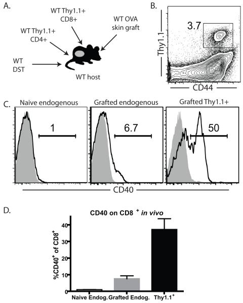
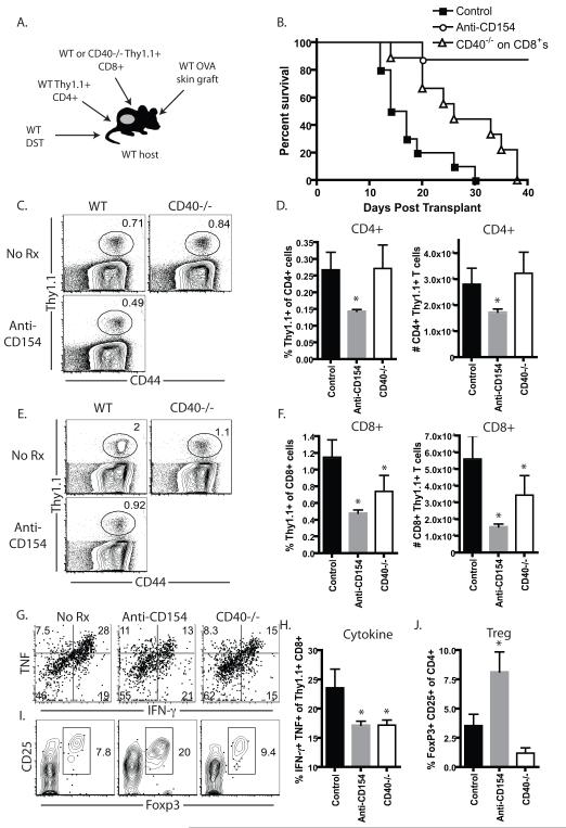
Current models of CD4(+) T cell help suggest a major role for CD154 binding to CD40 expressed on dendritic cells, with a lesser role for direct T:T interactions via CD40 expressed on CD8(+) T cells. However, the contribution of CD8(+) T cell-derived CD40 signals during the donor-reactive T cell response to a transplant has never been studied. In this study, we examined the graft-rejection kinetics and CD4(+) and CD8(+) donor-reactive T cell responses under conditions in which CD40 was genetically ablated only on APC, as well as under conditions in which CD40 was genetically ablated only on donor-reactive CD8(+) T cells. Our results revealed a significant role for CD8(+) T cell-expressed CD40 in the augmentation of donor-reactive CD8(+) T cell responses following transplantation and showed that CD40 expressed on CD8(+) T cells must be inhibited to allow conversion of CD4(+) T cells into induced regulatory T cells. Thus, this study identifies a major role for CD8(+) T cell-derived CD40 signals as a critical switch factor that both promotes optimal differentiation of cytokine-producing CD8(+) effector T cell responses and inhibits the differentiation of Ag-specific Foxp3(+) induced regulatory T cells in vivo.


Figures

Figure 1
Figure 1. Genetic deficiency of CD40 on both donor and recipient derived APCs fails to recapitulate the effects of CD154 blockade
A, 1.5 × 10⁶ WT OVA-specific CD4+ and CD8+ T cells were adoptively transferred into naïve WT or CD40−/− hosts, which then received a WT or CD40−/− OVA-expressing skin graft under the cover of WT or CD40−/− DST. WT recipients received WT graft and DST, while CD40−/− recipients received CD40−/− DST. Additional controls included WT recipients that were treated with anti-CD154. B, Skin graft survival data. Median survival times (MSTs) were 14, 100, and 20 days in untreated, anti-CD154-treated, and CD40−/− recipients, respectively. No Rx = WT animals that have received a skin graft and DST but no other treatment or manipulation of the CD40 pathway. C-D, Representative examples and summary data of frequency and absolute numbers (per spleen) of donor-reactive CD4+ T cells at day 10 post-transplant. E-F. Representative examples and summary data of frequency and absolute numbers (per spleen) of donor-reactive CD8+ T cells at day 10 post-transplant. G-H, Representative examples and summary data of frequency and absolute numbers (per spleen) of donor-reactive CD4+ Foxp3+ iTreg at day 10 post-transplant. Data shown are cumulative from four independent experiments, with a total of 12 animals per group. *p<0.05.
Figure 2
Figure 2. CD40 is upregulated on donor-reactive CD8+ T cells activated following activation
A, B. CD40−/− hosts received CD40−/− OVA-expressing skin grafts in the absence of any T cell adoptive transfer. Untreated WT animals receiving WT OVA-expressing skin grafts served as negative controls and anti-CD154 treated WT recipients of WT OVA-expressing skin grafts served as positive controls (A). While untreated recipients rejected their grafts with an MST of 16 d, CD40−/− recipients and those treated with anti-CD154 experienced increased graft survival that was comparable between the two groups (B). C, D. Splenocytes from OT-I × Thy1.1 or OT-II × Thy1.1 animals were incubated in vitro with cognate peptide antigen (OVA 257-264 and OVA323-339, respectively) for 4 d in vitro. Cultures without peptide antigen served as negative controls. Cells were then harvested and CD11c+ DC, CD4+ Thy1.1+ T cells, and CD8+ Thy1.1+ T cells were then stained for surface expression of CD40 or were stained with an isotype control. C, Representative histograms of CD40 staining (black histograms) or isotype control (grey shaded histograms) on DCs, CD4+ T cells, and CD8+ T cells from both unstimulated and peptide stimulated cultures. D, Summary data from three independent experiments. *p<0.05.
Figure 3
Figure 3. CD40 is upregulated on donor-reactive CD8+ T cells in vivo
1.5×10⁶ WT Thy1.1+ OVA-specific CD4+ and CD8+ T cells were adoptively transferred into naïve WT recipients that then received an OVA-expressing skin graft and no further treatment (A). DLN were harvested on day 10 post-transplant (B) and CD40 expression was assessed on activated graft-specific Thy1.1+ CD8+ T cells. C, Representative histograms of CD40 staining (black histograms) or isotype control (grey shaded histograms) on naïve endogenous CD8+ T cells isolated from naïve B6 animals, endogenous Thy1.1 CD8+ T cells isolated from grafted animals, and Thy1.1+ CD8+ donor-reactive T cells isolated from grafted animals. D, Summary data of frequencies of CD40+ cells among CD8+ T cells within the three groups. Data are cumulative of two independent experiments (n=8/group).
Figure 4
Figure 4. CD40 functions in a T cell intrinsic manner to enhance donor-reactive CD8+ T cell responses and inhibit Foxp3+ iTreg conversion
A, 1.5 × 10⁶ WT OVA-specific CD4+ and 1.5 × 10⁶ WT OVA-specific CD8+ or 1.5 × 10⁶ CD40−/− OVA-specific CD8+ T cells were adoptively transferred into naïve WT hosts, which then received a WT OVA-expressing skin graft under the cover of WT DST. Additional controls included WT recipients that were treated with anti-CD154. B, Skin graft survival data. MSTs were 15.5 days in WT controls vs. 26 days in recipients of CD40−/− T cells. No Rx = WT animals that have received a skin graft and DST but no other treatment or manipulation of the CD40 pathway. C-D, Representative examples and summary data of frequency and absolute numbers (per spleen) of donor-reactive WT CD4+ T cells at day 10 post-transplant in recipients containing either WT or CD40−/− T cells. E-F. Representative examples and summary data of frequency and absolute numbers (per spleen) of donor-reactive CD8+ T cells at day 10 post-transplant. G-H, Representative examples and summary data of frequency of donor-reactive CD8+ cytokine producing cells at day 10 post-transplant. I-J, Representative examples and summary data of frequency of donor-reactive CD4+ Foxp3+ iTreg at day 10 post-transplant. Data shown are cumulative from three independent experiments, with a total of 9 animals per group. *p<0.05.
Figure 5
Figure 5. OVA-specific CD8+ T cells can dispense with T cell-intrinsic CD40 signals in the context of pathogen infection
WT or CD40−/− Thy1.1+ CD8+ OVA-specific OT-I T cells (106) were adoptively transferred into naïve animals that were then infected with an OVA-expressing Listeria (A). Animals were sacrificed on day 10 and OVA-specific Thy1.1+ CD8+ T cell responses were analyzed in the spleen. Representative flow plots (B) and summary data (C) of frequencies of WT vs. CD40−/− OVA-specific Thy1.1+ T cells. Representative flow plots (D) and summary data (E) of frequencies of IFN-γ+ TNF+ producing cells in either WT or CD40−/− OT-I populations following ex vivo restimulation. Data shown are cumulative from two independent experiments (n=10/group).
Figure 6
Figure 6. Loss of CD40 signaling on both donor-reactive CD8+ T cells and APC synergize to promote Foxp3+ iTreg induction
A, 1.5 × 10⁶ WT OVA-specific CD4+ and 1.5 × 10⁶ CD40−/− CD8+ T cells were adoptively transferred into naïve CD40−/− hosts, which then received a CD40−/− OVA-expressing skin graft under the cover of CD40−/− DST. Negative controls were WT recipients that received WT graft and DST. Positive controls were WT recipients that were treated with anti-CD154. No Rx = WT animals that have received a skin graft and DST but no other treatment or manipulation of the CD40 pathway. B, Analysis of frequency and absolute numbers (per spleen) of donor-reactive CD4+ T cells at day 10 post-transplant. C, Analysis of frequency and absolute numbers (per spleen) of donor-reactive CD8+ T cells at day 10 post-transplant. D-E, Representative examples and summary data of frequency and absolute numbers (per spleen) of donor-reactive CD4+ Foxp3+ iTreg at day 10 post-transplant. Data shown are cumulative from two independent experiments, with a total of 6 animals per group. *p<0.05.

References

    1. Bennett SRM, Carbone FR, Karamalis F, Flavell RA, Miller JFAP, Heath WR. Help for cytotoxic-T-cell responses is mediated by CD40 signalling. Nature. 1998;393:478–480. - PubMed
    1. Schoenberger SP, Toes RE, van der Voort EI, Offringa R, Melief CJ. T-cell help for cytotoxic T lymphocytes is mediated by CD40-CD40L interactions. Nature. 1998;393:480–483. - PubMed
    1. Ridge JP, Di Rosa F, Matzinger P. A conditioned dendritic cell can be a temporal bridge between CD4+ T-helper and a T-killer cell. Nature. 1998;393:474–478. - PubMed
    1. Quezada SA, Jarvinen LZ, Lind EF, Noelle RJ. CD40/CD154 interactions at the interface of tolerance and immunity. Annu Rev Immunol. 2004;22:307–328. - PubMed
    1. Toes RE, Schoenberger SP, van der Voort EI, Offringa R, Melief CJ. CD40-CD40Ligand interactions and their role in cytotoxic T lymphocyte priming and anti-tumor immunity. Semin Immunol. 1998;10:443–448. - PubMed

Publication types

MeSH terms