. 2013 Aug 15;191(4):1586-93.
doi: 10.4049/jimmunol.1300575. Epub 2013 Jul 15.

Human CD1a deficiency is common and genetically regulated

Affiliations

Human CD1a deficiency is common and genetically regulated

Chetan Seshadri et al. J Immunol. .

Abstract

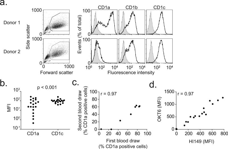
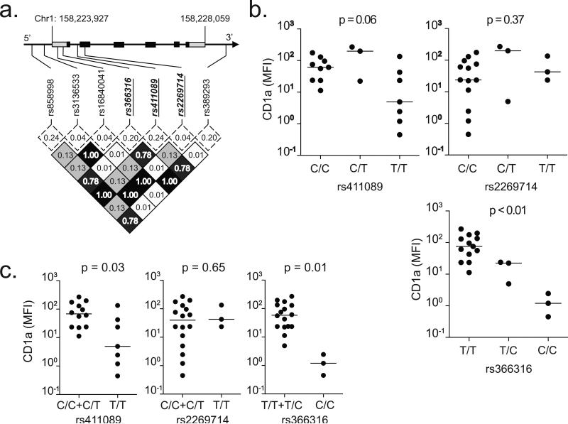
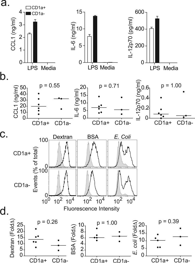
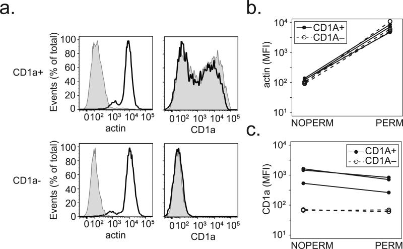
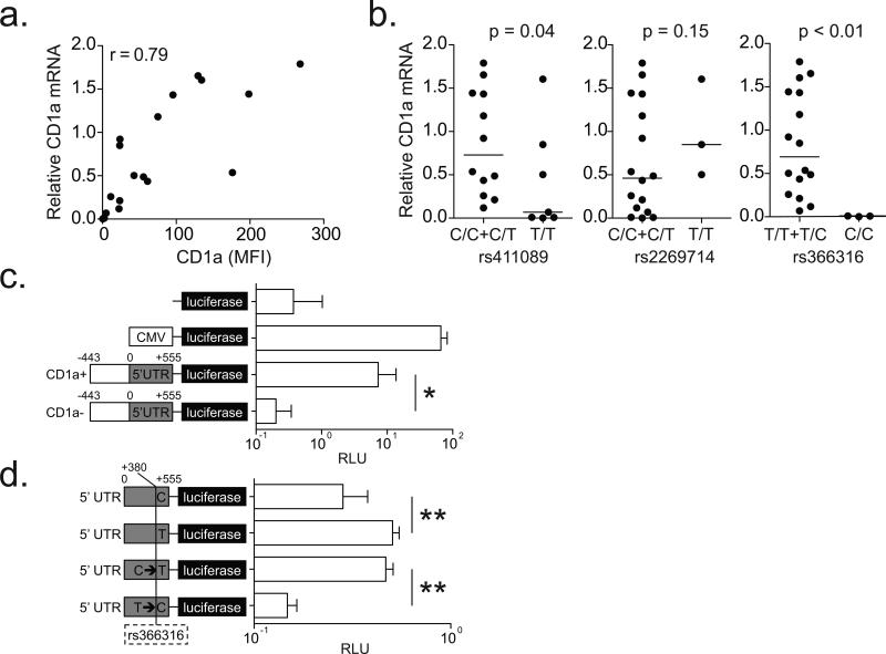
CD1 proteins evolved to present diverse lipid Ags to T cells. In comparison with MHC proteins, CD1 proteins exhibit minimal allelic diversity as a result of nonsynonymous single nucleotide polymorphisms (SNPs). However, it is unknown if common SNPs in gene regulatory regions affect CD1 expression and function. We report surprising diversity in patterns of inducible CD1a expression on human dendritic cells (DCs), spanning the full range from undetectable to high density, a finding not seen with other CD1 isoforms. CD1a-deficient DCs failed to present mycobacterial lipopeptide to T cells but had no defects in endocytosis, cytokine secretion, or expression of costimulatory molecules after LPS treatment. We identified an SNP in the 5' untranslated region (rs366316) that was common and strongly associated with low CD1a surface expression and mRNA levels (p = 0.03 and p = 0.001, respectively). Using a CD1a promoter-luciferase system in combination with mutagenesis studies, we found that the polymorphic allele reduced luciferase expression by 44% compared with the wild-type variant (p < 0.001). Genetic regulation of lipid Ag presentation by varying expression on human DCs provides a mechanism for achieving population level differences in immune responses despite limited structural variation in CD1a proteins.


Figures

Figure 1
Figure 1. Deficiency of CD1a on human dendritic cells
(a) Viable monocyte-derived dendritic cells, identified by high forward and side scatter profiles, were stained with fluorescently-conjugated antibodies against CD1a, CD1b, and CD1c (dark lines) as well as isotype control antibody (shaded histogram). Shown are representative plots from two donors. (b) Median fluorescence intensity (MFI) of CD1a and CD1c is shown for 19 healthy blood donors. In four donors, CD1a MFI is less than 10. P-value reflects Bartlett's test for non-homogeneity of variances. (c) Simple linear correlation between CD1a staining results of sequential blood draws for eight subjects. Data are represented as % positive cells rather than MFI to adjust for temporal variation in flow cytometry calibration. (d) Simple linear correlation of staining between two antibodies (OKT6 and HI149) that bind CD1a.
Figure 2
Figure 2. CD1a-deficiency does not reverse with increased IL-4 concentrations or extended time in culture
Human monocytes were cultured with IL-4 sourced from either Peprotech or R&D Systems at a concentration of either 20ng/ml or 100ng/ml for either three or six days. Cells were harvested and stained for flow cytometry with fluorescently conjugated antibodies against CD14, CD1a, or CD1c. (a) Shown are expression profiles from unstimulated monocytes (shaded histogram) or cells cultured for six days from a CD1a-sufficient donor (dark line), and a CD1a-deficient donor (dark histogram) for CD14. Results from five subjects are combined and shown as the median fluorescence intensity (MFI) of CD14 after (b) three days of culture or (c) six days of culture. (d) Representative CD1c expression from six day cultured cells or combined data showing CD1c MFI after (e) three days or (f) six days of culture. (g) Representative CD1a expression from six day cultured cells or combined data for CD1a after (h) three days or (i) six days of culture. Expression levels on unstimulated monocytes (dotted line) are shown for comparison. All data are representative of two independent experiments.
Figure 3
Figure 3. Association between CD1a SNPs and expression
(a) Linkage disequilibrium plot of SNPs in CD1a coding region ± 10kB flanking regions among CEU population from HapMap. CD1a spans 4132 bases on chromosome 1 and consists of six exons (black squares) and two untranslated regions (gray squares). Minor allele frequencies (dotted box) and linkage disequilibrium as measured by R2 values (shaded box) are indicated. Three haplotype-tagging SNPs selected for genotyping are emphasized in bold italic underline. (b) CD1a median fluorescence intensity (MFI) stratified by SNP genotypes. The bar indicates the median value. The nonparametric Kruskal-Wallis test was used to determine statistical significance for a genotypic model. (c) Recessive model analysis combines SNP genotypes ‘AA’ and ‘Aa.’
Figure 4
Figure 4. CD1a-deficient dendritic cells fail to present antigen to T cells but are able to induce expression of co-stimulatory molecules
(a) CD1a-sufficient (CD1a+) or CD1a-deficient (CD1a-) dendritic cells were co-incubated with T cell clone CD8-2 in the presence or absence of mycobacterial lipopeptide antigen dideoxymycobactin (DDM). IFN-γ levels were measured in overnight supernatants by ELISA for one donor per group. Shown are mean and standard deviation for triplicate measurements. (b) Shown are expression profiles for cells derived from two healthy blood donors and treated with media (shaded) or LPS (dark line) overnight and then stained with antibodies against co-stimulatory molecules and HLA-DR. (c) Results from nine subjects are combined and shown as fold change (LPS/Media) in median fluorescence intensity (MFI) for CD40, CD80, CD83, CD86, and HLA-DR. All data are representative of two or three independent experiments.
Figure 5
Figure 5. CD1a-deficient dendritic cells do not exhibit defects in cytokine production or endocytic capacity
(a) Supernatants from DCs exposed to media or LPS overnight were assessed for cytokines by ELISA. Histograms show mean and standard deviation of triplicate measurements from two healthy blood donors. (b) Results from nine subjects are combined and shown as mean values of triplicate measurements with media values subtracted. (c) DCs were exposed to FITC-conjugated dextran, bovine serum albumin (BSA), or E. coli for one hour at either 4°C (shaded histogram) or 37°C (dark line) to assess endocytic capacity. Shown are histograms from two healthy blood donors. (d) Results from nine subjects are combined and shown are fold change (37°C/4°C) in MFI for each particle. All data are representative of two or three independent experiments.
Figure 6
Figure 6. CD1a-deficiency is not the result of a defect in protein trafficking
DCs were permeabilized and stained for intracellular actin and CD1a. (a) Shown are expression profiles comparing permeabilized (dark line) and unpermeabilized (shaded histogram) cells from a CD1a-sufficient (CD1a+) and CD1a-deficient (CD1a-) donor. Results from three CD1a-sufficient and three CD1a-deficient donors are combined and shown as the difference in median fluorescence intensity (MFI) between permeabilized (PERM) and unpermeabilized (NOPERM) cells for (b) intracellular actin and (c) CD1a. All data are representative of two independent experiments.
Figure 7
Figure 7. CD1a-deficiency is the result of transcriptional regulation by a SNP in the 5’ UTR
(a) CD1a mRNA levels were normalized to that of the housekeeping gene glyceraldehyde 3-phosphate dehydrogenase (GAPDH). Simple linear correlation between relative CD1a mRNA levels and surface protein expression in DCs is shown. (b) Relative CD1a mRNA levels stratified by SNP genotypes are shown with results of one-sided hypothesis testing. (c) HEK293T cells were transfected with firefly luciferase-expressing plasmids (pGL4) under the control of various promoter sequences: none, cytomegalovirus (CMV), 998 bp from CD1a-sufficient (CD1a+), or CD1a-deficient (CD1a-). Cells were co-transfected with Renilla luciferase under a SV40 promoter, and data are reported as relative luciferase units (RLU) which is calculated by dividing the firefly luciferase signal by the Renilla luciferase signal. (d) HEK293T cells were transfected with firefly luciferase-expressing plasmids (pGL4) under the control of 555 bp CD1A 5’ UTR with variants of rs366316: natural C allele, natural T allele, C mutated to T (C→T), or T mutated to C (T→C). All data are representative of two or three independent experiments. * p=0.003 and ** p<0.001 by Welch two sample t-test.

References

    1. Robinson J, Halliwell JA, McWilliam H, Lopez R, Parham P, Marsh SG. The IMGT/HLA database. Nucleic Acids Res. 2013;41:D1222–D1227. - PMC - PubMed
    1. Dascher CC. Evolutionary biology of CD1. Curr Top Microbiol Immunol. 2007;314:3–26. - PubMed
    1. Kasmar A, Van Rhijn I, Moody DB. The evolved functions of CD1 during infection. Current opinion in immunology. 2009;21:397–403. - PMC - PubMed
    1. Van Rhijn I, Zajonc DM, Wilson IA, Moody DB. T-cell activation by lipopeptide antigens. Current opinion in immunology. 2005;17:222–229. - PubMed
    1. Martin LH, Calabi F, Milstein C. Isolation of CD1 genes: a family of major histocompatibility complex-related differentiation antigens. Proc Natl Acad Sci U S A. 1986;83:9154–9158. - PMC - PubMed

Publication types

MeSH terms