Review
. 2013 Jun;26(3):339-46.
doi: 10.1177/197140091302600315. Epub 2013 Jul 16.

Endovascular treatment of vertebro-vertebral arteriovenous fistula. A report of three cases and literature review

Affiliations
Review

Endovascular treatment of vertebro-vertebral arteriovenous fistula. A report of three cases and literature review

F Briganti et al. Neuroradiol J. 2013 Jun.

Abstract

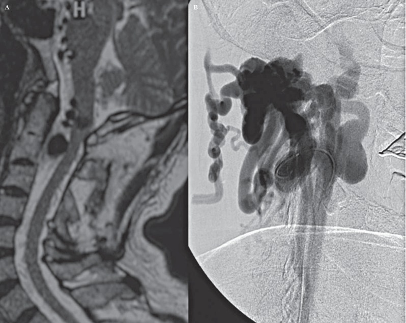
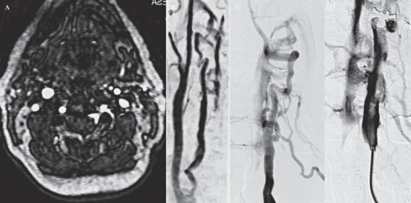
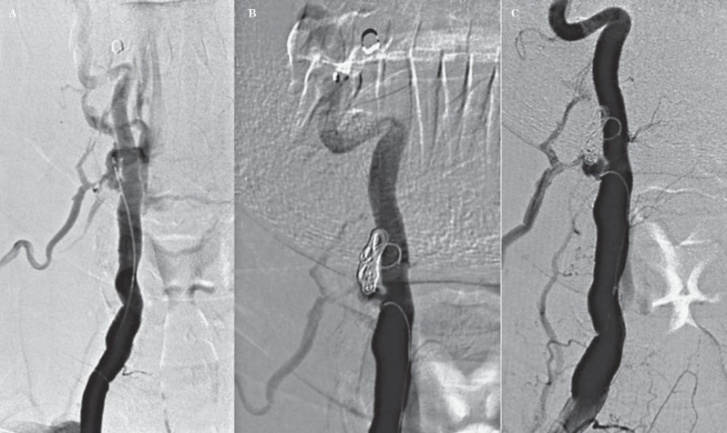
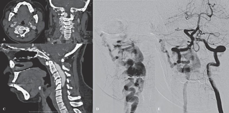
This report describes endovascular approaches for occlusion of vertebro-vertebral arteriovenous fistula (VV-AVF) in a series of three cases and a review of the literature. Complete neuroimaging assessment, including CT, MR and DSA was performed in three patients (two female, one male) with VV-AVF. Based on DSA findings, the VV-AVF were occluded by endovascular positioning of detachable balloons (case 1), coils (case 2), or a combination of both (case 3) with parent artery patency in two out of three cases. In this small series, endovascular techniques for occlusion of VV-AVF were safe and effective methods of treatment. To date, there are no guidelines on the best treatment for VV-AVF. Detachable balloons, endovascular coiling, combined embolization procedures could all be considered well-tolerated treatments.


Figures

Figure 1
Figure 1
A) Sagittal T2-w MRI shows a right ectasic vertebral artery at the anterior epidural level; B) Preoperative right vertebral artery DSA shows a direct VV-AVF between the right VA at C2 level and the periradicular venous plexus.
Figure 2
Figure 2
Operative DSA (A-C): the balloon is detached at the fistula point with preservation of parent vertebral artery patency.
Figure 3
Figure 3
Axial MRA TOF 3D (A) and MIP reconstruction (B) showing an extradural fistula of the vertebral artery involving the perivertebral venous plexus at C3-C4 level. Vertebral artery DSA (C, D) shows a dilated extracranial segment of the right vertebral artery between the muscular branches of the vertebral artery and a dilated periradicular venous plexus.
Figure 4
Figure 4
Vertebral artery operative DSA (A-C) shows complete occlusion of the fistula with coils and preservation of right vertebral artery patency.
Figure 5
Figure 5
Contrast-enhanced CT angiography, MPR reconstruction (A) axial (B) coronal (C) sagittal, shows an extradural fistula of the right VA with multiple arteriovenous high-flow shunts at C2-C5 level. Pre-operative DSA right (D) and left (E) VA injection: large VVAF in NF1 with flow steal.
Figure 6
Figure 6
Operative DSA: partial occlusion with detachable balloon (A). Complete VA and fistula occlusion with further coiling (B). C) Post-embolization DSA by controlateral VA injection shows patency of the right PICA.

References

    1. Deans WR Block S Leibrock L et al. Arteriovenous fistula in a patient with neurofibromatosis. Radiology. 1982; 144: 103–107. - PubMed
    1. Beaujeux RL Reizine DC Casasco DC et al. Endovascular treatment of vertebral arteriovenous fistula. Radiology; 1992; 183: 361–367. - PubMed
    1. Modi M Bapuraj JR Lal A et al. Vertebral arteriovenous fistula presenting as cervical myelopathy: a rapid recovery with balloon embolization. Cardiovasc Intervent Radiol. 2010; 33 (6): 1253–1256. doi: 10.1007/s00270-009-9708-2. Epub 2009 Sep 19. - DOI - PubMed
    1. Hori Y Goto K Ogata N et al. Diagnosis and endovascular treatment of vertebral arteriovenous fistulas in Neurofibromatosis Type 1. Interv Neuroradiol. 2000; 6 (3): 239–250. - PMC - PubMed
    1. Benndorf G Assmann U Bender A et al. Vertebral arteriovenous fistula associated with neurofibromatosis type I misdiagnosed as a giant aneurysm. Interv Neuroradiol. 2000. 6 (1): 67–74. - PMC - PubMed

LinkOut - more resources