Review
. 2014 Jan 1:84:1082-93.
doi: 10.1016/j.neuroimage.2013.07.014. Epub 2013 Jul 14.

The current state-of-the-art of spinal cord imaging: applications

Affiliations
Review

The current state-of-the-art of spinal cord imaging: applications

C A Wheeler-Kingshott et al. Neuroimage. .

Abstract

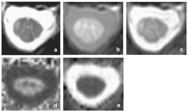
A first-ever spinal cord imaging meeting was sponsored by the International Spinal Research Trust and the Wings for Life Foundation with the aim of identifying the current state-of-the-art of spinal cord imaging, the current greatest challenges, and greatest needs for future development. This meeting was attended by a small group of invited experts spanning all aspects of spinal cord imaging from basic research to clinical practice. The greatest current challenges for spinal cord imaging were identified as arising from the imaging environment itself; difficult imaging environment created by the bone surrounding the spinal canal, physiological motion of the cord and adjacent tissues, and small crosssectional dimensions of the spinal cord, exacerbated by metallic implants often present in injured patients. Challenges were also identified as a result of a lack of "critical mass" of researchers taking on the development of spinal cord imaging, affecting both the rate of progress in the field, and the demand for equipment and software to manufacturers to produce the necessary tools. Here we define the current state-of-the-art of spinal cord imaging, discuss the underlying theory and challenges, and present the evidence for the current and potential power of these methods. In two review papers (part I and part II), we propose that the challenges can be overcome with advances in methods, improving availability and effectiveness of methods, and linking existing researchers to create the necessary scientific and clinical network to advance the rate of progress and impact of the research.

Keywords: Diffusion; Functional MRI; Magnetic resonance; Pathology; Spinal cord; Spinal cord injury.


Figures

Fig. 1
Fig. 1
Selective reconstruction of the corticospinal tracts with crossing in the medulla oblongata in a healthy subject. a) Coronal view of the non-diffusion weighted scan with the CSTs in red. b) axial view of the spinal cord FA, color coded with the direction of the principal eigenvector of the diffusion tensor, with the right and left CSTs in red and blue, coming out of the axial section and showing their crossing point.
Fig. 2
Fig. 2
(a–d) Sagittal T2, axial T2, DWI and 3D reconstruction of the spinal cord fibres in cervical spondylomyelopathy. Although one observes faint signal changes on sagittal T2 (a), 3D fiber tractography reconstruction (d) shows diffuse nerve fiber destruction, eventually explaining the clinical status of a patient.
Fig. 3
Fig. 3
(a–e) C6/C7 root compression due to herniated disk, and its effect on neural tissue integrity. DTI metrics allows us to observe increment in ADC and decrement in FA not only in the affected area but also in near proximity in patients with disk herniation in comparison with healthy subjects (mean ADC of healthy volunteer: 0.78 ± 0.0; mean FA of healthy volunteer: 61 ± 5).
Fig. 4
Fig. 4
Example of results obtained in a healthy female participant (top) and an age-matched participant with an incomplete (ASIA B) spinal cord injury (SCI) in the cervical level. Four dermatomes were stimulated, corresponding to the right and left C5 and C8 spinal cord segments, and results are shown in selected axial and sagittal slices showing the responses to right- and left-side stimulation of the little finger side of the palm (C8 dermatome). Results obtained in the healthy participant show activity in the ipsilateral dorsal horn of the spinal cord in the C8 segment, with a high degree of laterality corresponding to the side of the body being stimulated, as well as activity in the medulla of the brainstem. Results obtained in the participant with SCI show nearly normal responses on the left side of the spinal cord, and slightly altered responses on the right side, as well as corresponding activity in the medulla. In spite of an apparently extensive span of tissue damage in the cervical spinal cord, the neural activity detected is only slightly altered.
Fig. 5
Fig. 5
Spinal cord images acquired on a patient with multiple sclerosis. a) 3D Fast Field Echo (FFE) image acquired in the axial plane with the following parameters: 10 contiguous slices, FOV 240 × 180 mm2, TR 23 ms, TE 5 ms, flip angle α = 7°, number of averages = 8, voxel size = 0.5 × 0.5 × 5 mm3, total acquisition time = 13:34 min; b–c) Magnetization transfer imaging (b — MT pulse ‘ON’;c — MT pulse ‘OFF’) acquired with the following parameters: 3D slab-selective FFE sequence with two echoes (TR = 36 ms, TE1/TE2 = 3.5/5.9 ms, flip angle α = 9°), 22 axial slices, FOV = 240 × 180 mm2, voxel size 0.75 × 0.75 × 5 mm3 reconstructed to 0.5 × 0.5 × 5 mm3 to match the FFE scan resolution as in a), SENSE acceleration factor = 2, total acquisition time = 9 min. The MT pulse characteristics were: Sinc-Gaussian shaped MT saturating pulse of nominal α = 360º, offset frequency 1 kHz, duration 16 ms. d–e) Diffusion tensor imaging (DTI) derived maps (d — FA; e — MD). DW images were acquired axially with the following parameters: TE = 52 ms, TR = 12 RRs (cardiac gated), reduced FOV of 64 × 48 mm2, SENSE factor = 1.5, acquisition matrix 64 × 48, voxel size = 1 × 1 × 5 mm3. The DW imaging protocol consisted of 30 b = 1000 s mm−2 DWI volumes with gradient directions evenly distributed over the sphere and 3 non-DWI (b = 0) volumes.

References

    1. Agosta F, Filippi M. MRI of spinal cord in multiple sclerosis. J. Neuroimaging. 2007;17(Suppl. 1):46S–49S. - PubMed
    1. Agosta F, Benedetti B, Rocca MA, Valsasina P, Rovaris M, Comi G, Filippi M. Quantification of cervical cord pathology in primary progressive MS using diffusion tensor MRI. Neurology. 2005;64:631–635. - PubMed
    1. Agosta F, Absinta M, Sormani MP, Ghezzi A, Bertolotto A, Montanari E, Comi G, Filippi M. In vivo assessment of cervical cord damage in MS patients: a longitudinal diffusion tensor MRI study. Brain. 2007a;130:2211–2219. - PubMed
    1. Agosta F, Pagani E, Caputo D, Filippi M. Associations between cervical cord gray matter damage and disability in patients with multiple sclerosis. Arch. Neurol. 2007b;64:1302–1305. - PubMed
    1. Agosta F, Valsasina P, Caputo D, Stroman PW, Filippi M. Tactile-associated recruitment of the cervical cord is altered in patients with multiple sclerosis. NeuroImage. 2008a;39:1542–1548. - PubMed

MeSH terms