Review
. 2013 Oct;1829(10):1126-35.
doi: 10.1016/j.bbagrm.2013.07.003. Epub 2013 Jul 13.

Peptidylarginine deiminases in citrullination, gene regulation, health and pathogenesis

Affiliations
Review

Peptidylarginine deiminases in citrullination, gene regulation, health and pathogenesis

Shu Wang et al. Biochim Biophys Acta. 2013 Oct.

Abstract

Peptidylarginine deiminases are a family of enzymes that mediate post-translational modifications of protein arginine residues by deimination or demethylimination to produce citrulline. In vitro, the activity of PADs is dependent on calcium and reductive reagents carrying a free sulfhydryl group. The discovery that PAD4 can target both arginine and methyl-arginine for citrullination about 10years ago renewed our interest in studying this family of enzymes in gene regulation and their physiological functions. The deregulation of PADs is involved in the etiology of multiple human diseases, including cancers and autoimmune disorders. There is a growing effort to develop isoform specific PAD inhibitors for disease treatment. However, the regulation of the activity of PADs in vivo remains largely elusive, and we expect that much will be learned about the role of these enzymes in a normal life cycle and under pathology conditions.

Keywords: Autoimmunity; Cancer; Gene regulation; Histone; Peptidylarginine deiminase.


Figures

Fig 1
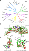
Fig 1. The PAD family of proteins and the effect of calcium on the PAD4 structure
(A) Phylogenetic analyses of 26 PAD proteins from several vertebrate species. Sequences of mammalian PADs were derived from databases for Human (Hs: Homo sapiens), mouse (Mm: Mus musculus), cow (Bt: Bos taurus), dog (Cf: Canis lupis familiaris). Also included are PADs from other species, such as chicken (Gg: Gallus gallus), trout (Om: Oncorhynchus mykiss) and frog (Xl: Xenopus laevis; Xt: Xenopus Silurana tropicalis). The unrooted phylogenetic tree was generated using ClustalW2 on EMBL-EBI website and visualized by MEGA 5.2. Scale bar represents 0.05 amino acid substitutions per site. Accession number for PADs in alignment are as follows: HsPAD1 (NP_037490.2), HsPAD2 (NP_031391.2), HsPAD3 (NP_057317.2), HsPAD4 (NP_036519.2), HsPAD6 (NP_997304.3), MmPAD1 (NP_035189.1), MmPAD2 (NP_032838.2), MmPAD3 (NP_035190.3), MmPAD4 (NP_035191.2), MmPAD6 (NP_694746.2), BtPAD1 (NP_001094742.1), BtPAD2 (NP_001098922.1), BtPAD3 (XP_003585850.1), BtPAD4 (NP_001179102.1), BtPAD6 (XP_002685843.1), CfPAD1 (XP_851932.1), CfPAD2 (XP_544539.2), CfPAD3 (XP_535391.2), CfPAD4 (XP_848494.1), CfPAD6 (NP_001091016), GgPAD1 (XP_425729.3), GgPAD2 (XP_425730.3), GgPAD3 (NP_990374), OmPAD1 (NP_001153973.1), XlPAD2 (NP_001080369), XtPAD2 (NP_001096490.1). (B) Ribbon presentation of superimposed structures of Ca2+ and substrate bound PAD4 (green, 1WDA) and free PAD4 (Red, 1WD8). Five Ca2+ ions bound to PAD4 are shown as purple spheres. Substrate BAA (benzoyl-L-arginine amide) is in a stick model. (C) Superimposed structure of Ca2+ and substrate bound PAD4 (green) and free PAD4 (red) in boxed region 1 (N-terminal amino acids 126–300). The arrow denotes the region with major conformational changes after calcium binding. (D) Superimposed structure of Ca2+ and substrate bound PAD4 (green) and free PAD4 (red) in boxed region 2 (C-terminal amino acids 301–420 and 624–663). Arrows denote the regions with major conformational changes.
Fig 2
Fig 2. A molecular switch operated on histone Arg and Lys residues of p53 target promoters for gene regulation
This molecular switch model predicts that p53 target genes can be activated through PAD4 and HDAC2 inhibition. (A) Before p53 target gene activation, PAD4 and HDAC2 function at the promoters to citrullinate Arg residues and deacetylate Lys residues on histone proteins to suppress gene expression. RNA Pol II is not recruited to the promoter or paused at the promoter under these conditions. (B) Upon activation signal such as DNA damage is sensed by p53, it switches its interaction partners from PAD4 and HDAC2 to PRMT1/4 and p300/CBP. These later two histonemodifying enzymes generate histone Arg methylation and Lys acetylation at target gene promoters, such as p21, GADD45 and PUMA to facilitate gene activation.
Fig 3
Fig 3. Cit-methyl and methyl-phos binary codes regulate the function of HP1
(A) The trimethylation of histone H3 Lys9 (K9me3) by the Suv39 histone methyltransferase leads to the binding of heterochromatin protein 1 (HP1) to regulate heterochromatin formation and/or to repress gene expression. (B) The citrullination of H3 Arg8 by PAD4 produces a Cit-methyl binary code to decrease the binding of HP1 to K9me3. (C) The phosphorylation of H3 Ser10 by aurora B or JIL-1 produces a methyl-phos binary code to decreases the binding of HP1 to K9me3.
Fig 4
Fig 4. An epigenetic view of tumorigenesis and cancer treatment
During the process of cancer initiation and progression, DNA methylation catalyzed by the DNA methyltransferases (DNMTs), histone deacetylation catalyzed by histone deacetylases (HDACs), and histone citrullination catalyzed by PAD4 work singularly or synergistically to epigenetically silence tumor suppressor genes thereby leading to untamed growth of cancer cells and tissues. Based on this theory, cancer treatment can be achieved with inhibitors targeting these epigenetic modifiers to restore histone modification patterns that are favorable for tumor suppressor gene expression thereby promoting cell cycle arrest and cell death.

Similar articles

Cited by

References

    1. Rogers GE. Occurrence of Citrulline in Proteins. Nature. 1962;194:1149–1151. - PubMed
    1. Rogers G, Taylor L. The enzymic derivation of citrulline residues from arginine residues in situ during the biosynthesis of hair proteins that are cross-linked by isopeptide bonds. 1977 - PubMed
    1. Vossenaar ER, Zendman AJW, van Venrooij WJ, Pruijn GJM. PADa, growing family of citrullinating enzymes: genes, features and involvement in disease. BioEssays. 2003;25:1106–1118. - PubMed
    1. Baka Z, Gyorgy B, Geher P, Buzas EI, Falus A, Nagy G. Citrullination under physiological and pathological conditions. Joint, bone, spine : revue du rhumatisme. 2012;79:431–436. - PubMed
    1. Horibata S, Coonrod SA, Cherrington BD. Role for peptidylarginine deiminase enzymes in disease and female reproduction. The Journal of reproduction and development. 2012;58:274–282. - PubMed

Publication types

MeSH terms