. 2013 Oct;33(10):1564-73.
doi: 10.1038/jcbfm.2013.109. Epub 2013 Jul 17.

TNF-α induces phenotypic modulation in cerebral vascular smooth muscle cells: implications for cerebral aneurysm pathology

Affiliations

TNF-α induces phenotypic modulation in cerebral vascular smooth muscle cells: implications for cerebral aneurysm pathology

Muhammad S Ali et al. J Cereb Blood Flow Metab. 2013 Oct.

Abstract

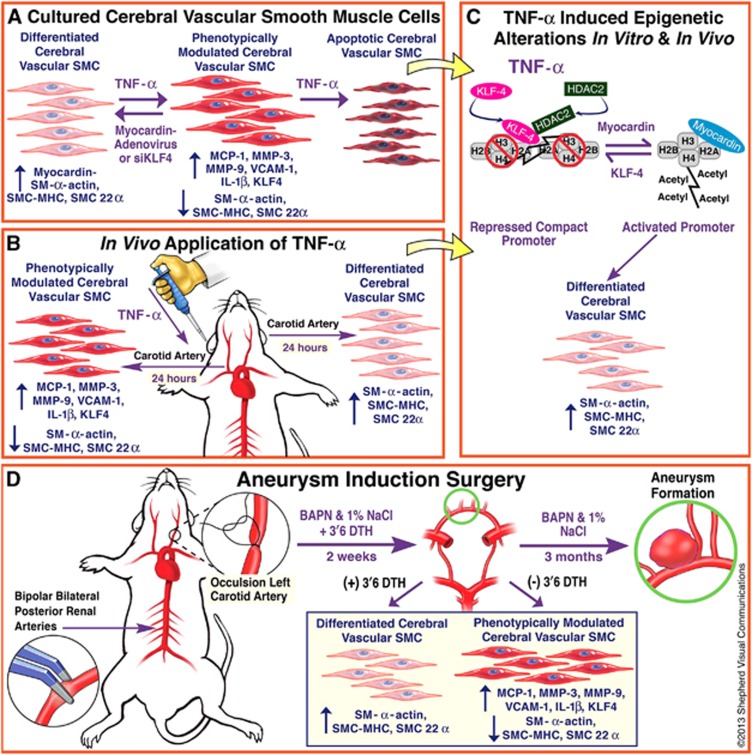
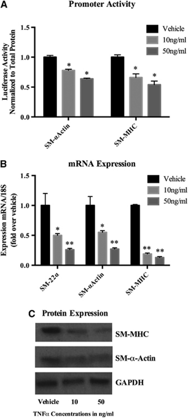
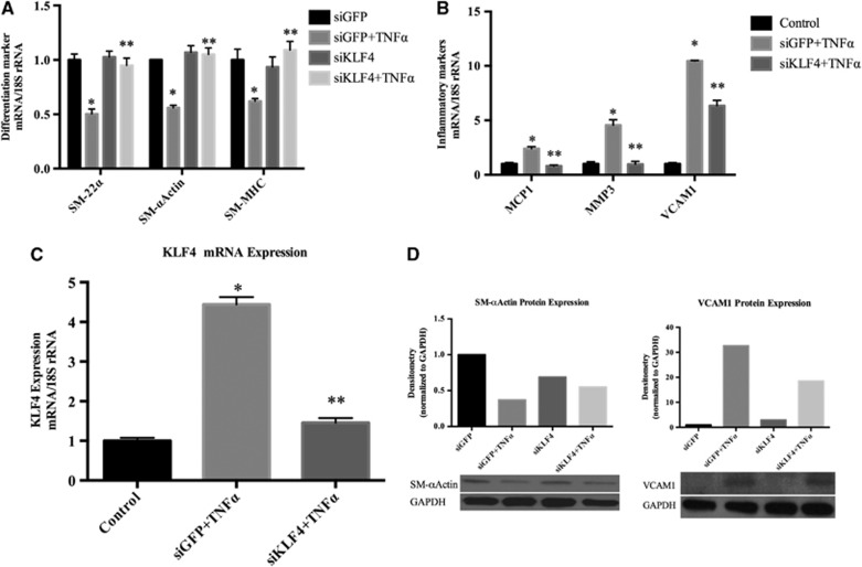
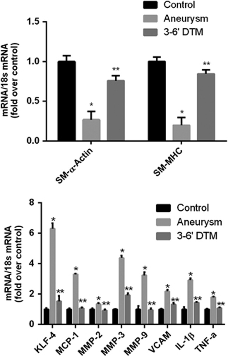
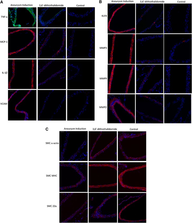
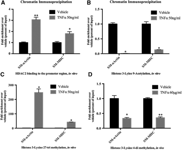
Little is known about vascular smooth muscle cell (SMC) phenotypic modulation in the cerebral circulation or pathogenesis of intracranial aneurysms. Tumor necrosis factor-alpha (TNF-α) has been associated with aneurysms, but potential mechanisms are unclear. Cultured rat cerebral SMCs overexpressing myocardin induced expression of key SMC contractile genes (SM-α-actin, SM-22α, smooth muscle myosin heavy chain), while dominant-negative cells suppressed expression. Tumor necrosis factor-alpha treatment inhibited this contractile phenotype and induced pro-inflammatory/matrix-remodeling genes (monocyte chemoattractant protein-1, matrix metalloproteinase-3, matrix metalloproteinase-9, vascular cell adhesion molecule-1, interleukin-1 beta). Tumor necrosis factor-alpha increased expression of KLF4, a known regulator of SMC differentiation. Kruppel-like transcription factor 4 (KLF4) small interfering RNA abrogated TNF-α activation of inflammatory genes and suppression of contractile genes. These mechanisms were confirmed in vivo after exposure of rat carotid arteries to TNF-α and early on in a model of cerebral aneurysm formation. Treatment with the synthesized TNF-α inhibitor 3,6-dithiothalidomide reversed pathologic vessel wall alterations after induced hypertension and hemodynamic stress. Chromatin immunoprecipitation assays in vivo and in vitro demonstrated that TNF-α promotes epigenetic changes through KLF4-dependent alterations in promoter regions of myocardin, SMCs, and inflammatory genes. In conclusion, TNF-α induces phenotypic modulation of cerebral SMCs through myocardin and KLF4-regulated pathways. These results demonstrate a novel role for TNF-α in promoting a pro-inflammatory/matrix-remodeling phenotype, which has important implications for the mechanisms behind intracranial aneurysm formation.


Figures

Figure 1
Figure 1
Time-line of experiments. (A) Cerebral blood vessels (circle of Willis) from rats were harvested for cerebral vascular smooth muscle cell (SMC) culture and treated with tumor necrosis factor-alpha (TNF-α) for quantitative polymerase chain reaction, western blot, evaluation of apoptosis, and assessment after adenovirus promoter transfection. (B) After experiments in vitro, experiments were carried out after application of pluronic gel containing TNF-α to the adventitial surface of rat carotid arteries to directly evaluate phenotypic modulation in vivo. (C) Additionally, chromatin immunoprecipitation assays were carried out to determine epigenetic alterations in vitro and in vivo. (D) Subsequently, the role of TNF-α and the TNF-α inhibitor 3,6′-dithiothalidomide was assessed early on in an established rodent cerebral aneurysm model induced by hypertension and hemodynamic stress. BAPN, β-aminopropionitrile; SMC-MHC, smooth muscle cell myosine heavy chain.
Figure 2
Figure 2
Tumor necrosis factor-alpha (TNF-α) suppressed promoter activity, messenger RNA (mRNA) levels, and protein expression of smooth muscle cell (SMC) marker genes. (A) Smooth muscle myosine heavy chain (SM-MHC)-luc and SM α-actin-luc promoter-luciferase constructs were transiently transfected into cerebral vascular SMCs for 48 hours and were treated with TNF-α for 24 hours. Luciferase activity was measured, normalized to total protein content, and then expressed as fold increase over vehicle. Values represent mean±s.e.m. *P<0.001 vs vehicle. (B) Cultured cerebral SMCs were treated for 24 hours with the indicated concentration of TNF-α or vehicle. Real-time polymerase chain reaction (RT–PCR) was performed, normalized to 18S ribosomal RNA (rRNA), and expressed as fold increase over vehicle. Values represent mean±s.e.m. *P<0.05; **P<0.005; ***P<0.0001 vs vehicle. (C) Cultured SMCs were starved for 72 hours and further treated with TNF-α with the indicated range of concentration for another 72 hours. Total protein lysate of SMCs (0.2 μg) were subjected to western blot analysis of SM-MHC and SM-α-actin protein expression. Glyceraldehyde 3-phosphate dehydrogenase (GAPDH) was used as a loading control.
Figure 3
Figure 3
Tumor necrosis factor-alpha (TNF-α)-induced messenger (mRNA) and protein expression of pro-inflammatory genes. (A) Cultured cerebral smooth muscle cells (SMCs) were treated with the indicated concentration of TNF-α for 24 hours. Real-time polymerase chain reaction was performed, normalized to 18S ribosomal RNA (rRNA), and expressed as fold increase over vehicle. Values represent mean±s.e.m. *P<0.001 vs vehicle. *P<0.0001 vs vehicle. (B) Cultured SMCs were starved for 72 hours and further treated with TNF-α with the indicated concentration for another 72 hours. Total protein lysate of SMCs (0.2 μg) were subjected to western blot analysis of monocyte chemoattractant protein-1 (MCP-1) and vascular cell adhesion molecule-1 (VCAM-1) protein expression. Glyceraldehyde 3-phosphate dehydrogenase (GAPDH) was used as a loading control.
Figure 4
Figure 4
Tumor necrosis factor-alpha (TNF-α) induced suppression of smooth muscle cell (SMC) marker genes and induction of pro-inflammatory genes was mediated by Kruppel-like transcription factor 4 (KLF4). (A-C) Cultured cerebral SMCs were transfected with KLF4 small interfering (siRNA), oligonucleotides (short interfering RNA specific to KLF4 (siKLF4)), or nonspecific control oligonucleotides (siGFP) with TNF-α (50 ng/mL) or vehicle treatment. Total RNA samples were isolated and differentiation marker genes (SM-22α, SM-α-actin and smooth muscle myosine heavy chain (SM-MHC)), inflammatory marker genes (MCP-1, VCAM-1, and MMP-3), and KLF4 messenger RNA (mRNA) expression were analyzed by real-time polymerase chain reaction. Values were normalized to 18S ribosomal RNA (rRNA) and represent mean±s.e.m. The experiment was repeated four times and the representative data are shown. (A) *P<0.05 vs vehicle; **P<0.005 vs siGFP. (B) *P<0.01 vs vehicle; **P<0.0001 vs siGFP. (C) *P<0.0001 vs vehicle; **P<0.0002 vs siGFP. (D) Three micrograms of total protein lysate of SMCs transfected with KLF4 small interfering RNA (siRNA) or green fluorescent protein (GFP) siRNA were subjected to western blot analysis using anti-SM-α- actin, anti-VCAM-1 (vascular cell adhesion molecule-1), and anti-GAPDH (glyceraldehyde 3-phosphate dehydrogenase) antibodies. The band intensity was quantified by densitometry. Relative band intensity was normalized to the band intensity of GAPDH and presented as fold increase over vehicle.
Figure 5
Figure 5
Aneurysm induction surgery is associated with a suppression of smooth muscle cell (SMC)-α-actin, smooth muscle cell myosine heavy chain (SM-MHC), and an increase in expression of TNF-α, Kruppel-like transcription factor 4 (KLF4), and pro-inflammatory/matrix-remodeling genes in vivo, which is reversed with treatment with the TNF-α synthesis inhibitor 3,6′-dithiothalidomide. Messenger RNA (mRNA) expression of anterior cerebral circulation 2 weeks after aneurysm induction surgery. Aneurysm induction surgeries were performed as detailed in Materials and Methods. Circle of Willis from animals undergoing aneurysm induction surgery and daily intraperitoneal injections of 3,6′-dithiothalidomide (N=6), aneurysm induction surgery and vehicle (N=6), or controls treated only with vehicle (N=6) were harvested after 14 days. RNA was extracted, real-time polymerase chain reaction was performed, and expression levels of SM-α-actin, SM-MHC, KLF4, TNF-α, and pro-inflammatory marker genes were normalized to 18S ribosomal RNA (rRNA) and represent fold increase over control. Messenger RNA (mRNA) expression of SM-α-actin, SM-MHC was decreased and expression of KLF4, TNF-α, and pro-inflammatory marker genes was increased after aneurysm induction surgery, as compared with controls. Treatment with daily intraperitoneal injections of 3,6′-dithiothalidomide significantly reversed the decreased expression of SM-α-actin and SM-MHC and the increase in expression of KLF4, TNF-α, and pro-inflammatory marker genes. Values represent mean±s.e.m. *P<0.001 vs control; **P<0.001 aneurysm induction versus 3,6′-dithiothalidomide. 3,6′DTM represents 3,6′-dithiothalidomide.
Figure 6
Figure 6
After aneurysm induction surgery, immunohistochemistry staining demonstrates decreased expression of SMC-α-actin and smooth muscle myosine heavy chain (SM-MHC) and increased expression of tumor necrosis factor-alpha (TNF-α,) Kruppel-like transcription factor 4 (KLF4), and pro-inflammatory/matrix-remodeling genes in vivo, which is reversed with treatment with the TNF-α synthesis inhibitor 3,6′-dithiothalidomide. Immunofluorescence staining of the Circle of Willis 2 weeks after aneurysm induction surgery (N=6) demonstrated increased expression of (A) TNF-α (green ), monocyte chemoattractant protein-1 (MCP-1) (red), interleukin-1 beta (IL-1β) (red ), vascular cell adhesion molecule-1 (VCAM) (red ), (B) KLF4 (red), matrix metalloproteinase (MMP-2) (red), MMP-3 (red ), MMP-9 (red ), and decreased expression of (C) SM-α-actin (red), SM-MHC (red) as compared with controls. Alterations in animals undergoing aneurysm induction surgery were reversed with daily intraperitoneal injections of 3,6′-dithiothalidomide (N=6). Expression of SM-22α (red) was decreased after aneurysm induction surgery, but changes in expression were not reversed with 3,6′-dithiothalidomide treatment. Nuclei (blue) are counter stained with 4',6-Diamidino-2-phenylindole (DAPI). 3,6′DTM represents 3,6′-dithiothalidomide. Scale bar represents 100 μm.
Figure 7
Figure 7
Tumor necrosis factor-alpha (TNF-α)-induced suppression of smooth muscle cell (SMC) marker genes was accompanied by recruitment of histone deacetylase 2 (HDAC2), promoter hypoacetylation, and changes in promoter methylation. (A) Cultured cerebral SMCs were treated with TNF-α (50 ng/mL) for 6 hours. Association of HDAC2 to the CArG-containing promoter region of SMC marker genes (SM-α-actin and smooth muscle myosine heavy chain (SM-MHC)) was determined with chromatin immunoprecipitation (ChIP) assay using anti-HDAC2 antibody. Values represent fold increase over vehicle and are expressed as mean±s.e.m. *P<0.01; **P<0.0007 vs vehicle. (B & C). Similar to the above, ChIP assays were performed with the following antibodies: anti-H3K9Ac and anti-H3K27triMe. Values represent fold increase over vehicle and represent mean±s.e.m. *P<0.0001 vs vehicle. (D): Pluronic gel containing TNF-α (60 μg/mL) was applied to the rat carotid arteries (n=12). Vessels were harvested and ChIP assay performed using anti-H3K4diMe. Values represent fold increase over vehicle and mean±s.e.m. *P<0.0001; **P<0.0005 vs vehicle. CArG, Cytosine Cytosine [Adenosine/Thymine]6 Guanine Guanine.

References

    1. Owens GK, Kumar MS, Wamhoff BR. Molecular regulation of vascular smooth muscle cell differentiation in development and disease. Physiol Rev. 2004;84:767–801. - PubMed
    1. Alexander MR, Owens GK. Epigenetic control of smooth muscle cell differentiation and phenotypic switching in vascular development and disease. Annu Rev Physiol. 2012;74:13–40. - PubMed
    1. Edvinsson LI, Povlsen GK. Vascular plasticity in cerebrovascular disorders. J Cereb Blood Flow Metab. 2011;31:1554–1571. - PMC - PubMed
    1. Yoshida T, Owens GK. Molecular determinants of vascular smooth muscle cell diversity. Circ Res. 2005;96:280–291. - PubMed
    1. Kilic T, Sohrabifar M, Kurtkaya O, Yildirim O, Elmaci I, Günel M, et al. Expression of structural proteins and angiogenic factors in normal arterial and unruptured and ruptured aneurysm walls Neurosurgery 200557997–1007.discussion 1997-1007. - PubMed

Publication types

MeSH terms