

Handheld shape discrimination hyperacuity test on a mobile device for remote monitoring of visual function in maculopathy
- PMID: 23860761
- PMCID: PMC3743459
- DOI: 10.1167/iovs.13-12037
Handheld shape discrimination hyperacuity test on a mobile device for remote monitoring of visual function in maculopathy
Abstract
Purpose: Frequency monitoring of age-related macular degeneration (AMD) and diabetic retinopathy (DR) is crucial for timely intervention. This study evaluated a handheld shape discrimination hyperacuity (hSDH) test iPhone app designed for visual function self-monitoring in patients with AMD and DR.
Methods: One hundred subjects (27 visually normal, 37 with AMD, and 36 with DR) were included based on clinical documentation and visual acuity of 20/100 or better. The hSDH test was implemented on the iOS platform. A cross-sectional study was conducted to compare the hSDH test with a previously established desktop SDH (dSDH) test and to assess the effect of disease severity on the hSDH test. A user survey was also conducted to assess the usability of the hSDH test on the mobile device.
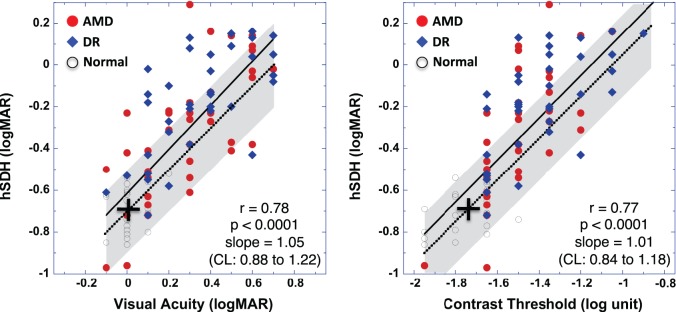
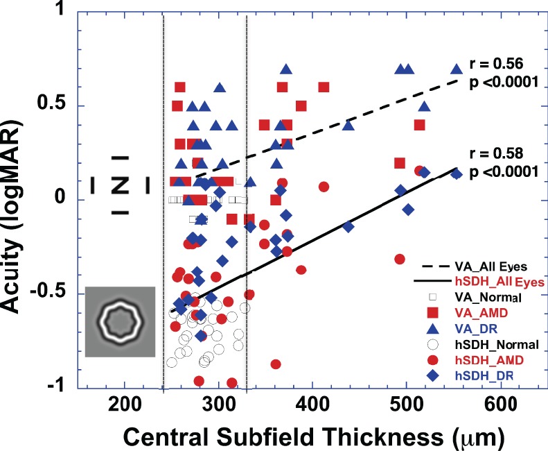
Results: The hSDH test and dSDH test were highly correlated (r = 0.88, P < 0.0001). Bland-Altman analysis indicated no significant difference in hSDH and dSDH measurements. One-way ANOVA indicated that the mean hSDH measurement of the eyes with advanced AMD (n = 16) or with severe to very severe nonproliferative DR (NPDR) (n = 12) was significantly worse than that of the eyes with intermediate AMD (n = 11) or with mild to moderate NPDR (n = 11) (P < 0.0001). Ninety-eight percent of 46 patients (10 with AMD and 36 with DR) who completed the usability survey reported that the hSDH test was easy to use.
Conclusions: This study demonstrated that the hSDH test on a mobile device is comparable to PC-based testing methods. As a mobile app, it is intuitive to use, readily accessible, and sensitive to the severity of maculopathy. It has the potential to provide patients having maculopathy with a new tool to monitor their vision at home.
Keywords: age-related macular degeneration; diabetic retinopathy; remote vision self-testing; shape discrimination; visual acuity.
Figures


References
-
- Brown DM, Kaiser PK, Michels M, et al. Ranibizumab versus verteporfin for neovascular age-related macular degeneration. N Engl J Med. 2006; 355: 1432–1444 - PubMed
-
- Rosenfeld PJ, Brown DM, Heier JS, et al. Ranibizumab for neovascular age-related macular degeneration. N Engl J Med. 2006; 355: 1419–1431 - PubMed
-
- Photocoagulation treatment of proliferative diabetic retinopathy: relationship of adverse treatment effects to retinopathy severity: Diabetic Retinopathy Study report No 5. Dev Ophthalmol. 1981; 2: 248–261 - PubMed
-
- Early Treatment Diabetic Retinopathy Study Research Group Early photocoagulation for diabetic retinopathy: ETDRS report number 9. Ophthalmology. 1991; 98: 766–785 - PubMed
Publication types
MeSH terms
Grants and funding
LinkOut - more resources
Full Text Sources
Other Literature Sources
Medical
Research Materials
