. 2013 Sep;41(17):8391-402.
doi: 10.1093/nar/gkt621. Epub 2013 Jul 16.

Dystrophin rescue by trans-splicing: a strategy for DMD genotypes not eligible for exon skipping approaches

Affiliations

Dystrophin rescue by trans-splicing: a strategy for DMD genotypes not eligible for exon skipping approaches

Stéphanie Lorain et al. Nucleic Acids Res. 2013 Sep.

Abstract

RNA-based therapeutic approaches using splice-switching oligonucleotides have been successfully applied to rescue dystrophin in Duchenne muscular dystrophy (DMD) preclinical models and are currently being evaluated in DMD patients. Although the modular structure of dystrophin protein tolerates internal deletions, many mutations that affect nondispensable domains of the protein require further strategies. Among these, trans-splicing technology is particularly attractive, as it allows the replacement of any mutated exon by its normal version as well as introducing missing exons or correcting duplication mutations. We have applied such a strategy in vitro by using cotransfection of pre-trans-splicing molecule (PTM) constructs along with a reporter minigene containing part of the dystrophin gene harboring the stop-codon mutation found in the mdx mouse model of DMD. Optimization of the different functional domains of the PTMs allowed achieving accurate and efficient trans-splicing of up to 30% of the transcript encoded by the cotransfected minigene. Optimized parameters included mRNA stabilization, choice of splice site sequence, inclusion of exon splice enhancers and artificial intronic sequence. Intramuscular delivery of adeno-associated virus vectors expressing PTMs allowed detectable levels of dystrophin in mdx and mdx4Cv, illustrating that a given PTM can be suitable for a variety of mutations.


Figures
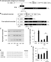
Figure 1.
Figure 1.
3′ replacement strategy by trans-splicing for dystrophin transcript repair. (A) Schematic representation of the dystrophin reporter minigene and PTM. The murine dystrophin premessenger consists of exons (boxes) and introns (lines with black balls illustrating the splice sites). The cross represents the nonsense mdx mutation in exon 23 (E23). The PTM is a transcript comprising a 150 nt AS complementary to intron 22 as well as a spacer, a strong conserved yeast BP, PPT, a 3′ acceptor site (3′SS, the three last elements are represented as blacks balls) and part of dystrophin wild-type coding sequence. (B) Expected dystrophin transcripts generated by cis- and trans-splicing. NIH3T3 cells were cotransfected with mdx dystrophin minigene and PTM expressing plasmids pSMD2-AS2-E23, pSMD2-AS2-E23-E59/70 and pSMD2-AS2-E23-E59/70opt. Arrows indicate the positions of forward E22-F and reverse E23-R, mdxE23-R and wtE23-R PCR primers in the cDNAs produced from those transcripts. (C) Detection of repaired dystrophin transcripts by trans-splicing. RT-PCR analysis was performed using PCR primers E22-F and E23-R (Total), mdxE23-R (mdx) and wtE23-R (wt) on NIH3T3 cells cotransfected with dystrophin minigene and pSMD2 (mdx), pSMD2-AS2-E23 (E23), pSMD2-AS2-E23-E59/70 (nonoptimized) and pSMD2-AS2-E23-E59/70opt (optimized). ‘wt’, transfection with the murine wild-type cDNA construct alone. ‘H2O’, PCR negative control. Representative results from three independent transfection experiments. (D) Quantification of dystrophin mRNA repair by trans-splicing. Absolute quantitative RT-PCR was used to evaluate the percentage of wild-type dystrophin transcripts on the total dystrophin transcripts in the experiment presented in (C). The data represent the mean values of three independent transfection experiments ± SD. *P ≤ 0.05, Student’s t-test. (E) Sequences of three different 3′ splice sites. Maximum entropy scores (MaxEnt) of the 3′ splice sites A, B and C of the PTMs (3′SSA, B and C) as well as the endogenous E23 3′SS were calculated with an algorithm developed by Burge and colleagues (http://genes.mit.edu/burgelab/maxent/Xmaxentscan_scoreseq_acc.html). (F) Quantification of dystrophin mRNA repair. NIH3T3 cells were cotransfected with mdx dystrophin minigene and PTM expressing plasmids pSMD2-AS2-3′SSA-E23-E59/70opt (3′SSA), pSMD2-AS2-3′SSB-E23-E59/70opt (3′SSB), pSMD2-AS2-3′SSC-E23-E59/70opt (3′SSC) and pSMD2-AS2-3′SSA-E23-E59/70opt-Intron (3′SSA Intron, pSMD2-AS2-3′SSA-E23-E59/70opt with an artificial intron inserted into the dystrophin cDNA). Absolute quantitative RT-PCR was used to evaluate the percentage of wild-type dystrophin transcripts in the total dystrophin transcripts. The data represent the mean values of three independent transfection experiments ± SD. *P ≤ 0.05, Student’s t-test.
Figure 2.
Figure 2.
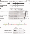
Dystrophin mRNA analysis after intramuscular injection of AAV1 expressing PTMs. (A) Expected dystrophin transcripts generated by cis- and trans-splicing. Arrows indicate E20ext-E26ext external primers and E20int and E26int internal primers used for nested PCR for detection of endogenous dystrophin transcripts (primers A/C) (10); E20ext and E59optext and E22-F and E59optint for trans-spliced dystrophin transcripts (primers A′/D); and E23-F and E59optext by single-round PCR for PTMs (primers B/D). The size in bp of expected amplicons is specified. (B) Detection of trans-spliced dystrophin transcripts in vivo. RT-PCR analysis was performed using PCR primers described in A on RNAs extracted from mdx and B6 muscles injected (+) or not (−) with AAV1-AS2-3′SSC-E23-E59/79opt (PTM), AAV1-ΔCMV-AS2-3′SSC-E23-E59/79opt (ΔCMV) and AAV1-Δlinker-E23-E59/79opt (Δlinker). AAV genomes were detected with primers B/D on genomic DNAs. ‘RT-’, PTM injected B6 sample without RT; ‘H2O’, PCR negative control. Analysis of three different injected TAs is presented. One of two representative experiments is shown. (C) Confirmation of in vivo trans-splicing events. An exact E22-E23 junction and wild-type E23 sequence were confirmed by sequencing of the A′/D amplicon obtained from injected mdx muscles. (D) Determination of the percentage of repaired dystrophin transcripts in vivo. Semiquantitative RT-PCR was used to amplify repaired dystrophin transcripts (upper panel) and total dystrophin transcripts (lower panel). Total dystrophin cDNAs were first amplified with E22-F/E23-R3 (15 cycles). The column-purified amplicons were used as matrix for a second PCR round (30 cycles) with E22-F/wtE23-R for repaired dystrophin transcripts (193 bp amplicon) and E22-F/E23-R for total dystrophin transcripts (308 bp amplicon). Band intensities from three PTM injected mdx TAs (+) (100 ng of RNAs) were compared with reference wild-type samples in which B6 RNAs were mixed to mdx RNAs (100, 10, 1 and 0% of B6 RNAs in 100 ng of total RNAs). ‘RT-’, PTM injected mdx sample without RT; ‘H2O’, PCR negative control. Analysis of three different injected TAs is presented. One of two representative experiments is shown.
Figure 3.
Figure 3.
Dystrophin mRNA analysis in mdx4Cv muscles after intramuscular injection of AAV1 expressing PTMs. (A) 3′ replacement strategy for mdx4Cv dystrophin transcript repair. Endogenous mdx4Cv dystrophin premessenger is represented with the nonsense mutation in E53 (black cross) as well as the PTM AS2-3′SSC-E23-E59/79opt targeting intron 22. (B) Detection of trans-spliced dystrophin transcripts in vivo. RT-PCR analysis was performed using PCR primers described in Figure 2A on RNAs extracted from mdx4Cv TA injected (+) or not (−) with AAV1-AS2-3′SSC-E23-E59/79opt. ‘RT-’, mdx4Cv injected sample without RT; ‘H2O’, PCR negative control. Analysis of two different injected TAs is presented. One of two representative experiments is shown. (C) Confirmation of in vivo trans-splicing events. An exact E22-E23 junction, wild-type E23 sequence and E23-E59opt junction were confirmed by sequencing of the A′/D amplicon obtained from injected mdx4Cv muscles.
Figure 4.
Figure 4.
Dystrophin rescue in mdx and mdx4Cv muscles after intramuscular injection of AAV1 expressing PTMs. Subsarcolemmal localization of the microdystrophin expressed from trans-spliced transcripts. Transversal sections from B6, mdx and mdx4Cv muscles injected or not with AAV1 expressing AS2-3′SSC-E23-pE59/79opt (+PTM) or Δlinker-E23-E59/79opt (+PTMΔlinker) were immunostained with MANEX1B monoclonal antibody recognizing the N-terminal moiety of the dystrophin.
Figure 5.
Figure 5.
Dystrophin mRNA analysis of human myoblasts transduced with lentivectors expressing PTMs. (A) 3′ replacement strategy for human dystrophin transcript repair. The endogenous human dystrophin premessenger is illustrated with E58 and E59 as white boxes and introns as black lines. The cross represents the HSK nonsense mutation in E71. PTMs comprise a 150 nt AS complementary to intron 58 as well as a spacer, a strong conserved yeast BP, a PPT, a 3′SS (the three last elements are represented as blacks balls) and the human dystrophin cDNA from E59 to E79 STOP codon. An EcoR1 restriction site (E1) was introduced in E60. Two PTM constructs were made with different ASs, AS1 and AS2. (B) Expected dystrophin transcripts generated by cis- and trans-splicing. Arrows indicate hE58ext-hE64ext external primers and hE58int and hE64int internal primers used for nested PCR for detection of total dystrophin transcripts (primers E/F), and E58ext and WPREext and hE58int and WPREint for trans-spliced dystrophin transcripts (primers E/H). The size in bp of expected amplicons is specified. (C) Detection of trans-spliced dystrophin transcripts. RT-PCR analysis using PCR primers E/F and E/H on total RNAs extracted from CHQ5B and HSK myotubes transduced or not (‘-’) with lentivectors expressing PTMs with AS1 and AS2. ‘H2O’, PCR negative control. Representative results from two independent transduction experiments. (D) Confirmation of trans-splicing events in human myotubes. An exact E58-E59 junction, the presence of the EcoR1 restriction site in E60 and wild-type E71 were confirmed by sequencing of the E/H amplicon obtained from transduced HSK myotubes.

References

    1. Pramono ZA, Takeshima Y, Alimsardjono H, Ishii A, Takeda S, Matsuo M. Induction of exon skipping of the dystrophin transcript in lymphoblastoid cells by transfecting an antisense oligodeoxynucleotide complementary to an exon recognition sequence. Biochem. Biophys. Res. Commun. 1996;226:445–449. - PubMed
    1. Wilton SD, Lloyd F, Carville K, Fletcher S, Honeyman K, Agrawal S, Kole R. Specific removal of the nonsense mutation from the mdx dystrophin mRNA using antisense oligonucleotides. Neuromuscul. Disord. 1999;9:330–338. - PubMed
    1. van Deutekom JC, Janson AA, Ginjaar IB, Frankhuizen WS, Aartsma-Rus A, Bremmer-Bout M, den Dunnen JT, Koop K, van der Kooi AJ, Goemans NM, et al. Local dystrophin restoration with antisense oligonucleotide PRO051. N. Engl. J. Med. 2007;357:2677–2686. - PubMed
    1. Kinali M, Arechavala-Gomeza V, Feng L, Cirak S, Hunt D, Adkin C, Guglieri M, Ashton E, Abbs S, Nihoyannopoulos P, et al. Local restoration of dystrophin expression with the morpholino oligomer AVI-4658 in Duchenne muscular dystrophy: a single-blind, placebo-controlled, dose-escalation, proof-of-concept study. Lancet Neurol. 2009;8:918–928. - PMC - PubMed
    1. Goemans NM, Tulinius M, van den Akker JT, Burm BE, Ekhart PF, Heuvelmans N, Holling T, Janson AA, Platenburg GJ, Sipkens JA, et al. Systemic administration of PRO051 in Duchenne's muscular dystrophy. N. Engl. J. Med. 2011;364:1513–1522. - PubMed

Publication types