

miR-26a and its target CKS2 modulate cell growth and tumorigenesis of papillary thyroid carcinoma
- PMID: 23861775
- PMCID: PMC3702500
- DOI: 10.1371/journal.pone.0067591
miR-26a and its target CKS2 modulate cell growth and tumorigenesis of papillary thyroid carcinoma
Retraction in
-
Retraction: miR-26a and its Target CKS2 Modulate Cell Growth and Tumorigenesis of Papillary Thyroid Carcinoma.PLoS One. 2021 Apr 13;16(4):e0250313. doi: 10.1371/journal.pone.0250313. eCollection 2021. PLoS One. 2021. PMID: 33848306 Free PMC article. No abstract available.
Abstract
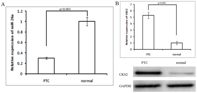
Background: While many studies have shown that levels of miR-26a are lower in papillary thyroid carcinoma (PTC), the role and mechanism of miR-26a in PTC are unclear.
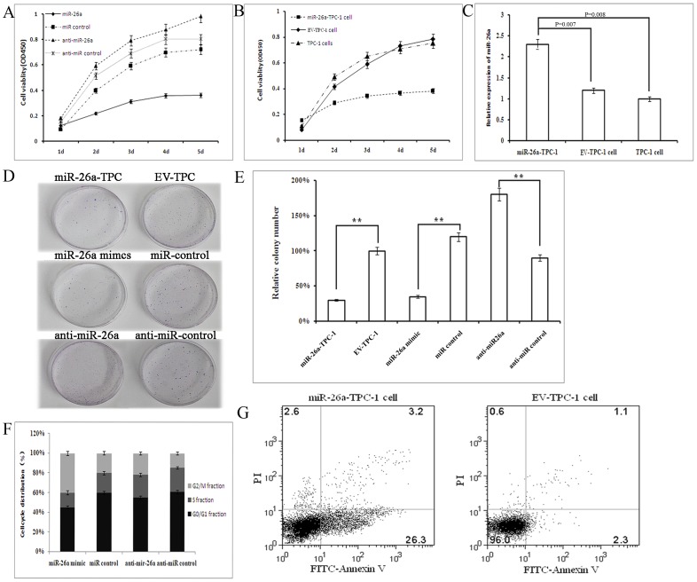
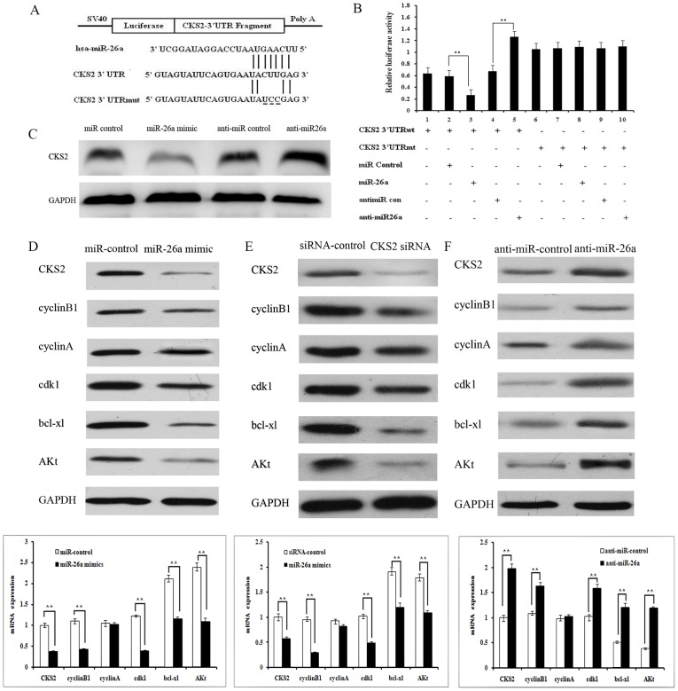
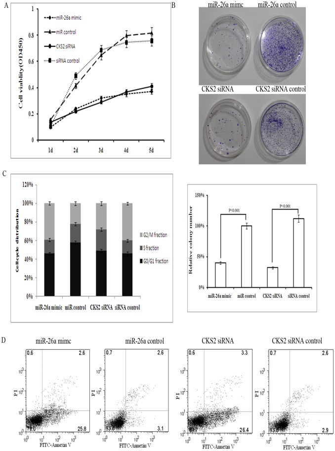
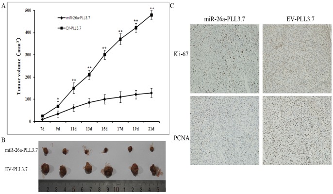
Method: We used database searches to select potential mRNA targets of miR-26a. Anti-miR-26a, miR-26a mimic, siRNA for CKS2 and their effects on cell growth, cell-cycle distribution and colony formation were evaluated. We also evaluate the over-expressed miR-26a in TPC-1 cells in severe combined immune-deficient mice. We used luciferase reporter assays, real-time PCR and western blot analysis to measure the expression and activity of miR-26a, CKS2, and related factors such as cyclin B1, cyclin A, cdk1, bcl-xl and Akt. Finally, we measured the relationship between the levels of miR-26a and CKS2 in PTC and normal thyroid tissues.
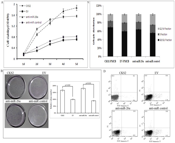
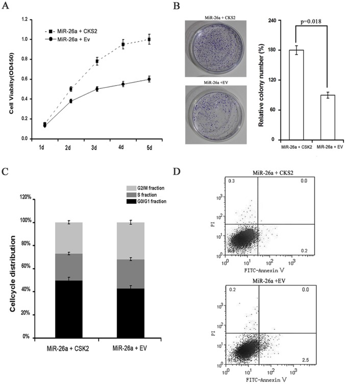
Results: Relative to normal thyroid tissues, miR-26a is consistently down-regulated in TPC specimens, and CKS2 was identified as a potential target. Up-regulated miR-26a expression or down-regulated CKS2 expression in TPC-1 and CGTH W3 cells lines caused G2 phase-arrest. Decreased miR-26a expression or increased CKS2 expression could have inverse function on PTC cell lines. CyclinB1, cyclinA, bcl-xl and AKt are indirectly regulated by miR-26a in a CKS2-dependent manner. Finally, CKS2 is overexpressed in PTC specimens relative to normal thyroid tissue, and a significant inverse correlation exists between miR-26a and CKS2 expression in clinical PTC specimens.
Conclusion: Our data indicate that miR-26a functions as a growth-suppressive miRNA in PTC, and that its suppressive effects are mediated mainly by repressing CKS2 expression.
Conflict of interest statement
Figures
Similar articles
-
MicroRNA-26a inhibits proliferation and tumorigenesis via targeting CKS2 in laryngeal squamous cell carcinoma.Clin Exp Pharmacol Physiol. 2018 May;45(5):444-451. doi: 10.1111/1440-1681.12890. Epub 2018 Jan 8. Clin Exp Pharmacol Physiol. 2018. PMID: 29143362
-
LINC00657/miR-26a-5p/CKS2 ceRNA network promotes the growth of esophageal cancer cells via the MDM2/p53/Bcl2/Bax pathway.Biosci Rep. 2020 Jun 26;40(6):BSR20200525. doi: 10.1042/BSR20200525. Biosci Rep. 2020. PMID: 32426838 Free PMC article.
-
MiR-34a targets GAS1 to promote cell proliferation and inhibit apoptosis in papillary thyroid carcinoma via PI3K/Akt/Bad pathway.Biochem Biophys Res Commun. 2013 Nov 29;441(4):958-63. doi: 10.1016/j.bbrc.2013.11.010. Epub 2013 Nov 9. Biochem Biophys Res Commun. 2013. PMID: 24220341
-
miR-451a is underexpressed and targets AKT/mTOR pathway in papillary thyroid carcinoma.Oncotarget. 2016 Mar 15;7(11):12731-47. doi: 10.18632/oncotarget.7262. Oncotarget. 2016. PMID: 26871295 Free PMC article. Review.
-
MicroRNA-146b: A Novel Biomarker and Therapeutic Target for Human Papillary Thyroid Cancer.Int J Mol Sci. 2017 Mar 15;18(3):636. doi: 10.3390/ijms18030636. Int J Mol Sci. 2017. PMID: 28294980 Free PMC article. Review.
Cited by
-
Up-regulated CKS2 promotes tumor progression and predicts a poor prognosis in human colorectal cancer.Am J Cancer Res. 2015 Aug 15;5(9):2708-18. eCollection 2015. Am J Cancer Res. 2015. PMID: 26609478 Free PMC article.
-
MiRNAs in Astrocyte-Derived Exosomes as Possible Mediators of Neuronal Plasticity.J Exp Neurosci. 2016 Aug 8;10(Suppl 1):1-9. doi: 10.4137/JEN.S39916. eCollection 2016. J Exp Neurosci. 2016. PMID: 27547038 Free PMC article.
-
High Expression of CKS2 Predicts Adverse Outcomes: A Potential Therapeutic Target for Glioma.Front Immunol. 2022 May 19;13:881453. doi: 10.3389/fimmu.2022.881453. eCollection 2022. Front Immunol. 2022. PMID: 35663965 Free PMC article.
-
Retraction: miR-26a and its Target CKS2 Modulate Cell Growth and Tumorigenesis of Papillary Thyroid Carcinoma.PLoS One. 2021 Apr 13;16(4):e0250313. doi: 10.1371/journal.pone.0250313. eCollection 2021. PLoS One. 2021. PMID: 33848306 Free PMC article. No abstract available.
-
The Cks1/Cks2 axis fine-tunes Mll1 expression and is crucial for MLL-rearranged leukaemia cell viability.Biochim Biophys Acta Mol Cell Res. 2018 Jan;1865(1):105-116. doi: 10.1016/j.bbamcr.2017.09.009. Epub 2017 Sep 20. Biochim Biophys Acta Mol Cell Res. 2018. PMID: 28939057 Free PMC article.
References
-
- Bartel DP, Chen CZ (2004) Micromanagers of gene expression: the potentially widespread influence of metazoan microRNAs. Nat Rev Genet 5: 396–400. - PubMed
-
- Schmittgen TD, Livak KJ (2008) Analyzing real-time PCR data by the comparative C(T) method. Nat Protoc 3: 1101–1108. - PubMed
-
- Pallante P, Visone R, Croce CM, Fusco A (2010) Deregulation of microRNA expression in follicular-cell-derived human thyroid carcinomas. Endocr Relat Cancer 17: F91–104. - PubMed
Publication types
MeSH terms
Substances
LinkOut - more resources
Full Text Sources
Other Literature Sources
Medical
Research Materials
Miscellaneous
