. 2013 Jul 5;8(7):e67976.
doi: 10.1371/journal.pone.0067976. Print 2013.

Measuring myeloperoxidase activity in biological samples

Affiliations

Measuring myeloperoxidase activity in biological samples

Benjamin Pulli et al. PLoS One. .

Abstract

Background: Enzymatic activity measurements of the highly oxidative enzyme myeloperoxidase (MPO), which is implicated in many diseases, are widely used in the literature, but often suffer from nonspecificity and lack of uniformity. Thus, validation and standardization are needed to establish a robust method that is highly specific, sensitive, and reproducible for assaying MPO activity in biological samples.

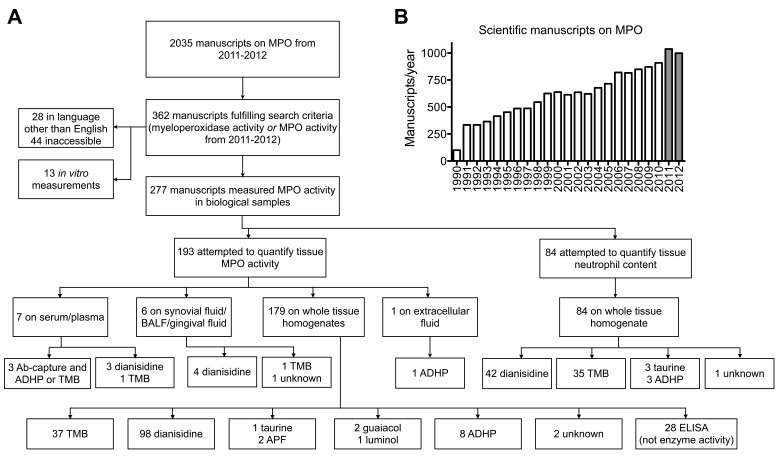
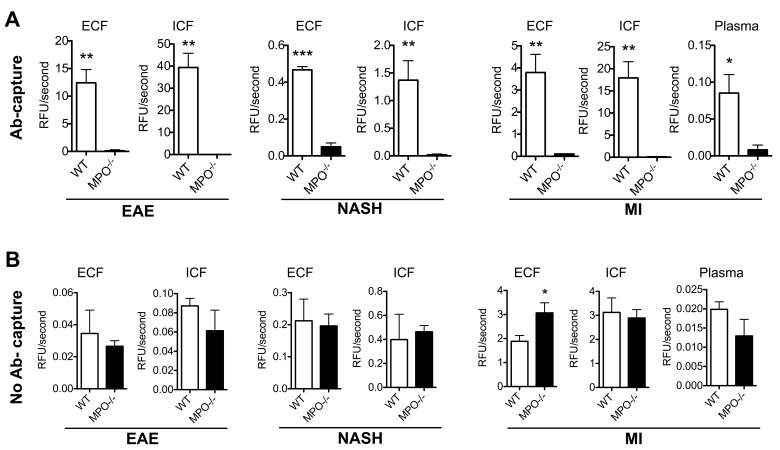
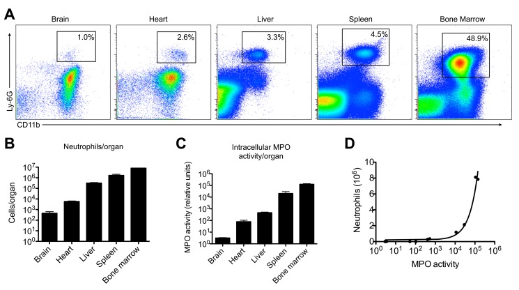
Principal findings: We found conflicting results between in vivo molecular MR imaging of MPO, which measures extracellular activity, and commonly used in vitro MPO activity assays. Thus, we established and validated a protocol to obtain extra- and intracellular MPO from murine organs. To validate the MPO activity assays, three different classes of MPO activity assays were used in spike and recovery experiments. However, these assay methods yielded inconsistent results, likely because of interfering substances and other peroxidases present in tissue extracts. To circumvent this, we first captured MPO with an antibody. The MPO activity of the resultant samples was assessed by ADHP and validated against samples from MPO-knockout mice in murine disease models of multiple sclerosis, steatohepatitis, and myocardial infarction. We found the measurements performed using this protocol to be highly specific and reproducible, and when performed using ADHP, to be highly sensitive over a broad range. In addition, we found that intracellular MPO activity correlated well with tissue neutrophil content, and can be used as a marker to assess neutrophil infiltration in the tissue.

Conclusion: We validated a highly specific and sensitive assay protocol that should be used as the standard method for all MPO activity assays in biological samples. We also established a method to obtain extra- and intracellular MPO from murine organs. Extracellular MPO activity gives an estimate of the oxidative stress in inflammatory diseases, while intracellular MPO activity correlates well with tissue neutrophil content. A detailed step-by-step protocol is provided.


Conflict of interest statement

Competing Interests: The authors have declared that no competing interests exist.

Figures

Figure 1
Figure 1. MPO in the literature.
(A) Usage of MPO activity assays in the Literature from 2011 to 2012. (B) Manuscripts published on MPO from 1990 to 2012; grey bars indicate manuscripts considered in (A). MPO = myeloperoxidase. TMB = 3,3′,5,5′-Tetramethylbenzidine. ADHP = 10-acetyl-3,7-dihydroxyphenoxazine. BALF = bronchoalveolar lavage fluid. Ab = antibody. APF = 3′-(p-aminophenyl) fluorescein. ELISA = enzyme-linked immunosorbent assay.
Figure 2
Figure 2. In vivo imaging and in vitro MPO activity assays demonstrate markedly different findings.
(A) MPO-Gd molecular MR imaging reveals MPO inhibition in vivo in mice with experimental autoimmune encephalomyelitis that were treated with ABAH. MPO activity maps are shown in 3D from two angles (left), as well as overlays of MPO activity maps over T1 images (right). (B) Quantification of imaging reveals significant difference in MPO activity in vivo (P = 0.03, n = 8 per group). (C) In vitro assays on whole tissue homogenates using ADHP or TMB do not confirm the in vivo imaging finding (P = 0.68 and 0.88, respectively, n = 4 per group). *: P<0.05, n.s. = not statistically significant. MPO = myeloperoxidase. TMB = 3,3′,5,5′-Tetramethylbenzidine. ADHP = 10-acetyl-3,7-dihydroxyphenoxazine. ABAH = 4-aminobenzoic acid hydrazide. Activation ratio = contrast-to-noise ratio 60 minutes over 15 minutes post MPO-Gd injection.
Figure 3
Figure 3. Validation of Extracellular Protein Isolation and MPO Protein Precipitation.
(A) LDH assay of intra- and extracellular protein fractions of different organs shows that the extracellular fraction only contains very low levels of LDH activity, while the intracellular fraction contains the majority of the LDH activity (left). LDH ratio shows a 90 or higher fold level of ICF LDH over ECF LDH activity (right). (B) Protein precipitation of MPO with acetone has no effect on its activity, as evaluated with ADHP (n = 2 per group). LDH = lactate dehydrogenase. BCA = bicinchoninic acid. MPO = myeloperoxidase.
Figure 4
Figure 4. Spike and recovery assay: tissue homogenates and extracellular fluid contain interfering substances.
(A) Extracellular protein fraction from different organs contains substances interfering with ADHP, luminol, and APF assays (n = 2 per group). (B) Intracellular protein fractions also contain interfering substances (n = 2 per group). MPO = myeloperoxidase. ADHP = 10-acetyl-3,7-dihydroxyphenoxazine. APF = 3′-(p-aminophenyl) fluorescein. HPF = 3′-(p-hydroxyphenyl) fluorescein.
Figure 5
Figure 5. Antibody capture improves the specificity of MPO activity assays on extra- and intracellular extracts in various models of inflammatory diseases.
(A) Antibody capture of MPO followed by activity detection with ADHP reveals high specificity towards MPO. This is shown in extra- and intracellular fractions in brains from EAE mice, livers from mice with NASH, and hearts and plasma from mice with myocardial infarction (n = 3 per group). (B) The same samples processed without antibody capture reveal poor specificity towards MPO, and no significant difference between WT and MPO-KO mice (n = 3 per group). * P<0.05. ** P<0.01. *** P<0.001. ADHP = 10-acetyl-3,7-dihydroxyphenoxazine. MPO = myeloperoxidase. EAE = experimental autoimmune encephalomyelitis. MI = myocardial infarction. NASH = non-alcoholic steatohepatitis. ECF = extracellular fraction. ICF = intracellular fraction. WT = wildtype C57BL/6. MPO−/− = MPO knockout.
Figure 6
Figure 6. Intracellular MPO activity correlates well with tissue neutrophil content.
(A) Flow cytometry demonstrates different neutrophil counts in brain, heart, liver, spleen, and bone marrow, as quantified in (B) (n = 2 per group). (C) Intracellular MPO activity was measured with the antibody-capture assay using ADHP, and shows a similar trend to neutrophil content per organ (n = 2 per group). (D) A close correlation was found between neutrophil content and intracellular MPO activity in these organs. MPO = myeloperoxidase. ADHP = 10-acetyl-3,7-dihydroxyphenoxazine.

References

    1. Schultz J, Kaminker K (1962) Myeloperoxidase of the leucocyte of normal human blood. I. Content and localization. Arch Biochem Biophys 96: 465–467. - PubMed
    1. Heinecke JW (1999) Mechanisms of oxidative damage by myeloperoxidase in atherosclerosis and other inflammatory disorders. J Lab Clin Med 133: 321–325. - PubMed
    1. Zhang R, Brennan ML, Fu X, Aviles RJ, Pearce GL, et al. (2001) Association between myeloperoxidase levels and risk of coronary artery disease. JAMA 286: 2136–2142. - PubMed
    1. Nahrendorf M, Sosnovik D, Chen JW, Panizzi P, Figueiredo JL, et al. (2008) Activatable magnetic resonance imaging agent reports myeloperoxidase activity in healing infarcts and noninvasively detects the antiinflammatory effects of atorvastatin on ischemia-reperfusion injury. Circulation 117: 1153–1160. - PMC - PubMed
    1. Brennan ML, Penn MS, Van Lente F, Nambi V, Shishehbor MH, et al. (2003) Prognostic value of myeloperoxidase in patients with chest pain. N Engl J Med 349: 1595–1604. - PubMed

Publication types