. 2013 Jul 5;8(7):e68328.
doi: 10.1371/journal.pone.0068328. Print 2013.

Sphingosine kinase activity is not required for tumor cell viability

Affiliations

Sphingosine kinase activity is not required for tumor cell viability

Karen Rex et al. PLoS One. .

Abstract

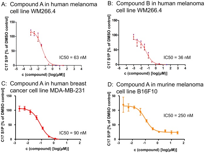
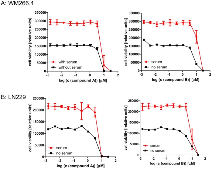
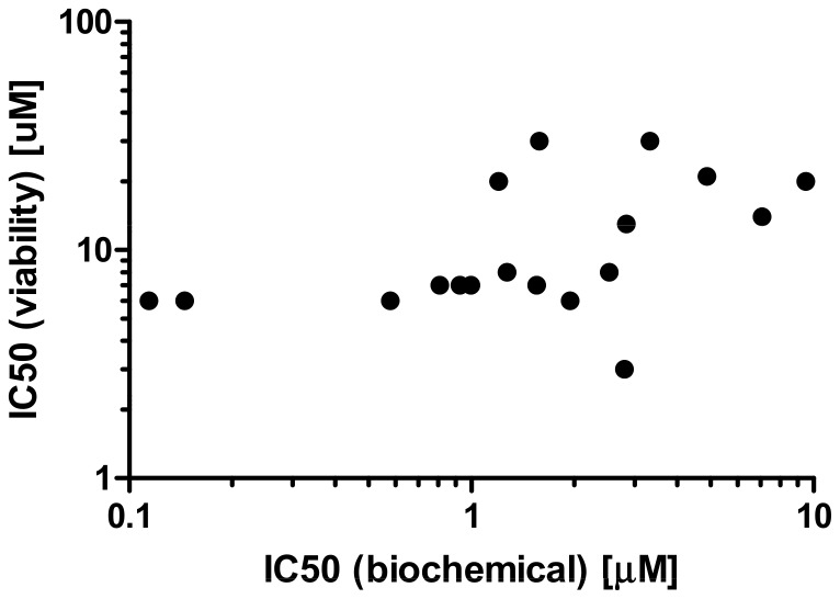
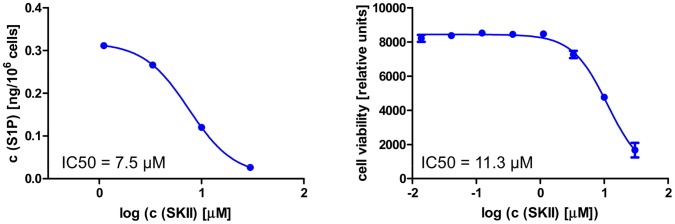
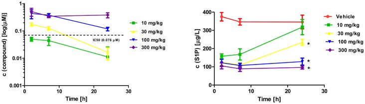
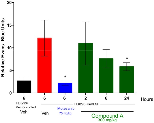
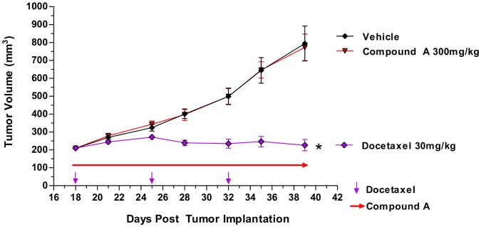
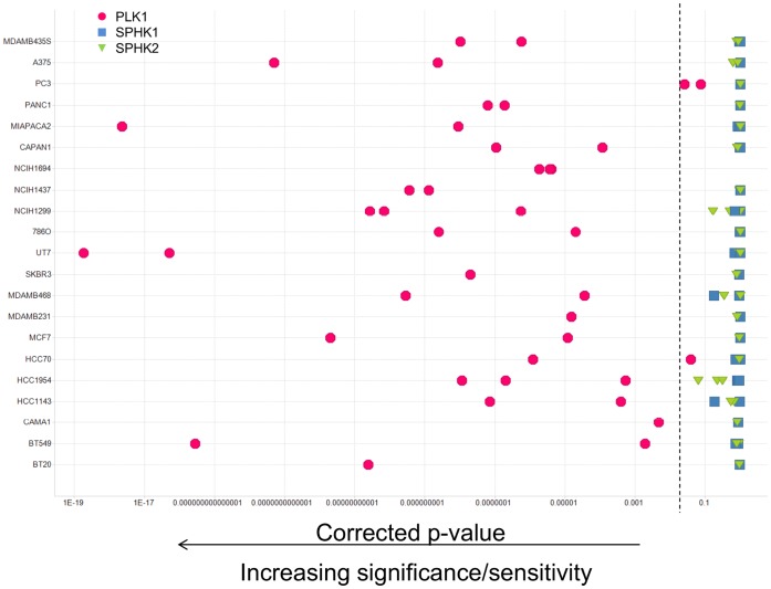
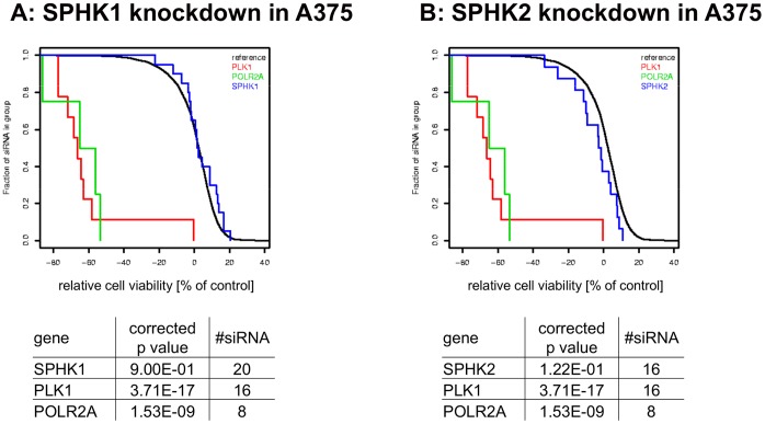
Sphingosine kinases (SPHKs) are enzymes that phosphorylate the lipid sphingosine, leading to the formation of sphingosine-1-phosphate (S1P). In addition to the well established role of extracellular S1P as a mitogen and potent chemoattractant, SPHK activity has been postulated to be an important intracellular regulator of apoptosis. According to the proposed rheostat theory, SPHK activity shifts the intracellular balance from the pro-apoptotic sphingolipids ceramide and sphingosine to the mitogenic S1P, thereby determining the susceptibility of a cell to apoptotic stress. Despite numerous publications with supporting evidence, a clear experimental confirmation of the impact of this mechanism on tumor cell viability in vitro and in vivo has been hampered by the lack of suitable tool reagents. Utilizing a structure based design approach, we developed potent and specific SPHK1/2 inhibitors. These compounds completely inhibited intracellular S1P production in human cells and attenuated vascular permeability in mice, but did not lead to reduced tumor cell growth in vitro or in vivo. In addition, siRNA experiments targeting either SPHK1 or SPHK2 in a large panel of cell lines failed to demonstrate any statistically significant effects on cell viability. These results show that the SPHK rheostat does not play a major role in tumor cell viability, and that SPHKs might not be attractive targets for pharmacological intervention in the area of oncology.


Conflict of interest statement

Competing Interests: All authors have an affiliation to the commercial funder of this research (Amgen Inc.). This does not alter the authors' adherence to all the PLOS ONE policies on sharing data and materials.

Figures

Figure 1
Figure 1. Structure of Compounds A and B.
Figure 2
Figure 2. Inhibition of cellular SPHK activity in tumor cell lines.
Two hours after addition of C17 sphingosine, cells were lysed and levels of C17 S1P were determined.
Figure 3
Figure 3. Effects of SPHK inhibition on cell viability.
The human melanoma cell line WM266.4 (panel A) and the human glioblastoma cell line LN229 (panel B) were treated for 72 h with the indicated concentrations of compound A (left panel) and compound B (right panel). Viability was assessed after 72 h.
Figure 4
Figure 4. Correlation between SPHK inhibition and cell viability.
A panel of 18 compounds structurally related to compounds A and B was tested in biochemical hSPHK1 assays (inflection point IC50s plotted on x-axis) and 72 h viability assays in WM266.4 cells (inflection point IC50s plotted on y-axis).
Figure 5
Figure 5. Effects of SPHK inhibition in colony formation assays.
LN229 cells were seeded at a density of 800 cells/well in a six well plate in the presence of the indicated concentrations of SPHK inhibitor. After 15 days, cells were fixed and stained with crystal violet.
Figure 6
Figure 6. Cellular activity of literature compound SKII.
The left panels shows levels of endogenous S1P 24 hours after compound treatment, the right panel depicts the result of 72 h viability assays performed in parallel in the human melanoma cell line LOX. The IC50s reflect the inflection point of the titration curve.
Figure 7
Figure 7. Phamacokinetic and pharmacodynamic properties of compound A in vivo.
Compound A was administered to athymic nude mice by oral gavage. At the indicated time points, blood was collected and plasma levels of compound A (left panel) as well as S1P (right panel) were determined. Compound levels were corrected for binding to murine plasma, and concentrations of free compound A is depicted in this graph. *P<0.05 compared to vehicle.
Figure 8
Figure 8. Inhibition of VEGF-induced vascular permeability in mice treated with Compound A.
HEK 293 cells transfected with murine VEGF or vector were mixed with Matrigel and injected s.c. into female C57Bl/6 mice. A single dose of compound A was given by oral gavage 24 hours after implantation of cells. At various time points after administration, vascular permeability in the skin overlying the Matrigel plug was measured by quantifying the extravasation of Evans blue dye. Columns, relative Evans blue units (n = 5) per group; bars, SE *, P<0.0001, significant difference from VEGF plus vehicle-injected control mice.
Figure 9
Figure 9. Effect of Compound A on the growth of MDA-MB-231 xenograft tumors.
Female athymic nude mice were injected with 5×10⁶ MDA-MB-231 cells on day 0. Treatment with compound A (300 mg/kg/dose QD) or Taxotere (30 mg/kg, once per week) was initiated on day 18 when tumors reached ∼ 200 mm3. (n = 7–10 per group); bars, SE. *, P<0.0001, compared to vehicle.
Figure 10
Figure 10. Reduction of SPHK expression by RNAi in a panel of cancer cell lines.
Multiple cancer cell lines were transfected in a high-throughput format with libraries of siRNA containing multiple triggers for each gene, and cell viability was determined 96 or 120 hours after transfection. The statistical significance of the observed effects was calculated (see materials and methods) and expressed as a p value. Polo like kinase 1 (PLK1) served as a positive control. Each symbol represents the result of one siRNA screen. Most cell lines were tested several times using different transfection conditions. Symbols to the left of the dashed line (p<0.05) indicate a statistically significant effect of gene knockdown on cell viability in a given experiment.
Figure 11
Figure 11. siRNA experiments in A375 cells.
SPHK1 (panel A) and SPHK2 (panel B) as well as the cytotoxic controls PLK1 and POLR2A were targeted with numerous siRNAs in the melanoma cell line A375. Each vertical line represents the effects of an individual siRNA transfection on relative cell viability, with negative values representing cell killing. Statistical significance was calculated as described in Materials and Methods.

References

    1. Maceyka M, Harikumar KB, Milstien S, Spiegel S (2012) Sphingosine-1-phosphate signaling and its role in disease. Trends Cell Biol 22: 50–60. - PMC - PubMed
    1. Orr Gandy KA, Obeid LM (2012) Targeting the sphingosine kinase/sphingosine 1-phosphate pathway in disease: Review of sphingosine kinase inhibitors. Biochim Biophys Acta. - PMC - PubMed
    1. Mizugishi K, Yamashita T, Olivera A, Miller GF, Spiegel S, et al. (2005) Essential role for sphingosine kinases in neural and vascular development. Mol Cell Biol 25: 11113–11121. - PMC - PubMed
    1. Brinkmann V, Billich A, Baumruker T, Heining P, Schmouder R, et al. (2010) Fingolimod (FTY720): discovery and development of an oral drug to treat multiple sclerosis. Nat Rev Drug Discov 9: 883–897. - PubMed
    1. English D, Kovala AT, Welch Z, Harvey KA, Siddiqui RA, et al. (1999) Induction of endothelial cell chemotaxis by sphingosine 1-phosphate and stabilization of endothelial monolayer barrier function by lysophosphatidic acid, potential mediators of hematopoietic angiogenesis. J Hematother Stem Cell Res 8: 627–634. - PubMed

MeSH terms

Substances